. 2020 Jun 9;4(2):026108.
doi: 10.1063/1.5140760. eCollection 2020 Jun.

An organosynthetic soft robotic respiratory simulator

An organosynthetic soft robotic respiratory simulator

Markus A Horvath et al. APL Bioeng. .

Abstract

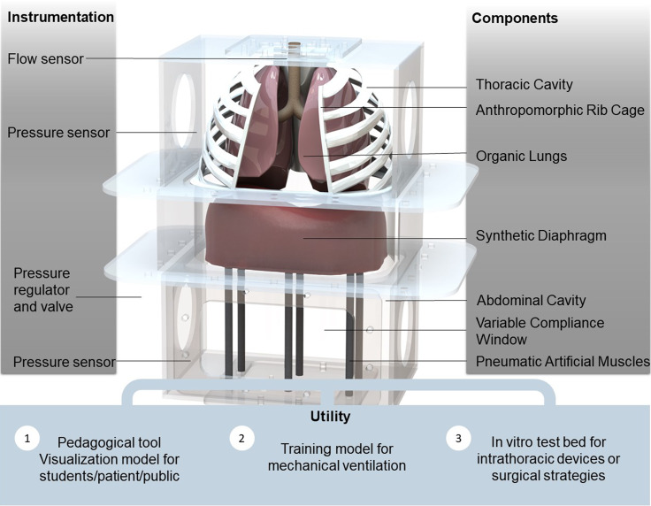
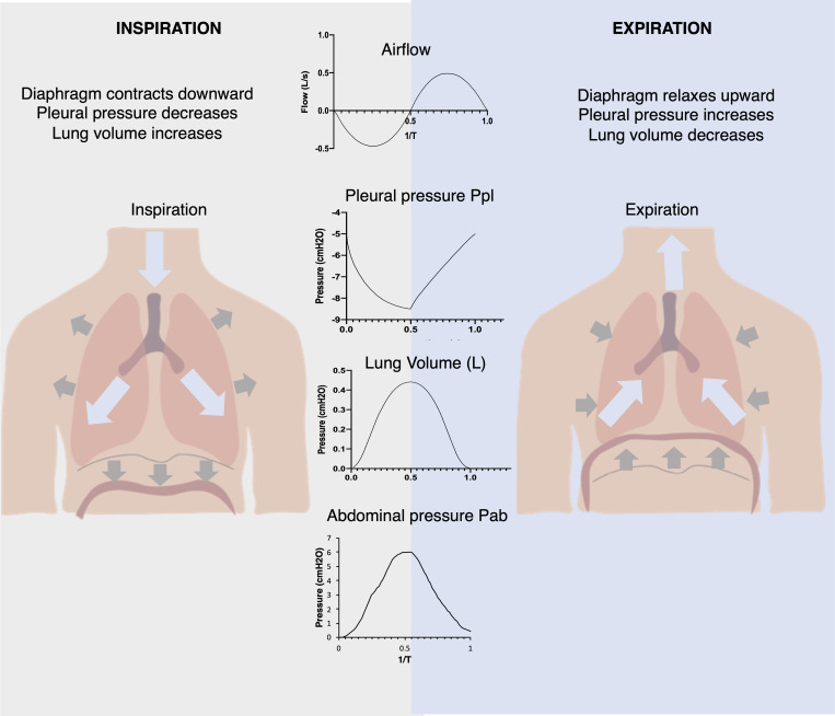
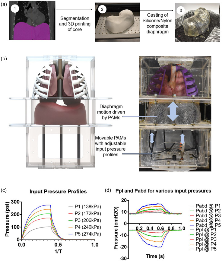
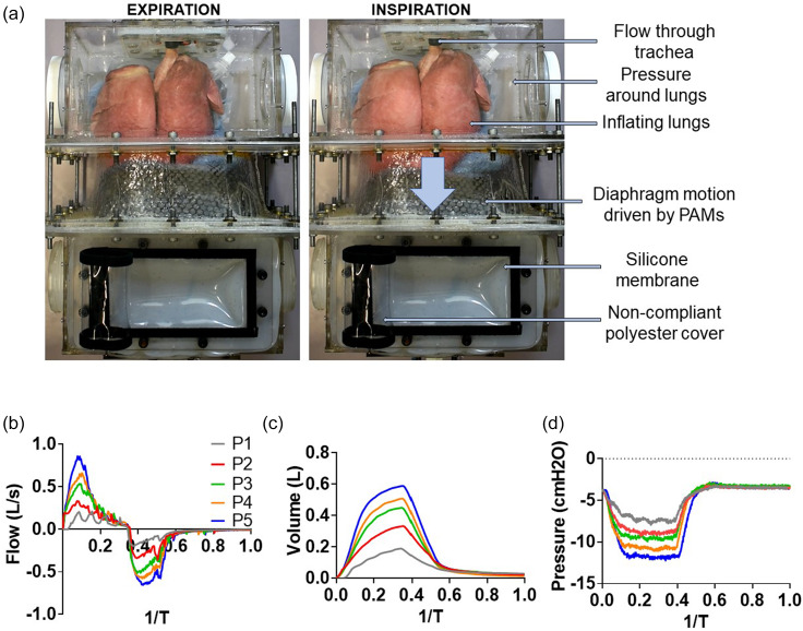
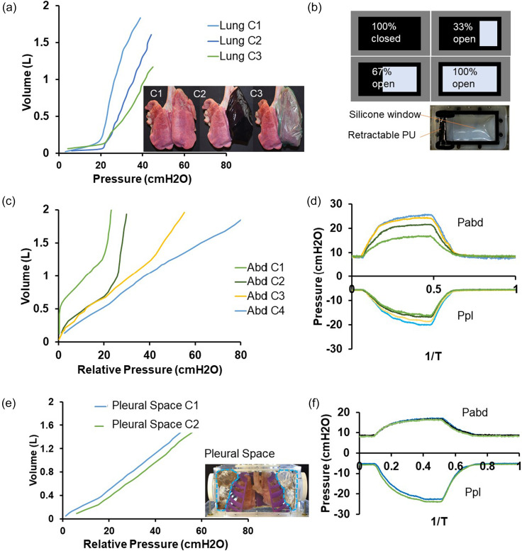
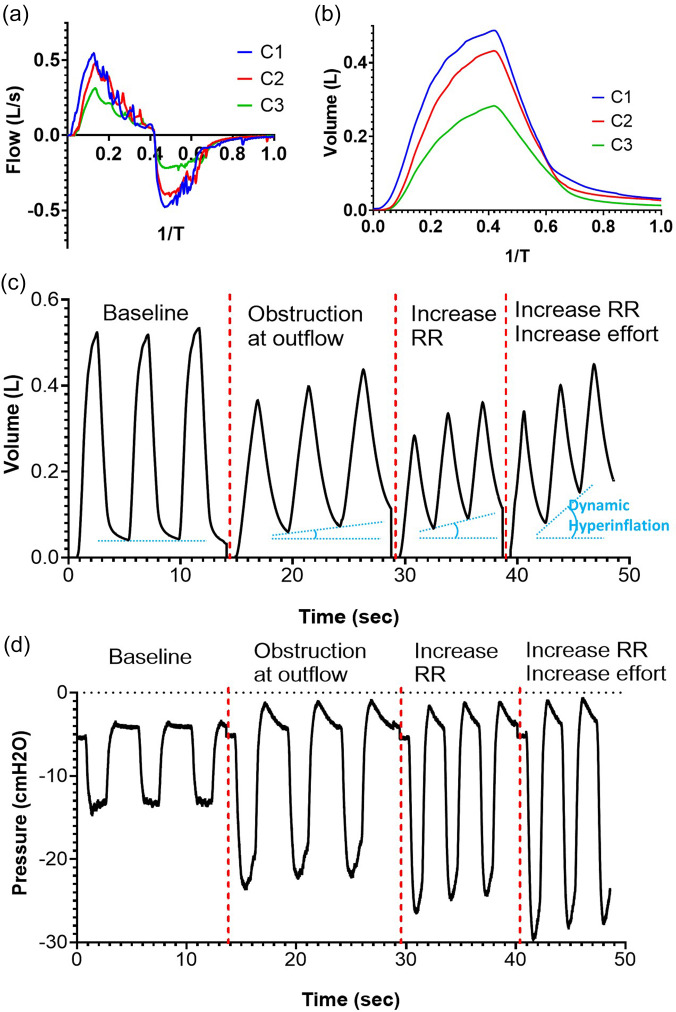
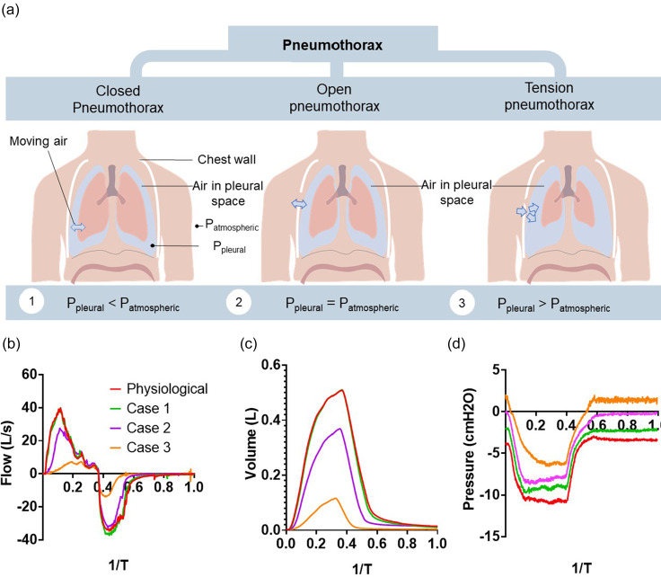
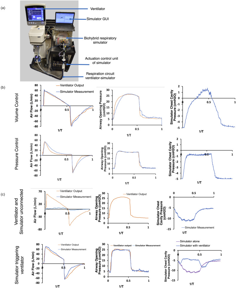
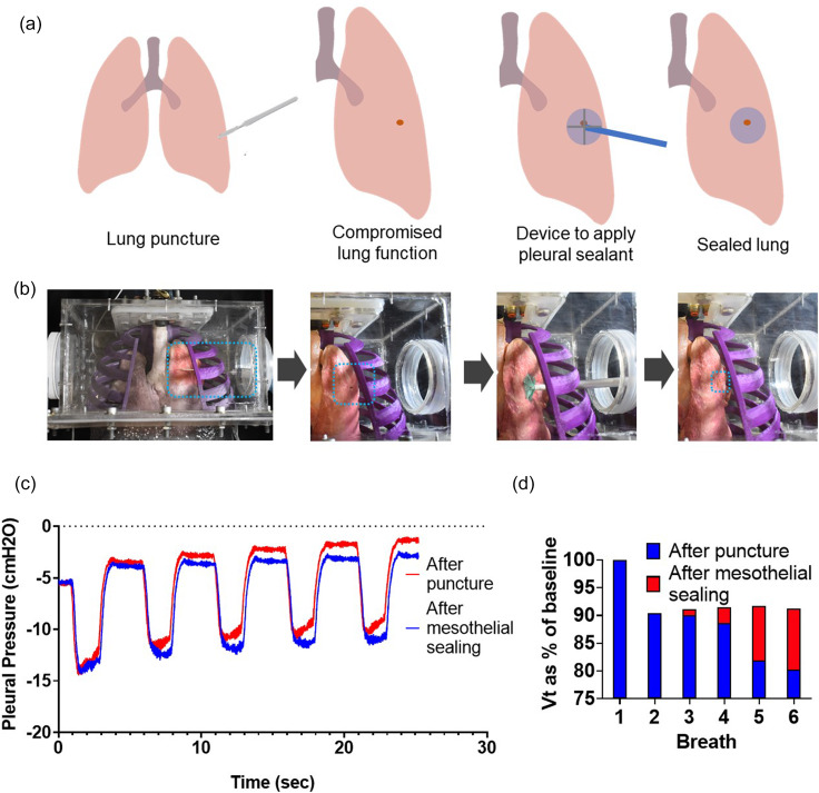
In this work, we describe a benchtop model that recreates the motion and function of the diaphragm using a combination of advanced robotic and organic tissue. First, we build a high-fidelity anthropomorphic model of the diaphragm using thermoplastic and elastomeric material based on clinical imaging data. We then attach pneumatic artificial muscles to this elastomeric diaphragm, pre-programmed to move in a clinically relevant manner when pressurized. By inserting this diaphragm as the divider between two chambers in a benchtop model-one representing the thorax and the other the abdomen-and subsequently activating the diaphragm, we can recreate the pressure changes that cause lungs to inflate and deflate during regular breathing. Insertion of organic lungs in the thoracic cavity demonstrates this inflation and deflation in response to the pressures generated by our robotic diaphragm. By tailoring the input pressures and timing, we can represent different breathing motions and disease states. We instrument the model with multiple sensors to measure pressures, volumes, and flows and display these data in real-time, allowing the user to vary inputs such as the breathing rate and compliance of various components, and so they can observe and measure the downstream effect of changing these parameters. In this way, the model elucidates fundamental physiological concepts and can demonstrate pathology and the interplay of components of the respiratory system. This model will serve as an innovative and effective pedagogical tool for educating students on respiratory physiology and pathology in a user-controlled, interactive manner. It will also serve as an anatomically and physiologically accurate testbed for devices or pleural sealants that reside in the thoracic cavity, representing a vast improvement over existing models and ultimately reducing the requirement for testing these technologies in animal models. Finally, it will act as an impactful visualization tool for educating and engaging the broader community.


Figures

FIG. 1.
FIG. 1.
A computational rendering of our biohybrid simulator focused on replicating respiratory mechanics. By tracking pressures and flows via included pressure sensors, the simulator may be utilized for a variety of education and training purposes.
FIG. 2.
FIG. 2.
Schematic of how diaphragm displacement affects the pressures in the respiratory system driving airflow generating changes in the volume of the lungs, providing for the gas exchange of inspiration and expiration.
FIG. 3.
FIG. 3.
(a) Physical diaphragm insert from 3D data derived from clinical imaging using Mimics software. (b) Rendering and physical realization of the biohybrid respiratory simulator. (c) Pneumatic artificial muscles with different input pressures generate different degrees of diaphragm displacement. (D) Measured outputs of abdominal and pleural pressure in response to varying actuator pressures.
FIG. 4.
FIG. 4.
Inclusion of organic lungs to visualize respiration and measure airflow. To visualize lung motion, the rib cage was removed. (a) When the artificial muscles are not contracted, the diaphragm is in its resting state. With downward displacement of the diaphragm, inspiration and lung expansion are observed. Spirometry readings replicate physiologic waveforms for flow (b), volume (c), and pleural pressure (d). Multimedia view: http://dx.doi.org/10.1063/1.5140760.1
FIG. 5.
FIG. 5.
Modifying the compliance of different elements. (a) Varying lung compliance. (b) Schematic of tunable compliance of the abdominal cavity via the silicone window. (c) Varying abdominal cavity compliance. (d) Effect of variable abdominal cavity compliance on abdominal and pleural pressures during 1 cycle of respiration. (e) Varying pleural cavity compliance. (f) Effect of variable pleural cavity compliance on abdominal and pleural pressures during 1 cycle of respiration.
FIG. 6.
FIG. 6.
Simulating restrictive and obstructive lung disease. (a) Decreased flow to the lungs during a breathing cycle in modeling restrictive lung disease. (b) Reduced tidal volumes during a breathing cycle in modeling restrictive lung disease. (c) Lung volume waveforms in simulating dynamic hyperinflation via increasing resistance, respiratory rate, and diaphragm effort. (d) Pleural pressure waveforms in simulating dynamic hyperinflation via increasing resistance, respiratory rate, and diaphragm effort.
FIG. 7.
FIG. 7.
Simulating the mechanical effects of a pneumothorax. (a) Schematic depicting the different categories of pneumothorax. (b) The ventilatory airflow measured for varying degrees of a pneumothorax. (c) The tidal volume generated for the varying degrees of a pneumothorax. (d) The measured pleural pressure in the varying degrees of a pneumothorax.
FIG. 8.
FIG. 8.
Integration of the respiratory simulator with a mechanical ventilator. (a) Image of our experimental setup combining the simulator with an existing mechanical ventilator. (b) Flow and pressure measurements in which respiration is driven only by the mechanical ventilator. (c) Flow and pressure measurements when the ventilator is disconnected, and the simulator is driving a low tidal volume breath. (d) Flow and pressure measurements when the simulator drives a low tidal volume breath that triggers a breath on the coupled ventilator.
FIG. 9.
FIG. 9.
Use of the respiratory simulator for medical device testing. (a) Schematic of the procedure to test the ability of a mesothelial patch to seal a lung puncture. (b) Images of the experimental procedure. (c) Pleural pressure over multiple breaths and (d) the tidal volume over multiple breaths.

References

    1. Ricotti L. et al., “ Biohybrid actuators for robotics: A review of devices actuated by living cells,” Sci. Rob. 2, eaaq0495 (2017).10.1126/scirobotics.aaq0495 - DOI - PubMed
    1. Webster-Wood V. A., Akkus O., Gurkan U. A., Chiel H. J., and Quinn R. D., “ Organismal engineering: Toward a robotic taxonomic key for devices using organic materials,” Sci. Rob. 2, eaap9281 (2017).10.1126/scirobotics.aap9281 - DOI - PMC - PubMed
    1. Euliano T. Y., “ Teaching respiratory physiology: Clinical correlation with a human patient simulator,” J. Clin. Monit. Comput. 16, 465–470 (2000).10.1023/A:1011401011887 - DOI - PubMed
    1. Cooper J. B. and Taqueti V. R., “ A brief history of the development of mannequin simulators for clinical education and training,” Postgrad. Med. J. 84, 563–570 (2008).10.1136/qshc.2004.009886 - DOI - PubMed
    1. Yip D. Y., “ An improved model for demonstrating the mechanism of breathing,” Am. Biol. Teach. 60, 528–530 (1998).10.2307/4450538 - DOI