

Differentiating pneumonia with and without COVID-19 using chest CT images: from qualitative to quantitative
- PMID: 32568167
- PMCID: PMC7505000
- DOI: 10.3233/XST-200689
Differentiating pneumonia with and without COVID-19 using chest CT images: from qualitative to quantitative
Abstract
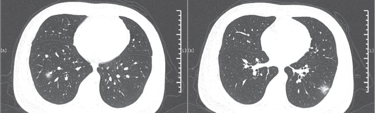
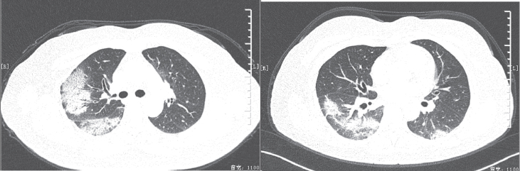
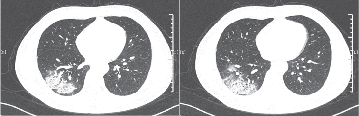
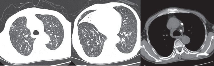
Background: Pneumonia caused by COVID-19 shares overlapping imaging manifestations with other types of pneumonia. How to objectively and quantitatively differentiate pneumonia patients with and without COVID-19 virus remains clinical challenge.
Objective: To formulate standardized scoring criteria and an objective quantization standard to guide decision making in detection and diagnosis of COVID-19 virus induced pneumonia in clinical practice.
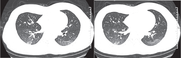
Methods: A retrospective dataset includes computed tomography (CT) images acquired from 43 pneumonia patients with COVID-19 virus detected by reverse transcription-polymerase chain reaction (RT-PCR) tests and 49 pneumonia patients without COVID-19 virus. All patients were treated during the same time period in two hospitals. Key indicators of differential diagnosis were identified in relevant literature and the scores were quantified namely, patients with more than 8 points were identified as high risk, those with 6-8 points as moderate risk, and those with fewer than 6 points as low risk for COVID-19 virus. In the study, 3 radiologists determined the scores for all patients. Diagnostic sensitivity and specificity were subsequently calculated.
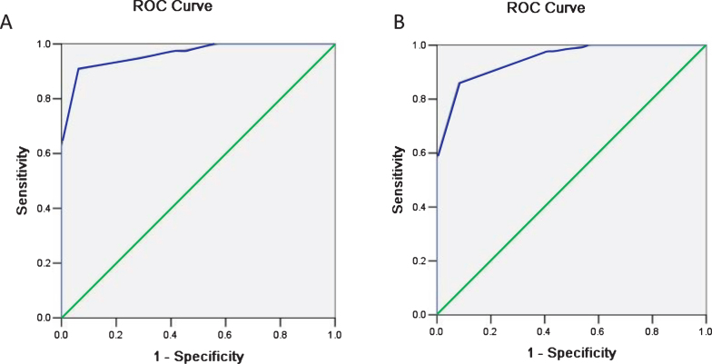
Results: A total of 61 patients were determined as high risk, among which 42 were COVID-19 positive by RT-PCR tests. Next, 9 were identified as moderate risk, one of whom was COVID-19 positive. Last, 22 were classified into the low-risk group, all of them are COVID-19 negative. Based on these results, the sensitivity of detection COVID-19 positive cases between the high-risk group and the non-high-risk group was 0.98 with 95% confidence interval [0.88, 1.00], and the specificity was 0.61 [0.46, 0.75]. The detection sensitivity between the moderate-/high-risk group and the low-risk group was 1.00 [0.92, 1.00], and the specificity was 0.45 [0.31, 0.60].
Conclusion: The proposed quantitative scoring criteria showed high sensitivity and moderate specificity in detecting COVID-19 using CT images, which indicates that these criteria may be beneficial for screening in real-world practice and helpful for long-term disease control.
Keywords: COVID-19; Coronavirus; Pneumonia; X-ray computed tomography.
Conflict of interest statement
All authors declare that they have no conflict of interest.
Figures
Similar articles
-
Performance of Radiologists in Differentiating COVID-19 from Non-COVID-19 Viral Pneumonia at Chest CT.Radiology. 2020 Aug;296(2):E46-E54. doi: 10.1148/radiol.2020200823. Epub 2020 Mar 10. Radiology. 2020. PMID: 32155105 Free PMC article.
-
Thoracic imaging tests for the diagnosis of COVID-19.Cochrane Database Syst Rev. 2020 Sep 30;9:CD013639. doi: 10.1002/14651858.CD013639.pub2. Cochrane Database Syst Rev. 2020. Update in: Cochrane Database Syst Rev. 2020 Nov 26;11:CD013639. doi: 10.1002/14651858.CD013639.pub3. PMID: 32997361 Updated.
-
Combination of CT and RT-PCR in the screening or diagnosis of COVID-19.J Glob Health. 2020 Jun;10(1):010347. doi: 10.7189/jogh.10.010347. J Glob Health. 2020. PMID: 32373325 Free PMC article. No abstract available.
-
Differential diagnosis of coronavirus disease 2019 from community-acquired-pneumonia by computed tomography scan and follow-up.Infect Dis Poverty. 2020 Aug 26;9(1):118. doi: 10.1186/s40249-020-00737-9. Infect Dis Poverty. 2020. PMID: 32843064 Free PMC article.
-
Diagnostic Tools for Coronavirus Disease (COVID-19): Comparing CT and RT-PCR Viral Nucleic Acid Testing.AJR Am J Roentgenol. 2020 Oct;215(4):834-838. doi: 10.2214/AJR.20.23418. Epub 2020 May 15. AJR Am J Roentgenol. 2020. PMID: 32412790 Review.
Cited by
-
Using artificial intelligence to assist radiologists in distinguishing COVID-19 from other pulmonary infections.J Xray Sci Technol. 2021;29(1):1-17. doi: 10.3233/XST-200735. J Xray Sci Technol. 2021. PMID: 33164982 Free PMC article.
-
RT-PCR Combined with CT Examination in the Diagnosis and Prognosis Evaluation of COVID-19 Patients in Fangcang Hospital: A Case Series.J Multidiscip Healthc. 2021 Jan 19;14:145-149. doi: 10.2147/JMDH.S293601. eCollection 2021. J Multidiscip Healthc. 2021. PMID: 33500623 Free PMC article.
-
Thoracic imaging tests for the diagnosis of COVID-19.Cochrane Database Syst Rev. 2021 Mar 16;3(3):CD013639. doi: 10.1002/14651858.CD013639.pub4. Cochrane Database Syst Rev. 2021. Update in: Cochrane Database Syst Rev. 2022 May 16;5:CD013639. doi: 10.1002/14651858.CD013639.pub5. PMID: 33724443 Free PMC article. Updated.
-
Diagnostic Performance of Antigen Rapid Diagnostic Tests, Chest Computed Tomography, and Lung Point-of-Care-Ultrasonography for SARS-CoV-2 Compared with RT-PCR Testing: A Systematic Review and Network Meta-Analysis.Diagnostics (Basel). 2022 May 24;12(6):1302. doi: 10.3390/diagnostics12061302. Diagnostics (Basel). 2022. PMID: 35741112 Free PMC article. Review.
-
Differential diagnosis of COVID-19 at the chest computed tomography scan: A review with special focus on cancer patients.World J Radiol. 2021 Aug 28;13(8):243-257. doi: 10.4329/wjr.v13.i8.243. World J Radiol. 2021. PMID: 34567434 Free PMC article.
References
-
- National Health Commission. Diagnosis and treatment of novel coronavirus pneumonia (Trial revision of the fifth edition) [EB/OL]. (2020-02-04) [2020-02-17]. http://www.gov.cn/zhengce/zhengceku/2020-02/05/content_5474791.html
MeSH terms
LinkOut - more resources
Full Text Sources
Medical
