

Exosomes released by breast cancer cells under mild hyperthermic stress possess immunogenic potential and modulate polarization in vitro in macrophages
- PMID: 32568583
- PMCID: PMC8694666
- DOI: 10.1080/02656736.2020.1778800
Exosomes released by breast cancer cells under mild hyperthermic stress possess immunogenic potential and modulate polarization in vitro in macrophages
Abstract
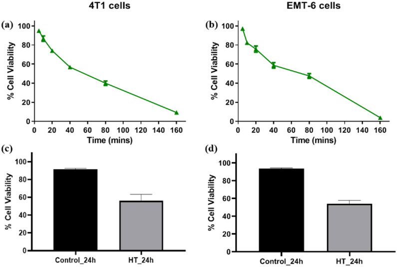
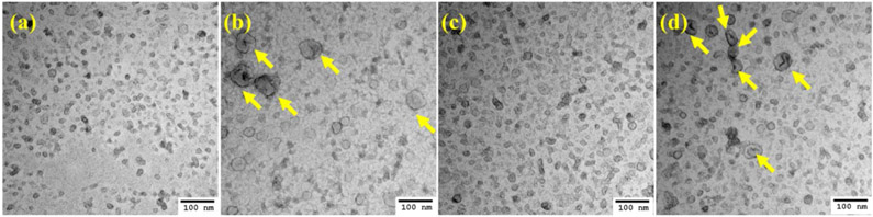
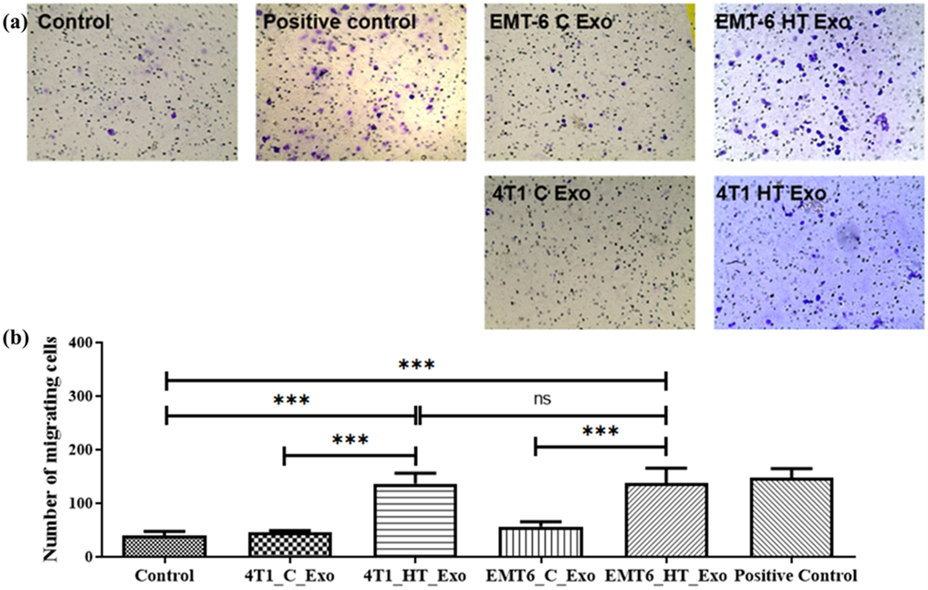
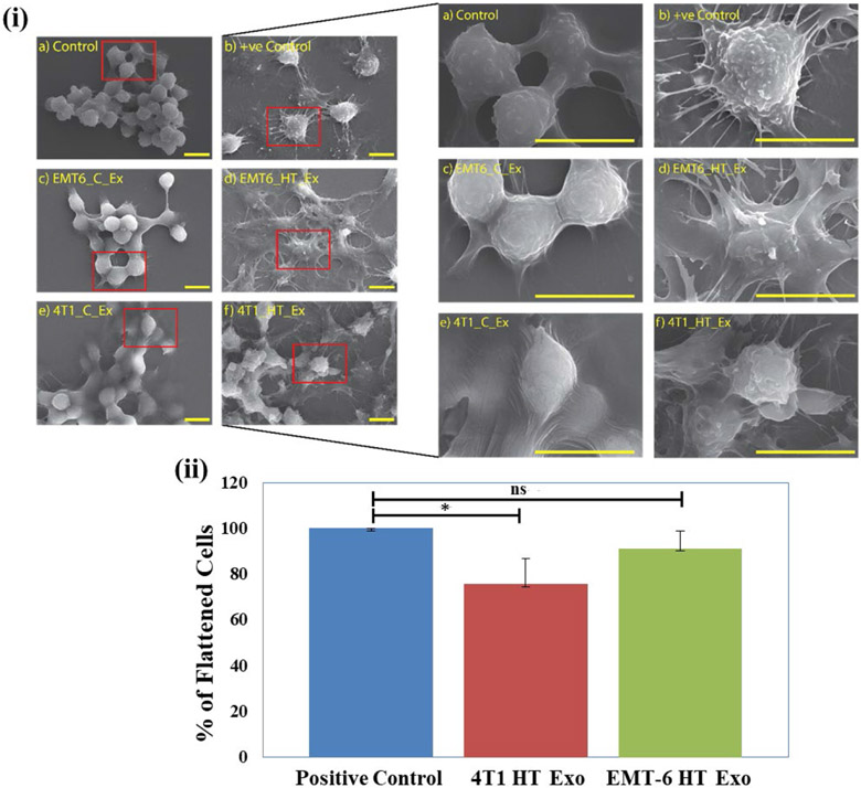
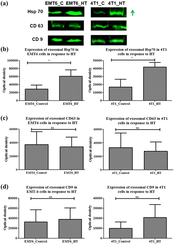
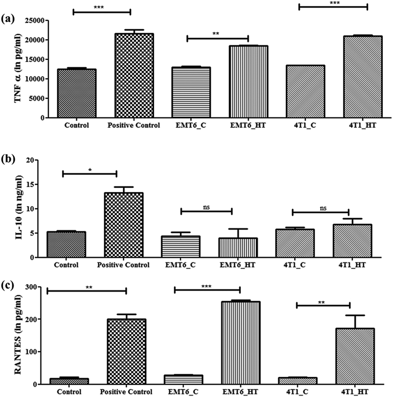
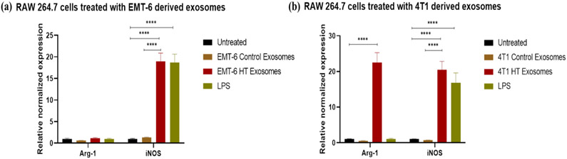
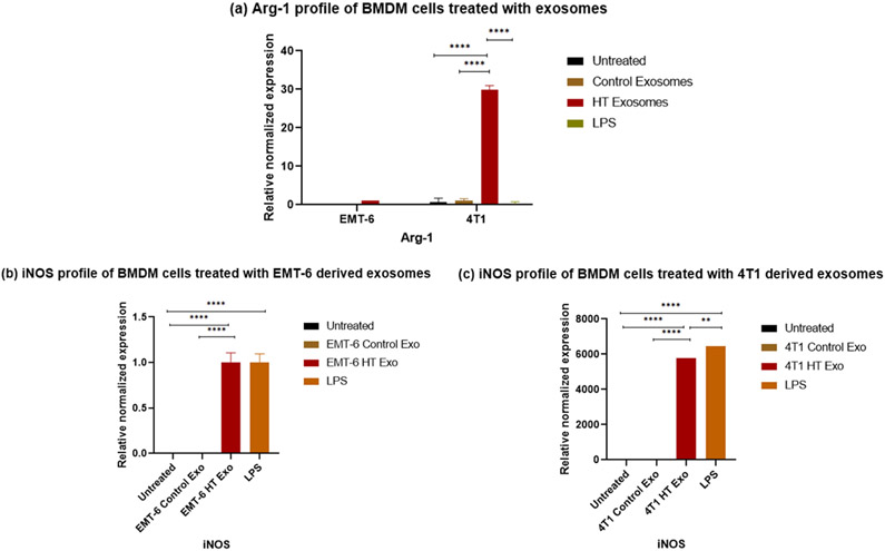
Macrophages play a dual role in tumor initiation and progression, with both tumor-promoting and tumor-suppressive effects; hence, it is essential to understand the distinct responses of macrophages to tumor progression and therapy. Mild hyperthermia has gained importance as a therapeutic regimen against cancer due to its immunogenic nature, efficacy, and potential synergy with other therapies, yet the response of macrophages to molecular signals from hyperthermic cancer cells has not yet been clearly defined. Due to limited response rate of breast cancer to conventional therapeutics the development, and understanding of alternative therapies like hyperthermia is pertinent. In order to determine conditions corresponding to mild thermal dose, cytotoxicity of different hyperthermic temperatures and treatment durations were tested in normal murine macrophages and breast cancer cell lines. Examination of exosome release in hyperthermia-treated cancer cells revealed enhanced efflux and a larger size of exosomes released under hyperthermic stress. Exposure of naïve murine macrophages to exosomes released from 4T1 and EMT-6 cells posthyperthermia treatment, led to an increased expression of specific macrophage activation markers. Further, exosomes released by hyperthermia-treated cancer cells had increased content of heat shock protein 70 (Hsp70). Together, these results suggest a potential immunogenic role for exosomes released from cancer cells treated with mild hyperthermia.
Keywords: Hyperthermia; breast cancer; exosome; immune system; macrophages.
Conflict of interest statement
Conflict of interest:
The authors declare no conflict of interest.
Figures

Similar articles
-
Hyperthermia promotes M1 polarization of macrophages via exosome-mediated HSPB8 transfer in triple negative breast cancer.Discov Oncol. 2023 May 26;14(1):81. doi: 10.1007/s12672-023-00697-0. Discov Oncol. 2023. PMID: 37233869 Free PMC article.
-
Exosome derived from epigallocatechin gallate treated breast cancer cells suppresses tumor growth by inhibiting tumor-associated macrophage infiltration and M2 polarization.BMC Cancer. 2013 Sep 17;13:421. doi: 10.1186/1471-2407-13-421. BMC Cancer. 2013. PMID: 24044575 Free PMC article.
-
Exosome-mediated miR-33 transfer induces M1 polarization in mouse macrophages and exerts antitumor effect in 4T1 breast cancer cell line.Int Immunopharmacol. 2021 Jan;90:107198. doi: 10.1016/j.intimp.2020.107198. Epub 2020 Nov 25. Int Immunopharmacol. 2021. PMID: 33249048
-
Tumor-derived exosomes in the regulation of macrophage polarization.Inflamm Res. 2020 May;69(5):435-451. doi: 10.1007/s00011-020-01318-0. Epub 2020 Mar 11. Inflamm Res. 2020. PMID: 32162012 Review.
-
Exosome: emerging biomarker in breast cancer.Oncotarget. 2017 Jun 20;8(25):41717-41733. doi: 10.18632/oncotarget.16684. Oncotarget. 2017. PMID: 28402944 Free PMC article. Review.
Cited by
-
The Role of Exosomes in the Female Reproductive System and Breast Cancers.Onco Targets Ther. 2020 Dec 8;13:12567-12586. doi: 10.2147/OTT.S281909. eCollection 2020. Onco Targets Ther. 2020. PMID: 33324075 Free PMC article. Review.
-
Airway acidification impaired host defense against Pseudomonas aeruginosa infection by promoting type 1 interferon β response.Emerg Microbes Infect. 2022 Dec;11(1):2132-2146. doi: 10.1080/22221751.2022.2110524. Emerg Microbes Infect. 2022. PMID: 35930458 Free PMC article.
-
Regenerative therapy by using mesenchymal stem cells-derived exosomes in COVID-19 treatment. The potential role and underlying mechanisms.Regen Ther. 2022 Jun;20:61-71. doi: 10.1016/j.reth.2022.03.006. Epub 2022 Mar 22. Regen Ther. 2022. PMID: 35340407 Free PMC article. Review.
-
Emerging role of exosomes in cancer progression and tumor microenvironment remodeling.J Hematol Oncol. 2022 Jun 28;15(1):83. doi: 10.1186/s13045-022-01305-4. J Hematol Oncol. 2022. PMID: 35765040 Free PMC article. Review.
-
Hyperthermia combined with immune checkpoint inhibitor therapy in the treatment of primary and metastatic tumors.Front Immunol. 2022 Aug 12;13:969447. doi: 10.3389/fimmu.2022.969447. eCollection 2022. Front Immunol. 2022. PMID: 36032103 Free PMC article. Review.
References
-
- Adachi S, et al. (2009). "Effect of hyperthermia combined with gemcitabine on apoptotic cell death in cultured human pancreatic cancer cell lines." International Journal of Hyperthermia 25(3): 210–219. - PubMed
Publication types
MeSH terms
Grants and funding
LinkOut - more resources
Full Text Sources
Other Literature Sources
Medical