

Application of deep learning technique to manage COVID-19 in routine clinical practice using CT images: Results of 10 convolutional neural networks
- PMID: 32568676
- PMCID: PMC7190523
- DOI: 10.1016/j.compbiomed.2020.103795
Application of deep learning technique to manage COVID-19 in routine clinical practice using CT images: Results of 10 convolutional neural networks
Abstract
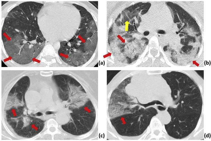
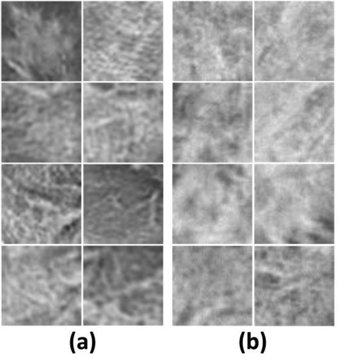
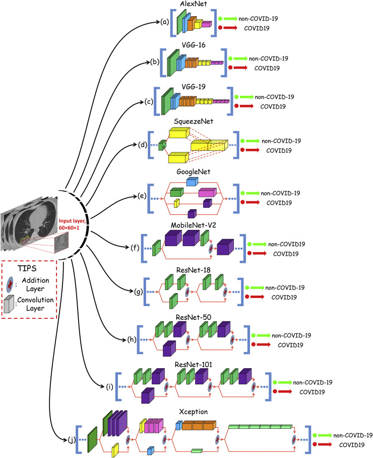
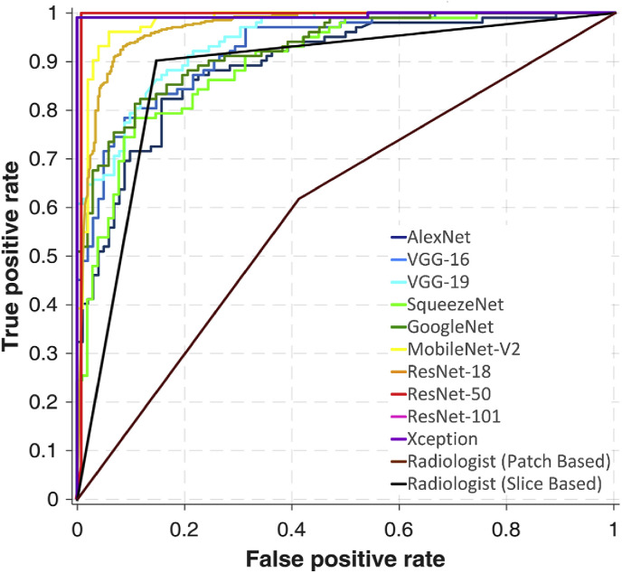
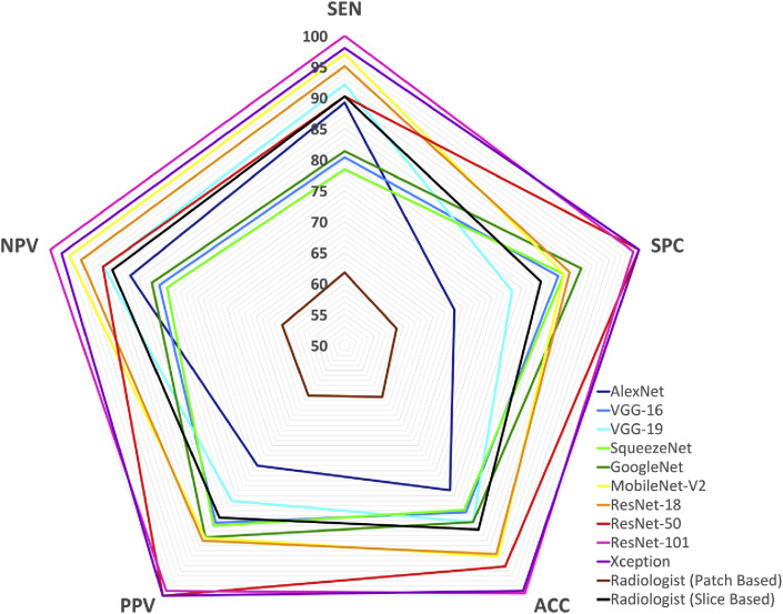
Fast diagnostic methods can control and prevent the spread of pandemic diseases like coronavirus disease 2019 (COVID-19) and assist physicians to better manage patients in high workload conditions. Although a laboratory test is the current routine diagnostic tool, it is time-consuming, imposing a high cost and requiring a well-equipped laboratory for analysis. Computed tomography (CT) has thus far become a fast method to diagnose patients with COVID-19. However, the performance of radiologists in diagnosis of COVID-19 was moderate. Accordingly, additional investigations are needed to improve the performance in diagnosing COVID-19. In this study is suggested a rapid and valid method for COVID-19 diagnosis using an artificial intelligence technique based. 1020 CT slices from 108 patients with laboratory proven COVID-19 (the COVID-19 group) and 86 patients with other atypical and viral pneumonia diseases (the non-COVID-19 group) were included. Ten well-known convolutional neural networks were used to distinguish infection of COVID-19 from non-COVID-19 groups: AlexNet, VGG-16, VGG-19, SqueezeNet, GoogleNet, MobileNet-V2, ResNet-18, ResNet-50, ResNet-101, and Xception. Among all networks, the best performance was achieved by ResNet-101 and Xception. ResNet-101 could distinguish COVID-19 from non-COVID-19 cases with an AUC of 0.994 (sensitivity, 100%; specificity, 99.02%; accuracy, 99.51%). Xception achieved an AUC of 0.994 (sensitivity, 98.04%; specificity, 100%; accuracy, 99.02%). However, the performance of the radiologist was moderate with an AUC of 0.873 (sensitivity, 89.21%; specificity, 83.33%; accuracy, 86.27%). ResNet-101 can be considered as a high sensitivity model to characterize and diagnose COVID-19 infections, and can be used as an adjuvant tool in radiology departments.
Keywords: COVID-19; Computed tomography; Coronavirus infections; Deep learning; Lung diseases; Machine learning; Pneumonia.
Copyright © 2020 Elsevier Ltd. All rights reserved.
Conflict of interest statement
Declaration of competing interest None.
Figures
References
-
- Huang C., Wang Y., Li X., Ren L., Zhao J., Hu Y., Zhang L., Fan G., Xu J., Gu X., Cheng Z., Yu T., Xia J., Wei Y., Wu W., Xie X., Yin W., Li H., Liu M., Xiao Y., Gao H., Guo L., Xie J., Wang G., Jiang R., Gao Z., Jin Q., Wang J., Cao B. Clinical features of patients infected with 2019 novel coronavirus in Wuhan, China. Lancet. 2020;395:497–506. - PMC - PubMed
-
- World Health Organization (WHO) Novel Coronavirus. (2019-nCoV) Technical Guidance: Laboratory Guidance. Geneva: WHO. https://www.who.int/emergencies/diseases/novel-coronavirus-2019/technica... Available from:
MeSH terms
LinkOut - more resources
Full Text Sources
Medical
