. 2020 Nov;161(11):2629-2651.
doi: 10.1097/j.pain.0000000000001951.

Studies on CRMP2 SUMOylation-deficient transgenic mice identify sex-specific Nav1.7 regulation in the pathogenesis of chronic neuropathic pain

Affiliations

Studies on CRMP2 SUMOylation-deficient transgenic mice identify sex-specific Nav1.7 regulation in the pathogenesis of chronic neuropathic pain

Aubin Moutal et al. Pain. 2020 Nov.

Abstract

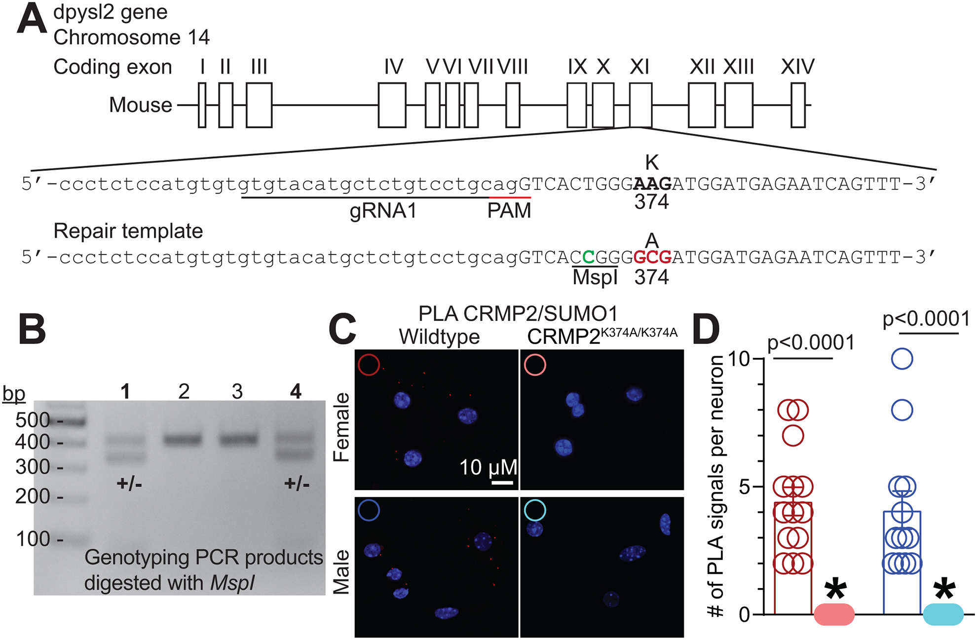
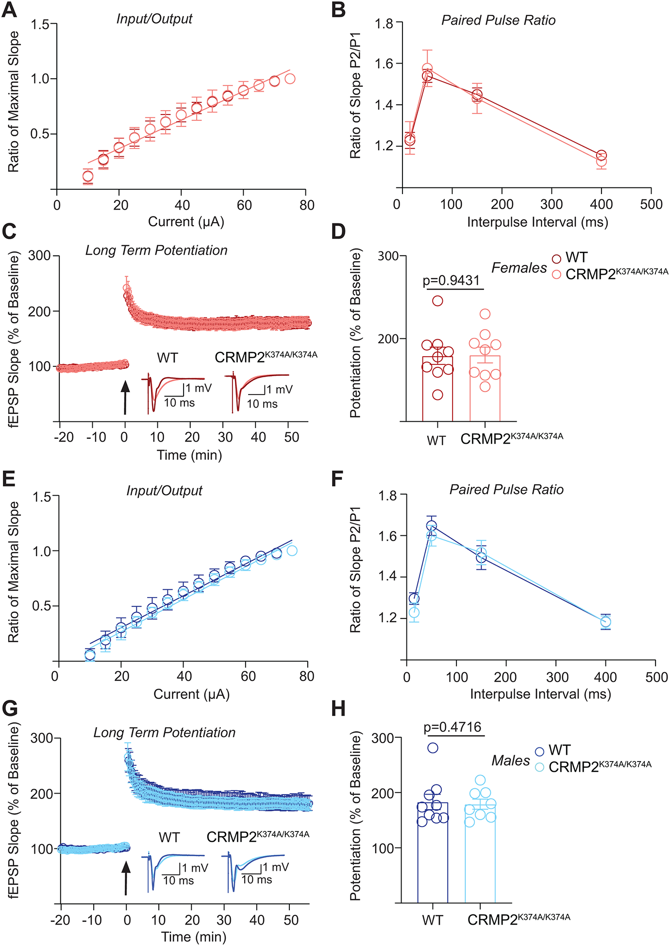
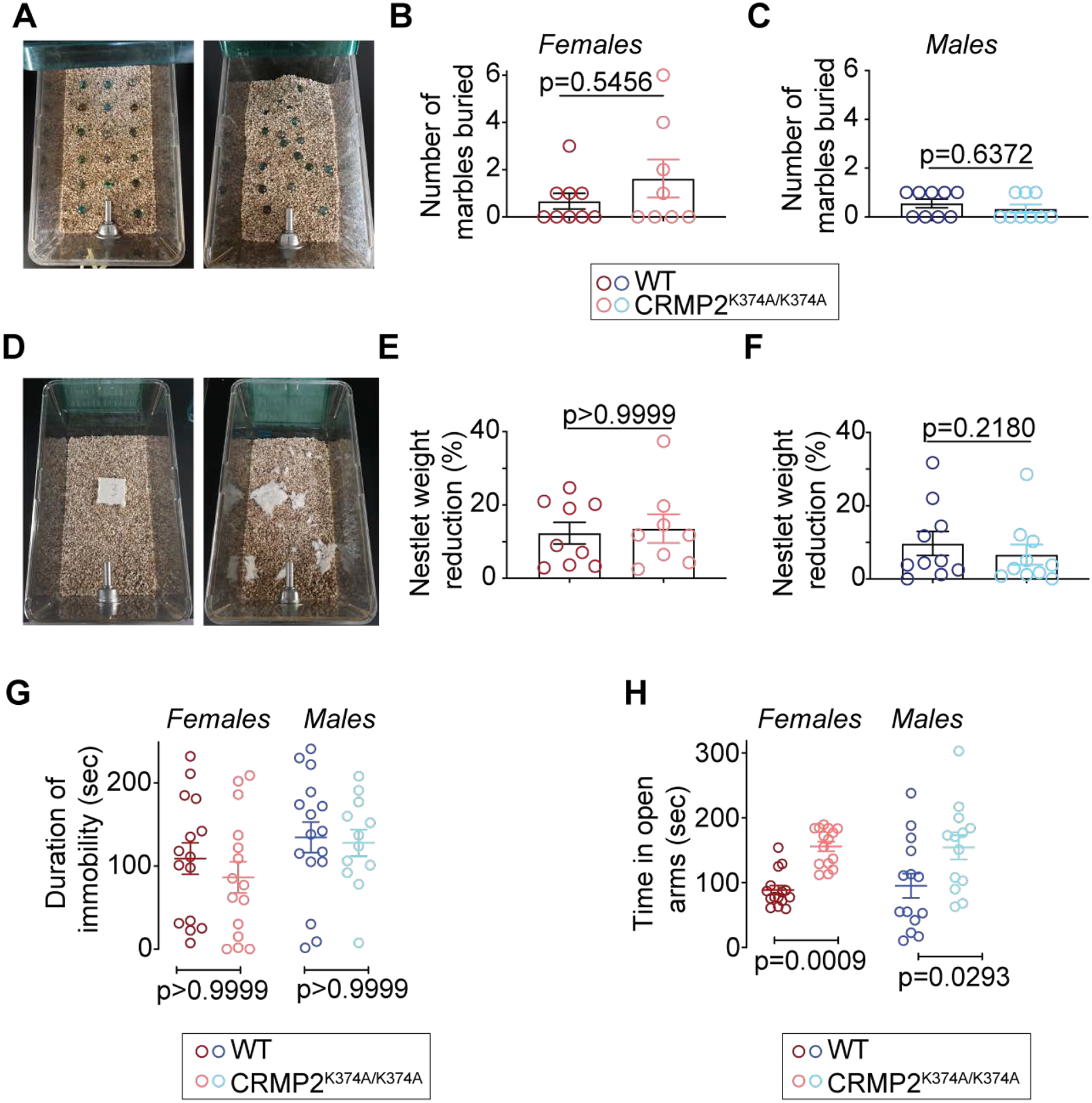
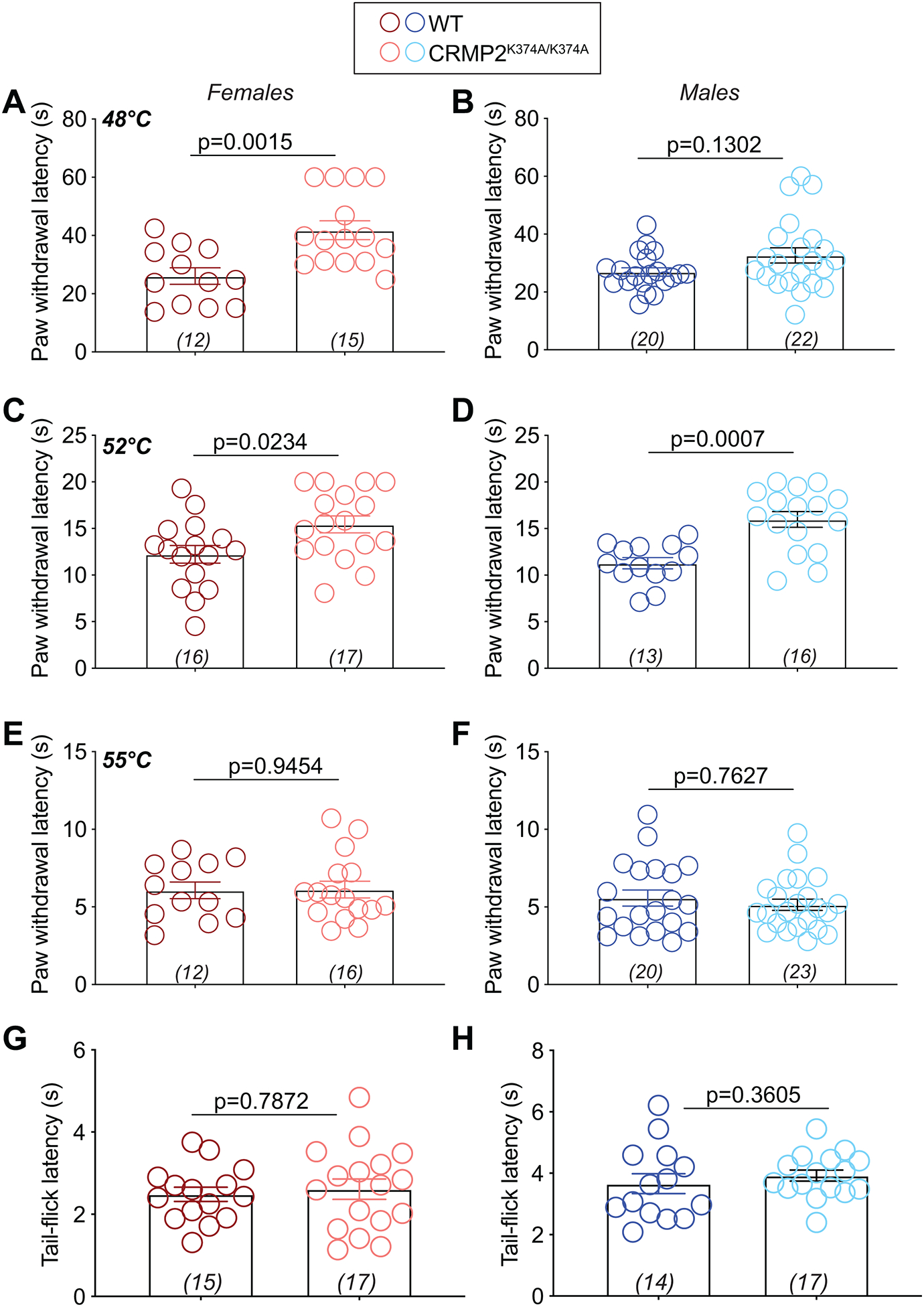
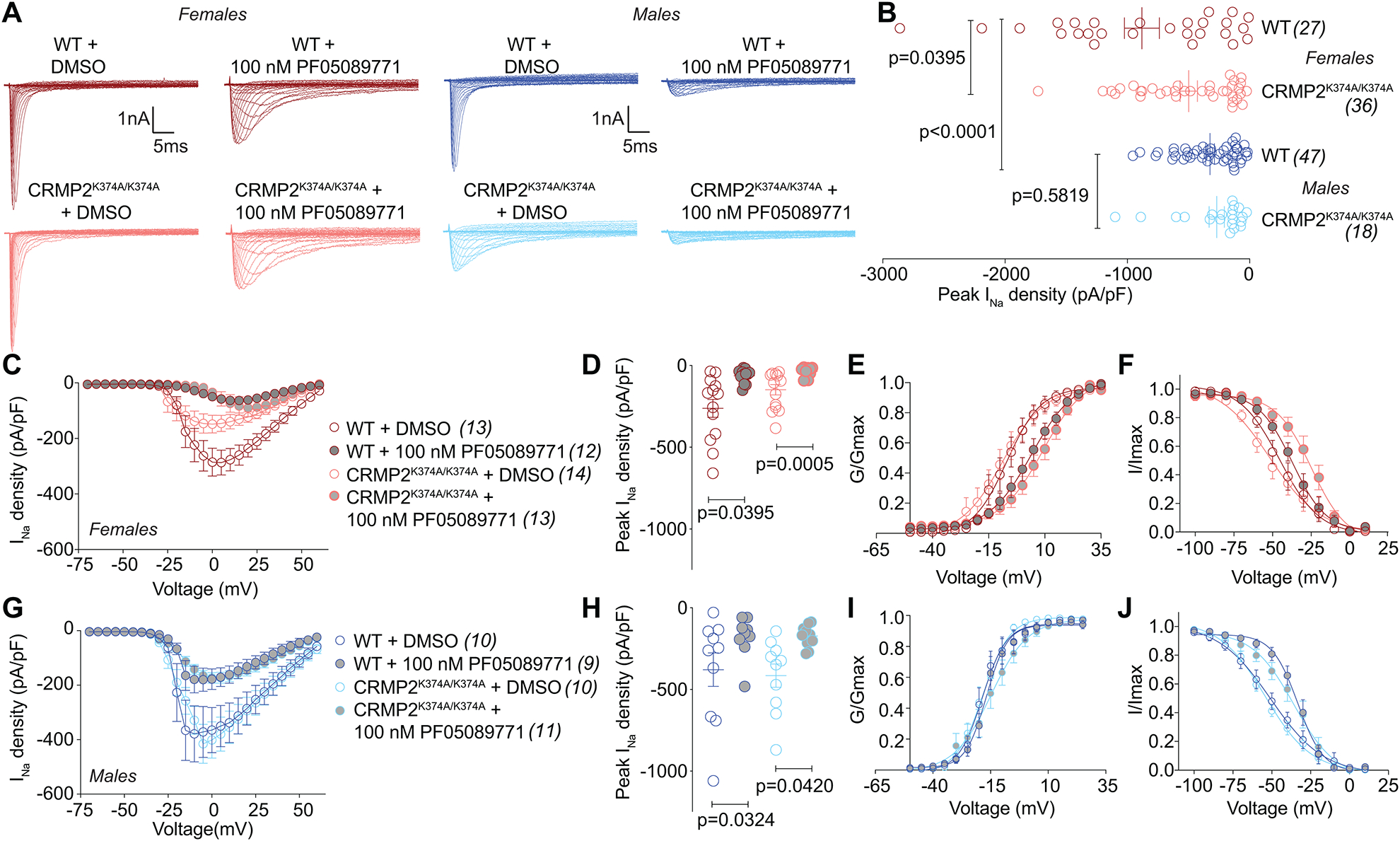
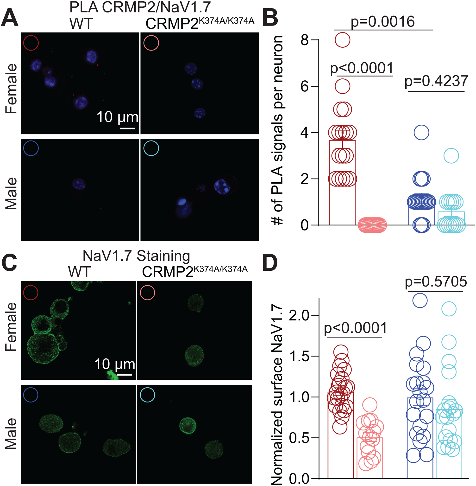
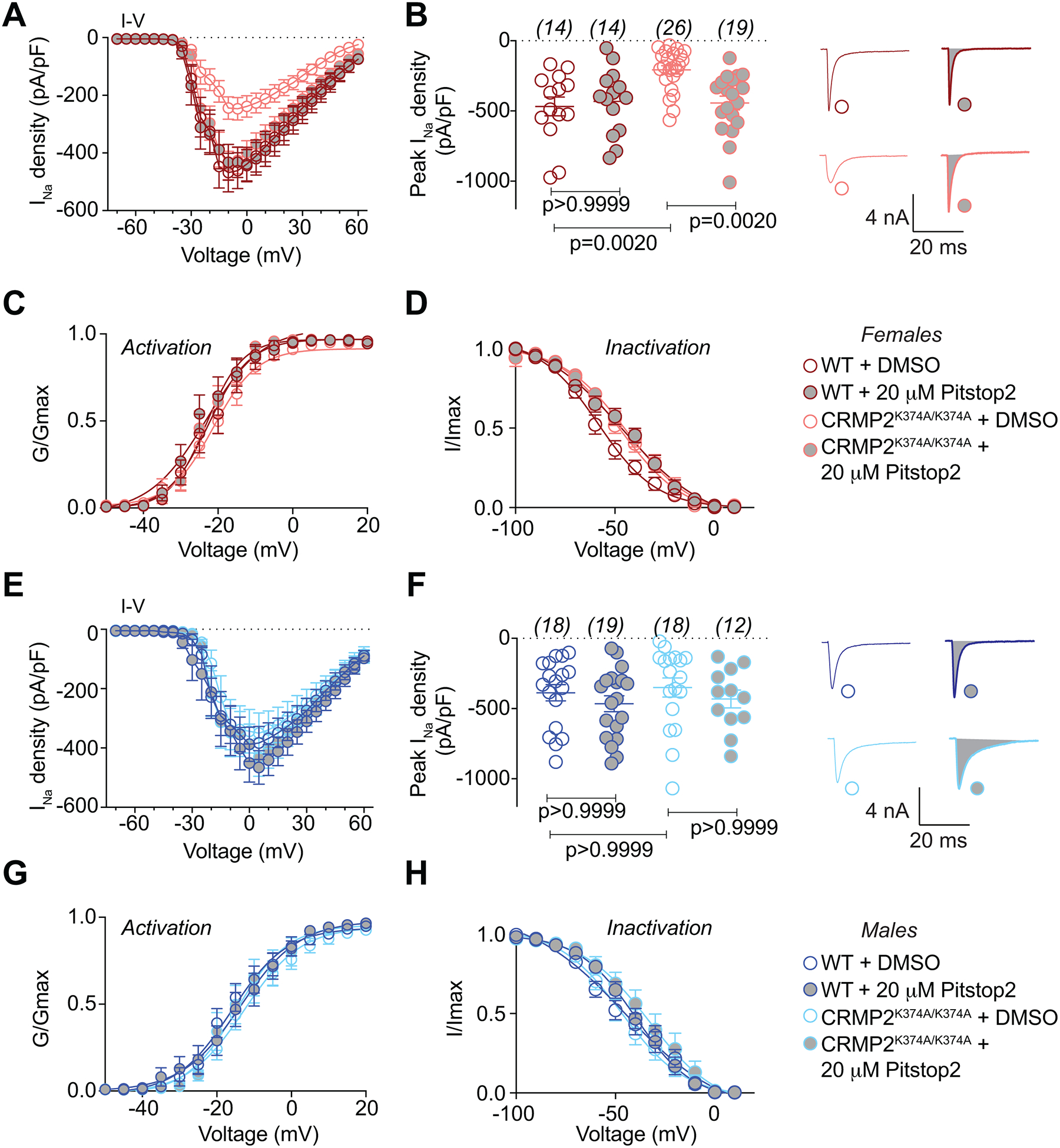
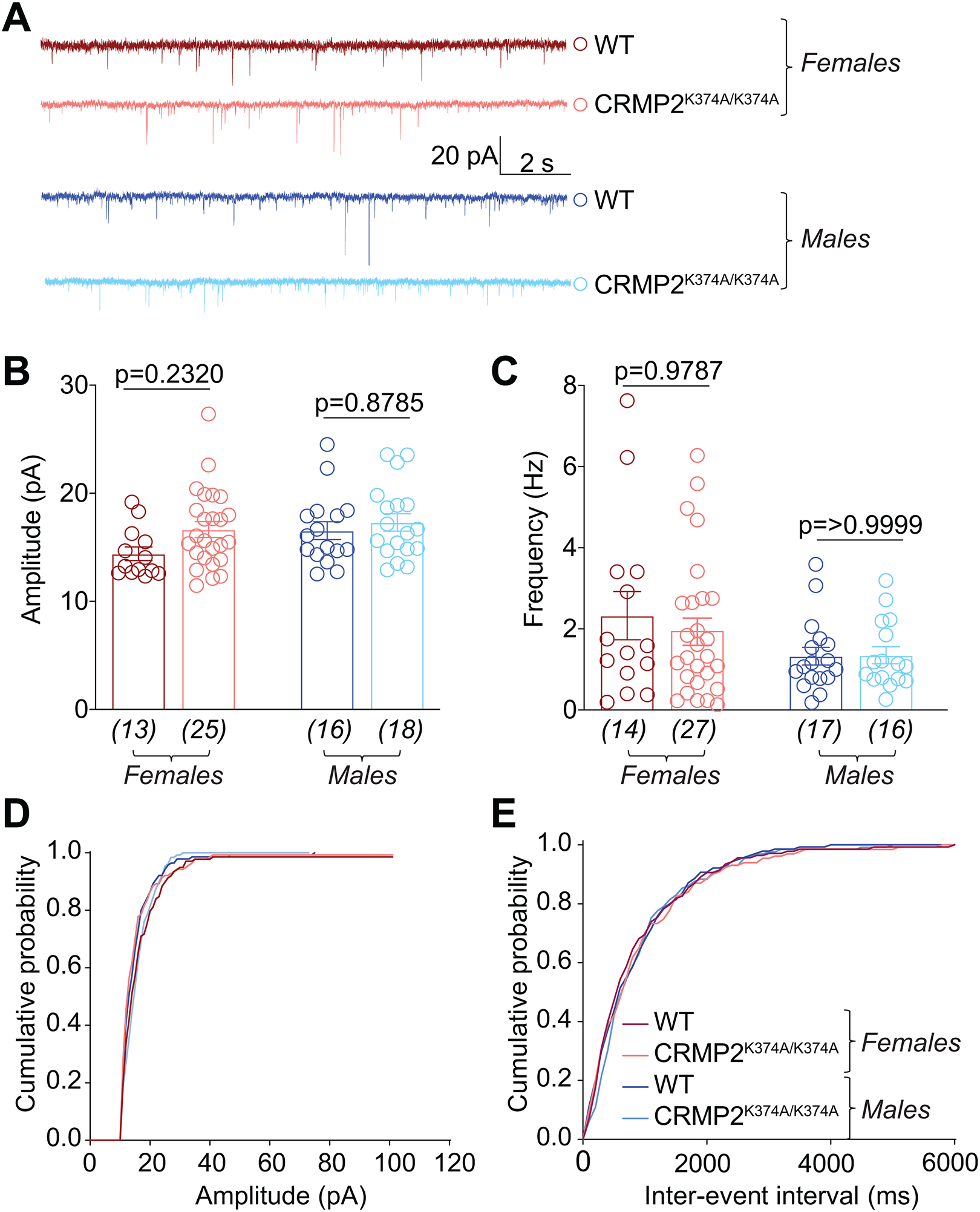
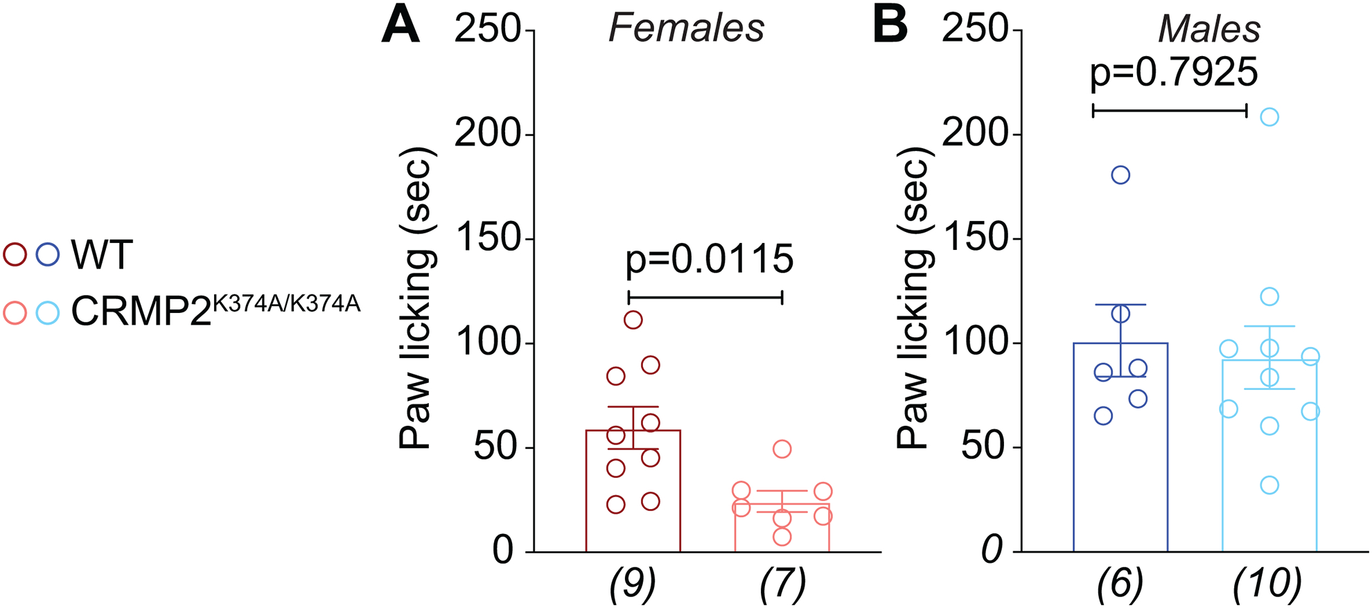
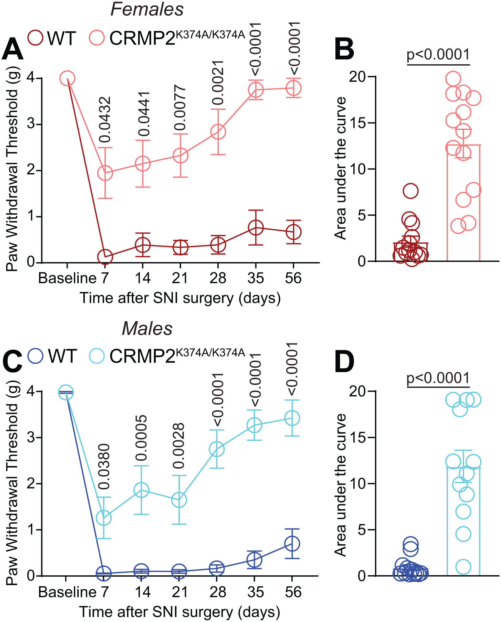
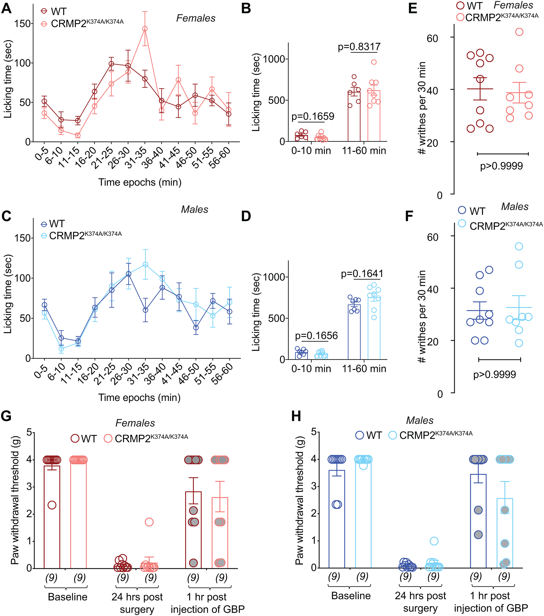
The sodium channel Nav1.7 is a master regulator of nociceptive input into the central nervous system. Mutations in this channel can result in painful conditions and produce insensitivity to pain. Despite being recognized as a "poster child" for nociceptive signaling and human pain, targeting Nav1.7 has not yet produced a clinical drug. Recent work has illuminated the Nav1.7 interactome, offering insights into the regulation of these channels and identifying potentially new druggable targets. Among the regulators of Nav1.7 is the cytosolic collapsin response mediator protein 2 (CRMP2). CRMP2, modified at lysine 374 (K374) by addition of a small ubiquitin-like modifier (SUMO), bound Nav1.7 to regulate its membrane localization and function. Corollary to this, preventing CRMP2 SUMOylation was sufficient to reverse mechanical allodynia in rats with neuropathic pain. Notably, loss of CRMP2 SUMOylation did not compromise other innate functions of CRMP2. To further elucidate the in vivo role of CRMP2 SUMOylation in pain, we generated CRMP2 K374A knock-in (CRMP2) mice in which Lys374 was replaced with Ala. CRMP2 mice had reduced Nav1.7 membrane localization and function in female, but not male, sensory neurons. Behavioral appraisal of CRMP2 mice demonstrated no changes in depressive or repetitive, compulsive-like behaviors and a decrease in noxious thermal sensitivity. No changes were observed in CRMP2 mice to inflammatory, acute, or visceral pain. By contrast, in a neuropathic model, CRMP2 mice failed to develop persistent mechanical allodynia. Our study suggests that CRMP2 SUMOylation-dependent control of peripheral Nav1.7 is a hallmark of chronic, but not physiological, neuropathic pain.


Conflict of interest statement

CONFLICT OF INTERESTS STATEMENT

R. Khanna is the co-founder of Regulonix LLC, a company developing non-opioids drugs for chronic pain. In addition, R. Khanna has patents US10287334 and US10441586 issued to Regulonix LLC. The other authors declare no competing financial interest.

Figures

Figure 1.
Figure 1.. Generation of CRMP2K374A/K374A knock-in mice.
(A) Strategy for the generation of the CRMP2–K374A knock-in mutation in mouse ES cells. Guide RNA (gRNA) design for targeting the mouse dpysl2 locus. The protospacer-adjacent motif (PAM) sequence is in underlined in red. The AAG to GCG mutation is in bold font. (B) PCR results of founder mice. Mice from two litters were genotyped by PCR amplification and MspI digestion. MspI digestion products were electrophoresed on a 1.5% agarose gel. Wild type mouse genomic DNA was used as negative control (not shown). (C) Representative images of mouse DRG cultures following proximity ligation assay (PLA) between CRMP2 and SUMO1. The PLA immunofluorescence labeled sites of interaction between CRMP2 and SUMO1 (red puncta). Additionally, nuclei are labeled with the nuclear labeling dye 4’,6-diamidino-2-phenylindole (DAPI). Scale bar: 10 μm. (D) Quantification of PLA puncta per neuron show that in DRGs from CRMP2K374A/K374A mice, the number of CRMP2-SUMO1 interactions are significantly reduced compared to wildtype DRGs (Kruskal-Wallis test with Dunn’s multiple comparison post-hoc test: p<0.0001 comparing female WT vs. female CRMP2K374A/K374A; p<0.0001 comparing male WT vs. male CRMP2K374A/K374A; and p>0.9999 comparing male WT vs. female WT). Error bars indicate mean ± SEM from 12–16 cells.
Figure 2.
Figure 2.. Basal excitability, presynaptic plasticity, and long-term potentiation in hippocampal neurons are not changed between wildtype and CRMP2K374A/K374A knock-in mice.
(A) Input output relationship of K374A wild type (WT) and homozygous (CRMP2K374A/K374A) groups obtained from female mice. Data was fit using nonlinear regression and a linear approximation of the slope that best fit the data from each group was obtained. Analysis of covariance was used to determine whether the slope of each group was significantly different. The line of best fit for both groups has the same slope [P = 0.9699, F(2, 290) = 0.03061]. WT N = 9 slices (3 animals), CRMP2K374A/K374A N = 12 slices (4 animals). (B) Paired pulse facilitation ratio obtained from K374A WT and CRMP2K374A/K374A female mice to assess changes in presynaptic plasticity. Pulse intervals were 15, 50, 150 and 400 ms. Two-way ANOVA was performed to identify significant differences between groups, and none were found [P = 0.9939, F(1,76) = 5.885 × 10⁻⁵]. WT N = 9 slices (3 animals), CRMP2K374A/K374A N = 12 slices (4 animals). (C) Long term potentiation of K374A WT and CRMP2K374A/K374A female mice represented as a percentage of averaged 20-minute baseline response. Arrow indicates time 0 when the theta burst stimulation (TBS) protocol was applied to the stimulation electrode positioned in the Schaffer Collateral projection from CA3 to CA1 region of the hippocampus. Inset shows representative fEPSP traces before (Gray) and after (Black) TBS application for both WT and CRMP2K374A/K374A groups. WT N = 9 slices (3 animals), CRMP2K374A/K374A N = 9 slices (4 animals). (D) Averaged level of potentiation during the last 5 minutes of the recording period for comparison between WT and CRMP2K374A/K374A groups in females. Averaged potentiation was compared between groups using a two-tailed unpaired t-test, which did not reach significance [P = 0.9431, t = 0.07245, df = 16]. WT N = 9 slices (3 animals), CRMP2K374A/K374A N = 9 slices (4 animals). (E) Input output relationship of WT and CRMP2K374A/K374A obtained from male mice. Data was fit using nonlinear regression and a linear approximation of the slope that best fit the data from each group was obtained. Analysis of covariance was used to determine whether the slope of each group was significantly different. The line of best fit for both groups has the same slope [P = 0.5191, F(1,276) = 0.4167]. WT N = 11 slices (3 animals), CRMP2K374A/K374A N = 9 slices (3 animals). (F) Paired pulse facilitation ratio obtained from WT and CRMP2K374A/K374A male mice to assess changes in presynaptic plasticity. Pulse intervals were 15, 50, 150 and 400 ms. Two-way ANOVA was performed to identify significant differences between groups, and none were found [P = 0.4716, F(1,72) = 0.5238]. WT N = 11 slices (3 animals), CRMP2K374A/K374A N = 9 slices (3 animals). (G) Long term potentiation of WT and CRMP2K374A/K374A male mice represented as a percentage of averaged 20-minute baseline response. Arrow indicates time 0 when the theta burst stimulation (TBS) protocol was applied to the stimulation electrode positioned in the Schaffer Collateral projection from CA3 to CA1 region of the hippocampus. Inset shows representative fEPSP traces before (Gray) and after (Black) TBS application for both WT and CRMP2K374A/K374A groups. WT N = 9 slices (3 animals), CRMP2K374A/K374A N = 8 slices (3 animals). (H) Averaged level of potentiation during the last 5 minutes of the recording period for comparison between WT and CRMP2K374A/K374A groups in males. Averaged potentiation was compared between groups using a two-tailed unpaired t-test, which did not reach significance [P = 0.8167, t = 0.2359, df = 15]. WT N = 9 slices (3 animals), CRMP2K374A/K374A N = 8 slices (3 animals). The experiments were conducted by investigators blinded to the genotype. Error bars indicate mean ± SEM.
Figure 3.
Figure 3.. CRMP2K374A/K374A knock-in mice do not exhibit compulsive-like or depression-associated but have reduced anxiety-associated behaviors.
(A) Each cage was filled with 5cm of bedding chips lightly pressed to create a flat, even surface. 17 glass marbles (1.5cm in diameter) were distributed on the bedding surface. Mice were placed in the cages (1 mouse/cage) and allowed to behave freely for 30 minutes. After 30 minutes the number of marbles at least at least 2/3rds buried/covered with bedding were counted. No differences between genotypes was observed in females (B) or males (C). (D) For the nestlet test of compulsive-like behaviors, a nestlet was weighed and placed in the center of the cage. Mice were placed in the cages (1 mouse/cage) and allowed to behave freely for 30 minutes. Afterward, the largest remaining piece of nestlet was weighed. No differences between genotypes were observed in females (E) or males (F). (G) Immobility time in the tail suspension test was no different between sexes or genotypes. (H) Time spent in the open arms of the elevated plus-maze was reduced in the female and male CRMP2K374A/K374A mice compared to their WT counterparts. See statistical analysis described in Table 2. Error bars indicate mean ± SEM.
Figure 4.
Figure 4.. Preventing CRMP2 SUMOylation decreases thermal sensitivity in males and females.
Latencies to respond to noxious heat (48˚C, 52˚C, or 55˚C) in the hot plate (A-F) or tail-flick (52˚C) (G, H) tests in female and male wildtype (WT) and CRMP2K374A/K374A mice. Paw withdrawal latency was increased in heterozygous males at 48°C vs. wildtype males and increased at 52°C between wildtype and homozygotes males. There were no changes in latencies at the highest temperature. At 48°C, female CRMP2K374A/K374A mice exhibited a higher latency to respond to the nociceptive stimulus compared to wildtype mice. There were no changes in latencies at 52°C or 55°C. There were no changes in the hot-plate test under any of the conditions; the test was stopped at the cutoff time of 10-s. See statistical analysis described in Table 2. Error bars indicate mean ± SEM.
Figure 5.
Figure 5.. Total sodium currents and PF-05089771-sensitive NaV1.7 currents are reduced in female, but not male, DRGs from CRMP2K374A/K374A knock-in mice.
(A) Representative current traces recorded from small-sized DRGs neurons isolated from female and male wildtype (WT) and CRMP2K374A/K374A knock-in mice in response to depolarization steps from −70 to +60 mV from a holding potential of −60 mV. (B) Summary of normalized peak currents (in picoamperes/picofarads, pA/pF) from DRG neurons cultured from female and male wildtype (WT) and CRMP2K374A/K374A knock-in mice. Total sodium current was similar between wildtype and homozygous male mice (n=18–47 cells/condition, p = 0.5819, Kruskal-Wallis test with Dunn’s post hoc). NaV1.7 currents were significantly smaller in DRGs from homozygous female mice vs. DRG neurons from wildtype female (n=27–36 cells/condition, p = 0.03959, Kruskal-Wallis test with Dunn’s post hoc) or the both genotypes of male DRGs (p <0.0001, Kruskal-Wallis test with Dunn’s post hoc). The NaV1.7-selective inhibitor PF-05089771 [2] (100 nM, 5–15 min) was used to assess the extent of sodium currents carried by NaV1.7 in DRGs neurons isolated from female and male WT and CRMP2K374A/K374A knock-in mice. Summary of current-voltage curves (Cfemales; Gmales) and normalized peak (Dfemales; Hmales) currents (pA/pF) from DRG neurons of WT and CRMP2K374A/K374A knock-in mice. Boltzmann fits for normalized conductance G/Gmax voltage relations for voltage dependent activation (E, I) and inactivation (F, J) of the sensory neurons as indicated. Half-maximal activation and inactivation (V1/2) and slope values (k) for activation and inactivation are presented in Table 1. Error bars indicate mean ± SEM.
Figure 6.
Figure 6.. Surface NaV1.7 expression and binding of NaV1.7 to CRMP2 are reduced in female, but not male, DRGs from CRMP2K374A/K374A knock-in mice.
(A) Representative images of mouse DRG cultures following proximity ligation assay (PLA) between CRMP2 and NaV1.7. The PLA immunofluorescence labeled sites of interaction between CRMP2 and NaV1.7 (red puncta). Additionally, nuclei are labeled with the nuclear labeling dye 4’,6-diamidino-2-phenylindole (DAPI). Scale bar: 10 μm. (B) Quantification of PLA puncta per neuron shows that in DRGs from CRMP2K374A/K374A mice, the number of NaV1.7-CRMP2 interactions was significantly reduced compared to wildtype DRGs from female mice (Kruskal-Wallis test with Dunn’s multiple comparison post-hoc test: p<0.0001 comparing female WT vs. female CRMP2K374A/K374A; p=0.4237 comparing male WT vs. male CRMP2K374A/K374A; and p=0.0016 comparing male WT vs. female WT; n=13–18 cells). (C) Representative confocal images of mouse DRG cultures labeled with an antibody against NaV1.7. (D) Quantification of normalized surface expression of NaV1.7 per neuron shows that in DRGs from CRMP2K374A/K374A mice, the surface expression of NaV1.7 was significantly reduced compared to wildtype DRGs from female mice, whereas in male mouse DRGs there was no difference in expression between the genotypes (Kruskal-Wallis test with Dunn’s multiple comparison post-hoc test: p<0.0001 comparing female WT vs. female CRMP2K374A/K374A; p=0.5705 comparing male WT vs. male CRMP2K374A/K374A; and p=0.8994 comparing male WT vs. female WT; n=14–22 cells). Error bars indicate mean ± SEM.
Figure 7.
Figure 7.. Reduction of NaV1.7 currents in DRGs from female but not male CRMP2K374A/K374A knock-in mice are normalized by inhibition of clathrin coated endocytosis with Pitstop2.
Summary of current-voltage curves (A – female, Emale) and normalized peak (B – female, Fmale) currents (pA/pF) from small-to-medium diameter DRG neurons of WT and CRMP2K374A/K374A knock-in mice (n = 14–26). In some experiments, currents were recorded following 30 minute application of 20 μM of Pitstop2, a clathrin-mediated endocytosis inhibitor [71]. Representative traces from female DRGs are displayed adjacent to the scatter graphs in B. Boltzmann fits for normalized conductance G/Gmax voltage relations for voltage dependent activation (C – female, Gmale) and inactivation (D – female, Hmale) of the sensory neurons as indicated. Half-maximal activation and inactivation (V1/2) and slope values (k) for activation and inactivation are presented in Table 1. There were no significant differences in V1/2 and k values of activation and inactivation in either sex or genotype with and without treatment with Pitstop2 (One-way ANOVA with Tukey’s post hoc test). Error bars indicate mean ± SEM.
Figure 8.
Figure 8.. Excitatory neurotransmission is not affected in the lumbar dorsal horn of CRMP2K374A/K374A knock-in mice.
(A) Representative traces of cells from both sexes and genotypes. Bar graph with scatter plot showing the summary of amplitudes (B) and frequencies (C) of spontaneous excitatory post-synaptic currents (sEPSCs) for the indicated groups are shown. The cumulative probability of amplitude (D) and inter-event interval (E) are indicated. No significant change was observed in either parameter across any of the conditions tested. Data are shown as mean ± S.E.M., n=13–27 cells from at least 4 mice per experimental condition. p > 0.05, one-way ANOVA followed by Tukey’s post hoc test. Error bars indicate mean ± SEM. The experiments were conducted by investigators blinded to the genotype.
Figure 9.
Figure 9.. Female CRMP2K374A/K374A knock-in mice have increased pain-like behaviors in response to injection of the sodium channel activator veratridine.
Licking in the indicated groups of mice in response to an intraplantar dose of 1-mg veratridine. At this dose, veratridine caused a statistically significant increase in paw licking and lifting behaviors compared to saline. A change was seen in CRMP2K374A/K374A female mice compared to wildtype counterparts (A), while no differences between genotypes were observed in males (B). See statistical analysis described in Table 2. Error bars indicate mean ± SEM. The experiments were conducted by investigators blinded to the genotype.
Figure 10.
Figure 10.. CRMP2K374A/K374A knock-in mice do not develop persistent mechanical allodynia in the spared nerve injury (SNI) model of neuropathic pain.
Paw withdrawal thresholds of age-matched and genotyped WT and CRMP2K374A/K374A mice were measured at baseline and for five weeks following SNI. Post SNI, von Frey testing was confined to the sural nerve innervating region of the paw. Time course (Afemales; Cmales) and area under the curve (Bfemales; Dmales) are shown. Area under the curve for paw withdrawal thresholds was derived using the trapezoid method. von Frey mechanical thresholds indicating that loss of CRMP2 SUMOylation prevented the development of mechanical allodynia after SNI in both male and female CRMP2K374A/K374A mice. See statistical analysis described in Table 2. Error bars indicate mean ± SEM. The experiments were conducted by investigators blinded to the genotype.
Figure 11.
Figure 11.. CRMP2K374A/K374A knock-in mice do not display increased responsiveness to chemical, surgical, or visceral models of pain.
Time course of number of flinches following subcutaneous (dorsal surface of the paw) injection of formalin (2.5% in 50 μl saline) in female (A) and male (C) wildtype (WT) and CRMP2K374A/K374A mice. The total number of flinches in formalin-induced phase 1 (0–10 min) and phase 2 (11–60 min) (B, D). No significant differences were detected between the both sexes or genotypes. Writhing, characterized by abdominal stretching combined with an exaggerated extension of the hind limbs, was induced by intraperitoneal injection of acetic acid. No significant differences were detected between the both sexes or genotypes (E, F). Mice received a plantar incision on the left hind paw. Paw withdrawal thresholds were significantly decreased 24 hours after incision in both sexes and genotypes (G, H). Gabapentin (GBP), at a dose of 100 mg/kg (i.p.), reversed the mechanical allodynia 1-hour post administration. See statistical analysis described in Table 2. Error bars indicate mean ± SEM. The experiments were conducted by investigators blinded to the genotype.

References

    1. Abrahamsen B, Zhao J, Asante CO, Cendan CM, Marsh S, Martinez-Barbera JP, Nassar MA, Dickenson AH, Wood JN. The cell and molecular basis of mechanical, cold, and inflammatory pain. Science 2008;321(5889):702–705. - PubMed
    1. Alexandrou AJ, Brown AR, Chapman ML, Estacion M, Turner J, Mis MA, Wilbrey A, Payne EC, Gutteridge A, Cox PJ, Doyle R, Printzenhoff D, Lin Z, Marron BE, West C, Swain NA, Storer RI, Stupple PA, Castle NA, Hounshell JA, Rivara M, Randall A, Dib-Hajj SD, Krafte D, Waxman SG, Patel MK, Butt RP, Stevens EB. Subtype-Selective Small Molecule Inhibitors Reveal a Fundamental Role for Nav1.7 in Nociceptor Electrogenesis, Axonal Conduction and Presynaptic Release. PloS one 2016;11(4):e0152405. - PMC - PubMed
    1. Alles SRA, Nascimento F, Lujan R, Luiz AP, Millet Q, Bangash MA, Santana-Varela S, Zhou X, Cox JJ, Okorokov AL, Beato M, Zhao J, Wood JN. Sensory neuron-derived NaV1.7 contributes to dorsal horn neuron excitability. Sci Adv 2020;6(8):eaax4568. - PMC - PubMed
    1. Angoa-Perez M, Kane MJ, Briggs DI, Francescutti DM, Kuhn DM. Marble burying and nestlet shredding as tests of repetitive, compulsive-like behaviors in mice. J Vis Exp 2013(82):50978. - PMC - PubMed
    1. Berta T, Poirot O, Pertin M, Ji RR, Kellenberger S, Decosterd I. Transcriptional and functional profiles of voltage-gated Na(+) channels in injured and non-injured DRG neurons in the SNI model of neuropathic pain. Mol Cell Neurosci 2008;37(2):196–208. - PubMed

Publication types

Substances