. 2020 Oct 15;202(8):1133-1145.
doi: 10.1164/rccm.202001-0180OC.

Recombinant Acid Ceramidase Reduces Inflammation and Infection in Cystic Fibrosis

Affiliations

Recombinant Acid Ceramidase Reduces Inflammation and Infection in Cystic Fibrosis

Aaron I Gardner et al. Am J Respir Crit Care Med. .

Abstract

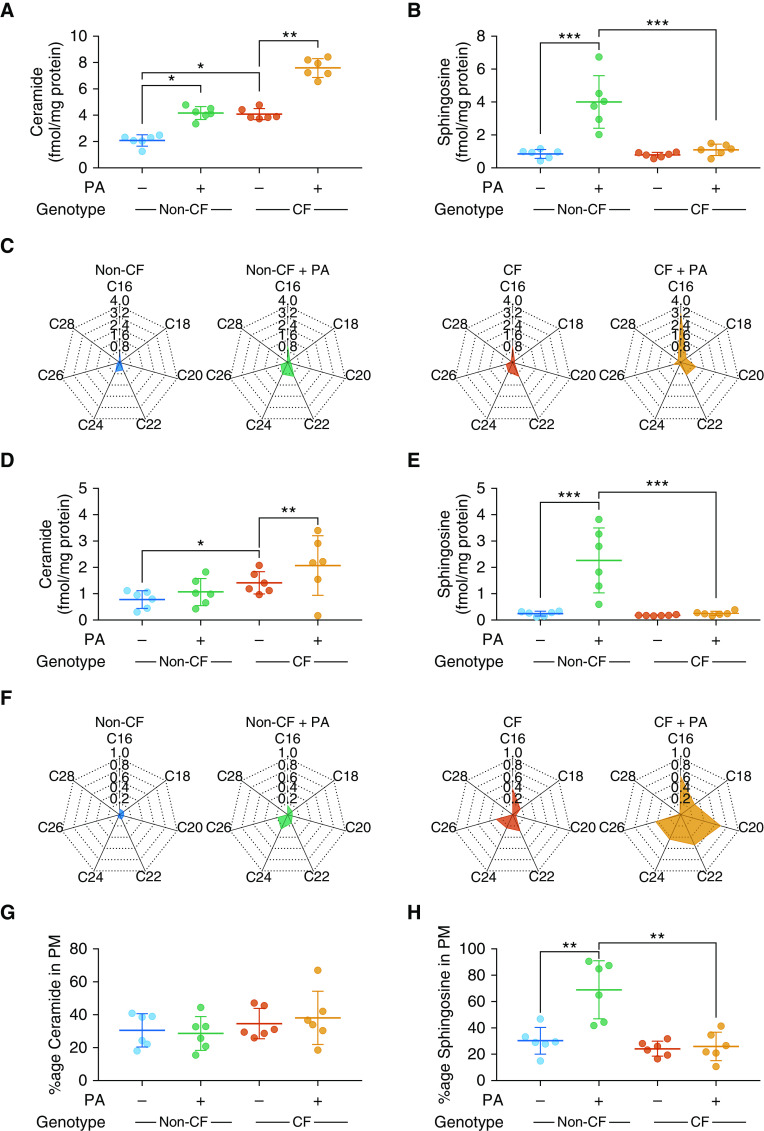
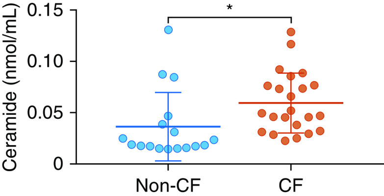
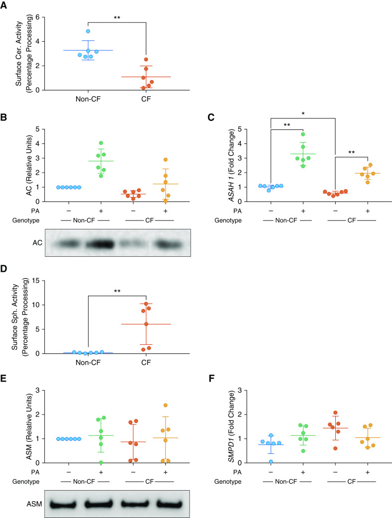
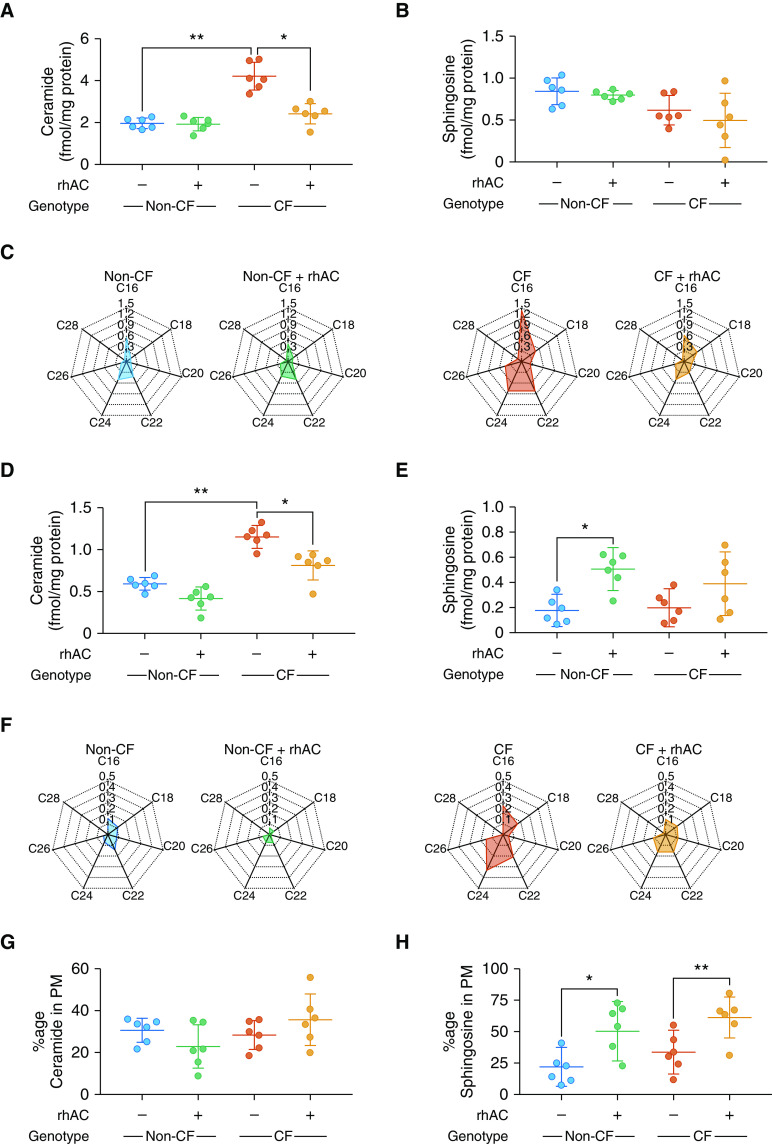
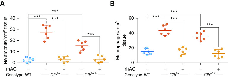
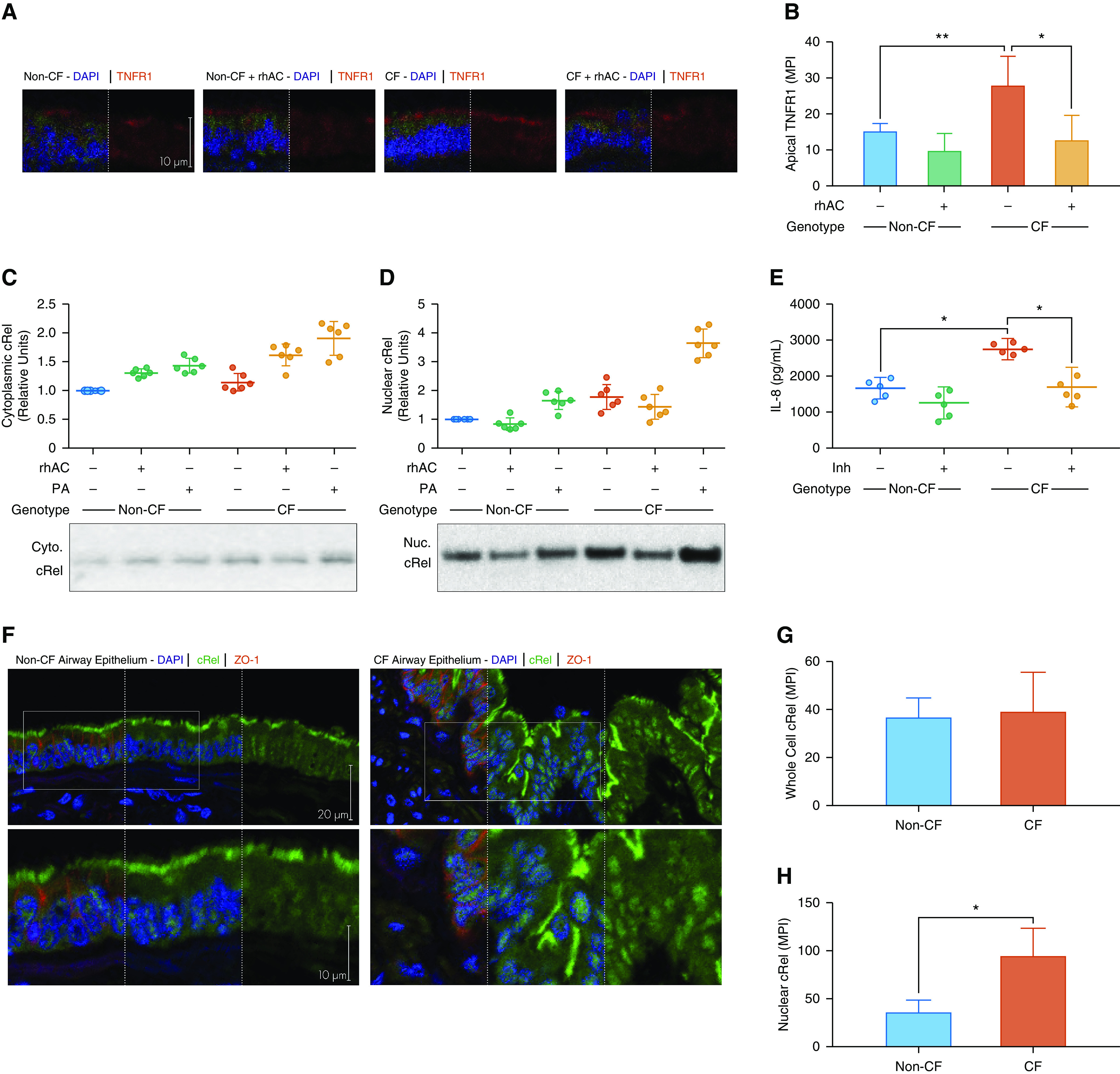
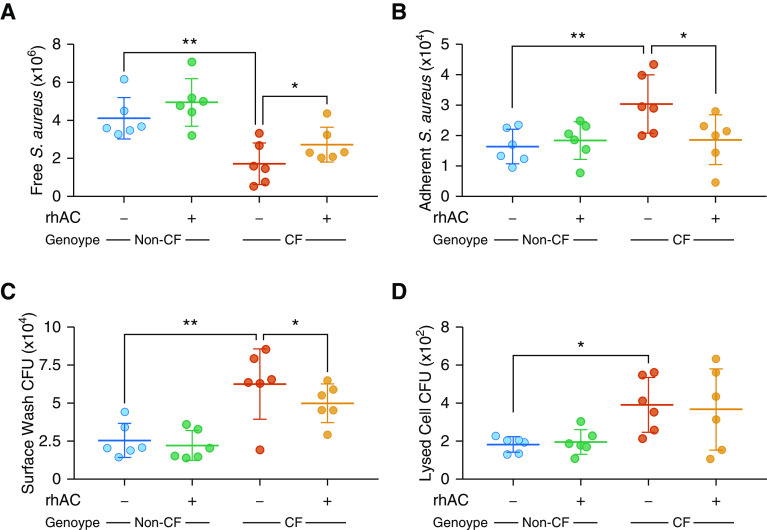
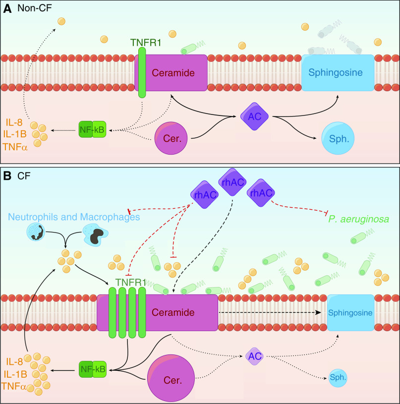
Rationale: In cystic fibrosis the major cause of morbidity and mortality is lung disease characterized by inflammation and infection. The influence of sphingolipid metabolism is poorly understood with a lack of studies using human airway model systems.Objectives: To investigate sphingolipid metabolism in cystic fibrosis and the effects of treatment with recombinant human acid ceramidase on inflammation and infection.Methods: Sphingolipids were measured using mass spectrometry in fully differentiated cultures of primary human airway epithelial cells and cocultures with Pseudomonas aeruginosa. In situ activity assays, Western blotting, and quantitative PCR were used to investigate function and expression of ceramidase and sphingomyelinase. Effects of treatment with recombinant human acid ceramidase on sphingolipid profile and inflammatory mediator production were assessed in cell cultures and murine models.Measurements and Main Results: Ceramide is increased in cystic fibrosis airway epithelium owing to differential function of enzymes regulating sphingolipid metabolism. Sphingosine, a metabolite of ceramide with antimicrobial properties, is not upregulated in response to P. aeruginosa by cystic fibrosis airway epithelia. Tumor necrosis factor receptor 1 is increased in cystic fibrosis epithelia and activates NF-κB signaling, generating inflammation. Treatment with recombinant human acid ceramidase, to decrease ceramide, reduced both inflammatory mediator production and susceptibility to infection.Conclusions: Sphingolipid metabolism is altered in airway epithelial cells cultured from people with cystic fibrosis. Treatment with recombinant acid ceramidase ameliorates the two pivotal features of cystic fibrosis lung disease, inflammation and infection, and thus represents a therapeutic approach worthy of further exploration.

Keywords: ceramide; lung; sphingolipid; sphingosine.


Figures

Figure 1.
Figure 1.
Ceramide and sphingosine levels in cystic fibrosis (CF) and non-CF fully differentiated primary human airway epithelial cell cultures at baseline and in response to Pseudomonas aeruginosa. (A and B) Levels of total ceramide (A) and sphingosine (B) from whole-cell lysates of CF and non-CF cultures at baseline and after coculture with P. aeruginosa (PA). (C) Radar charts of individual ceramide species (fmol/mg protein). (D and E) Plasma membrane (PM) fractions of cultures were isolated at baseline and after coculture with PA, allowing determination of total ceramide (D) and sphingosine (E) levels. (F) Individual ceramide species, displayed as radar charts (fmol/mg protein). (G and H) The proportion of ceramide in PMs (as a fraction of total cellular ceramide) (G) and equivalent for sphingosine (H). Throughout, n = 6 separate experiments from individual donors. Cultures were lysed and fractionated into whole-cell and plasma membrane fractions after 28 days at air–liquid interface and full differentiation. Individual data points are presented along with the mean (horizontal line) ± SD (error bars). For statistical tests used, see the online supplement. *P < 0.05, **P ≤ 0.01, and ***P ≤ 0.001.
Figure 2.
Figure 2.
Ceramide levels in BAL fluid from children and young people. Shown are levels of ceramide in BAL fluid collected during clinically indicated bronchoscopies from children and young people with cystic fibrosis (CF) and children who do not have CF but underwent a bronchoscopy for investigation of respiratory problems. Groups were matched for age (see Table E1). Individual data points are presented along with the mean (horizontal line) ± SD (error bars). An unpaired t test was used to determine significance. *P < 0.05.
Figure 3.
Figure 3.
AC (acid ceramidase) and ASM (acid sphingomyelinase) expression and function in cystic fibrosis (CF) and non-CF airway epithelial cell cultures. (A) Apical surface activity of ceramidase, as determined by percentage of fluorescently labeled ceramide processed into sphingosine. (B) Levels of AC protein in CF and non-CF cultures at baseline and after coculture with Pseudomonas aeruginosa (PA), displayed as change relative to untreated non-CF cultures. Representative blots are shown for AC (methods for full-length blots are in the online supplement, with details of loading controls shown in Figure E1). (C) Gene expression of ASAH1 (coding for acid ceramidase) at baseline in CF and non-CF cultures and after coculture with PA, displayed as fold change relative to untreated non-CF cultures. (D) Apical surface activity of sphingomyelinase, as determined by percentage of fluorescently labeled sphingomyelin processed into ceramide. (E) Levels of ASM protein, displayed as change relative to untreated non-CF cultures. Representative blots are shown for ASM (methods for full-length blots are in the online supplement, with details of loading controls shown in Figure E1). (F) Gene expression of SMPD1 (coding for acid sphingomyelinase), displayed as fold change relative to untreated non-CF cultures. For loading controls, antibody, and primer and reaction details, see Figure E1 and Tables E2–E4. Throughout, n = 6 separate experiments from individual donors. Individual data points are presented along with the mean (horizontal line) ± SD (error bars). For statistical tests used, see the online supplement. *P < 0.05 and **P ≤ 0.01. Cer = ceramidase; Sph = sphingomyelinase.
Figure 4.
Figure 4.
Effect of rhAC (recombinant human acid ceramidase) treatment on the ceramide and sphingosine profile of cystic fibrosis (CF) and non-CF airway epithelial cell cultures. (A and B) Levels of total ceramide (A) and sphingosine (B) from whole-cell lysates of CF and non-CF cultures at baseline and after treatment with rhAC. (C) Individual ceramide species, displayed as radar charts (fmol/mg protein). (D and E) Plasma membrane (PM) fractions of total ceramide (D) and sphingosine (E) in CF and non-CF cultures at baseline and after treatment with rhAC. (F) Individual ceramide species, displayed as radar charts (fmol/mg protein). (G and H) Proportion of ceramide in PMs (as a fraction of total cellular ceramide) (G) and the equivalent for sphingosine (H). Throughout, n = 6 separate experiments from individual donors. Cultures were lysed and fractionated into whole-cell and plasma membrane fractions after 28 days at air–liquid interface and full differentiation. Individual data points are presented along with the mean (horizontal line) ± SD (error bars). For statistical tests used, see the online supplement. *P < 0.05 and **P ≤ 0.01.
Figure 5.
Figure 5.
Effect of rhAC (recombinant human acid ceramidase) treatment on inflammatory mediator production by cystic fibrosis (CF) and non-CF airway epithelial cell cultures. (A–C) Time course of apical secretion of IL-8 (A), IL-1β (B), and TNFα (C) from CF and non-CF cultures at baseline and after a single treatment with rhAC. (D) Apical secretion of IL-8 from cultures after pretreatment with combinations of ivacaftor, tezacaftor–ivacaftor, and rhAC. (AC) n = 6 separate experiments; (D) n = 4, all from individual donors. (D) CF group all F508del/F508del genotype. Data are presented as mean ± SD for AC; for D, individual data points are presented along with the mean (horizontal line) ± SD (error bars). For statistical tests used, see the online supplement. *P < 0.05 and ***P ≤ 0.001. LLOD = lower limit of detection; ns = nonsignificant (P ≥ 0.05); TNF = tumor necrosis factor.
Figure 6.
Figure 6.
Effect of rhAC (recombinant human acid ceramidase) treatment on lung inflammation in murine models. (A and B) Number of neutrophils (A) and macrophages (B) in the submucosa of distal large bronchi in lung sections from wild-type, Cftrko, and CftrMHH mice at baseline and after nebulization daily for 3 days with rhAC. Throughout, n = 6 mice in each group. Individual data points are presented along with the mean (horizontal line) ± SD (error bars). For statistical tests used, see the online supplement. ***P ≤ 0.001. WT = wild type.
Figure 7.
Figure 7.
TNFR1 (tumor necrosis factor receptor 1) expression and cRel localization in cystic fibrosis (CF) airway epithelial cell cultures and lung tissue sections. (A and B) Expression of TNFR1 in CF and non-CF cultures with and without rhAC (recombinant human acid ceramidase) treatment as assessed by immunohistochemistry (A) with quantification of apical mean pixel intensity (B). (C and D) cRel expression in CF and non-CF cultures in response to Pseudomonas aeruginosa coculture in the presence or absence of rhAC in cytoplasmic (C) and nuclear (D) fractions. Representative blots are shown for both (methods are in the online supplement, with details of loading controls shown in Figure E1). (E) Apical IL-8 secretion in the presence or absence of a specific cRel inhibitor (Inh). (F–H) Expression and localization of cRel in airway epithelium in airway tissue sections from people with advanced CF lung disease and unused donor lungs (non-CF) (F) with quantification of whole-cell (G) and nuclear (H) localization. (AD) n = 6 separate experiments; (E) n = 5; (FH) n = 4, all from individual donors; see Table E1 for clinical details. Data are presented as mean ± SD for B, G, and H; for CE, individual data points are presented along with the mean (horizontal line) ± SD (error bars). For statistical tests used, see the online supplement. *P < 0.05 and **P ≤ 0.01. Cyto. = cytoplasmic; MPI = mean pixel intensity; Nuc. = nuclear; PA = Pseudomonas aeruginosa.
Figure 8.
Figure 8.
Effect of rhAC (recombinant human acid ceramidase) treatment on infection in cystic fibrosis (CF) airway epithelial cell cultures. (A and B) Number of fluorescently labeled heat-killed Staphylococcus aureus retrieved from apical surface washes (A) and adherent to apical surface (B) in CF and non-CF fully differentiated cultures with and without prior rhAC treatment. For representative images, see Figure E6 in the online supplement. (C and D) Colony-forming unit counts of Pseudomonas aeruginosa isolated from apical surface washes (C) and whole-cell lysates (D) (after washing, suggesting internalization) from CF and non-CF cultures with and without prior rhAC treatment. Live P. aeruginosa were added to the apical surface of cultures and allowed to proliferate for 24 hours. Throughout, n = 6 separate experiments. Individual data points are presented along with the mean (horizontal line) ± SD (error bars). For statistical tests used, see the online supplement. *P < 0.05 and **P ≤ 0.01.
Figure 9.
Figure 9.
Proposed model of how altered sphingolipid metabolism in cystic fibrosis (CF) airway epithelia may result in increased inflammation and susceptibility to infection. (A) In non-CF epithelia, AC maintains the balance of ceramide and sphingosine. Normal levels of ceramide do not promote a proinflammatory environment, and in response to Pseudomonas aeruginosa, levels of sphingosine are upregulated. (B) In CF epithelia, AC activity is deficient in both expression and activity, which, in combination with alterations in acid sphingomyelinase activity, leads to the accumulation of ceramide. Raised ceramide is associated with increased TNFR1 expression, enhanced NF-κB activation, and nuclear localization of cRel. This promotes the secretion of proinflammatory cytokines such as IL-8, IL-1β, and TNFα. In conjunction with excessive recruitment of immune cells, which also produce proinflammatory mediators, a positive feedback loop emerges in the CF airway. CF epithelia do not upregulate sphingosine in response to P. aeruginosa, increasing susceptibility to infection, which further contributes to the proinflammatory environment. Treatment with rhAC reduces ceramide and increases sphingosine, ameliorating these effects. AC = acid ceramidase; Cer = ceramide; rhAC = recombinant human acid ceramidase; Sph = sphingosine; TNFR1 = tumor necrosis factor receptor 1.

Comment in

References

    1. Elborn JS. Cystic fibrosis. Lancet. 2016;388:2519–2531. - PubMed
    1. Ramsey BW, Downey GP, Goss CH. Update in cystic fibrosis 2018. Am J Respir Crit Care Med. 2019;199:1188–1194. - PMC - PubMed
    1. Haq IJ, Gray MA, Garnett JP, Ward C, Brodlie M. Airway surface liquid homeostasis in cystic fibrosis: pathophysiology and therapeutic targets. Thorax. 2016;71:284–287. - PubMed
    1. Pezzulo AA, Tang XX, Hoegger MJ, Abou Alaiwa MH, Ramachandran S, Moninger TO, et al. Reduced airway surface pH impairs bacterial killing in the porcine cystic fibrosis lung. Nature. 2012;487:109–113. - PMC - PubMed
    1. Hoegger MJ, Fischer AJ, McMenimen JD, Ostedgaard LS, Tucker AJ, Awadalla MA, et al. Impaired mucus detachment disrupts mucociliary transport in a piglet model of cystic fibrosis. Science. 2014;345:818–822. - PMC - PubMed

Publication types

MeSH terms