Review
. 2020 Sep;159(3):849-863.
doi: 10.1053/j.gastro.2020.04.077. Epub 2020 Jun 20.

Contribution of the Intestinal Microbiome and Gut Barrier to Hepatic Disorders

Affiliations
Review

Contribution of the Intestinal Microbiome and Gut Barrier to Hepatic Disorders

Daniel M Chopyk et al. Gastroenterology. 2020 Sep.

Abstract

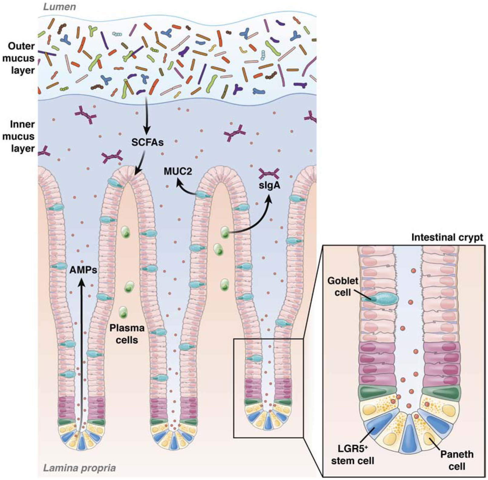
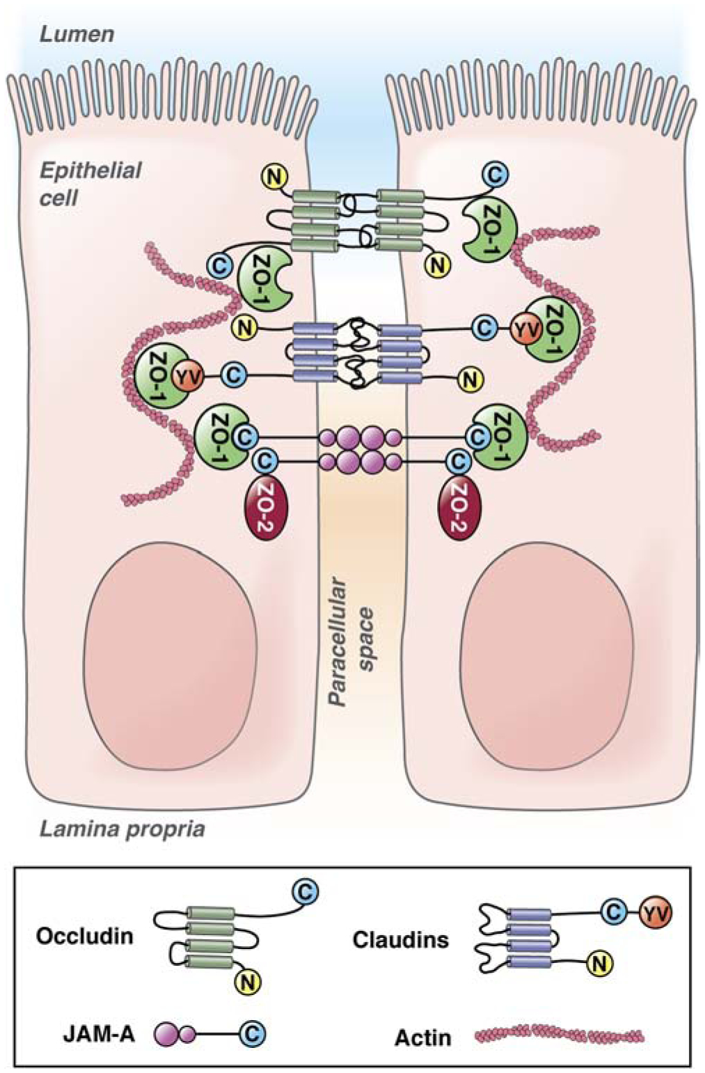
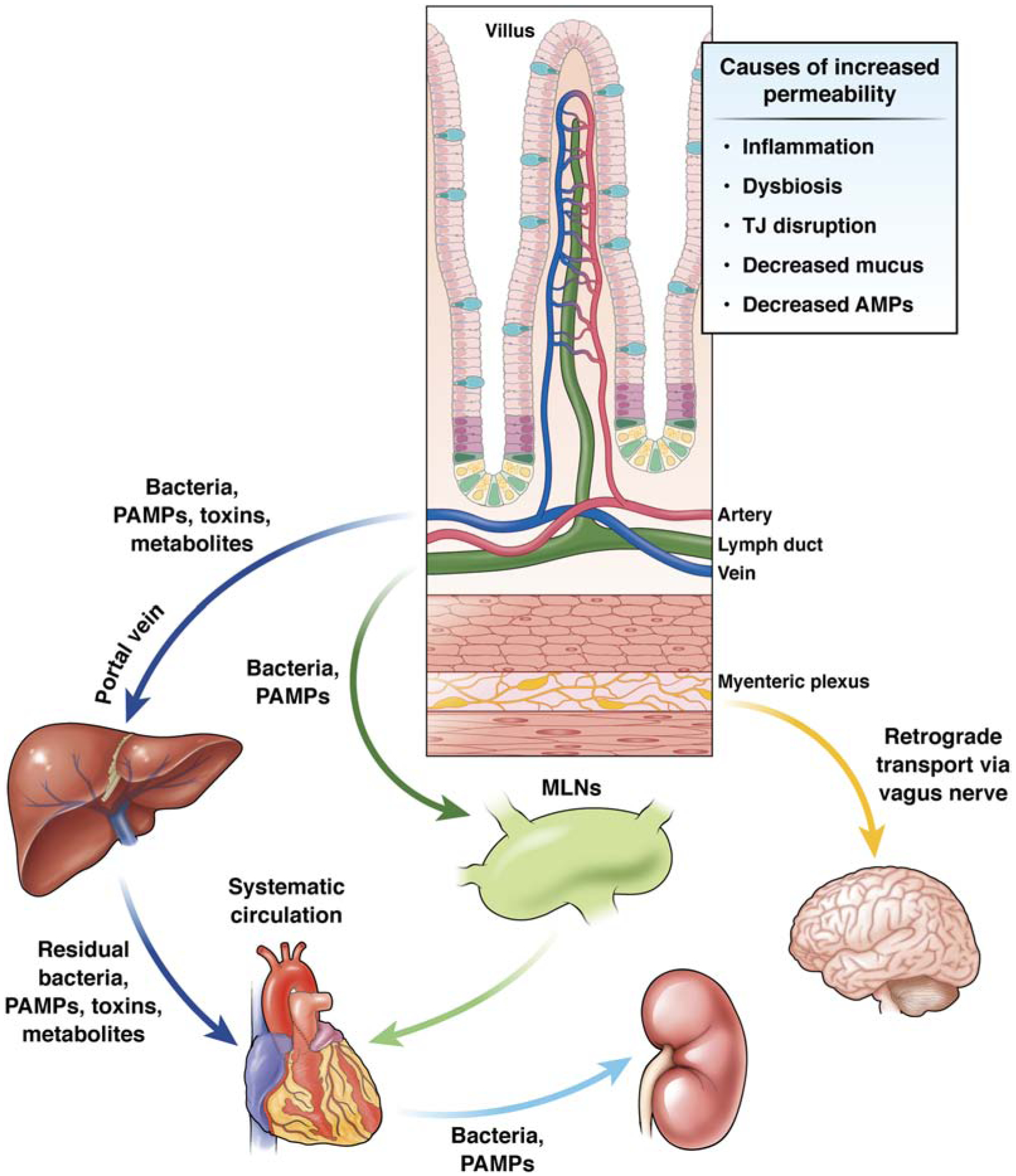
Intestinal barrier dysfunction and dysbiosis contribute to development of diseases in liver and other organs. Physical, immunologic, and microbiologic (bacterial, fungal, archaeal, viral, and protozoal) features of the intestine separate its nearly 100 trillion microbes from the rest of the human body. Failure of any aspect of this barrier can result in translocation of microbes into the blood and sustained inflammatory response that promote liver injury, fibrosis, cirrhosis, and oncogenic transformation. Alterations in intestinal microbial populations or their functions can also affect health. We review the mechanisms that regulate intestinal permeability and how changes in the intestinal microbiome contribute to development of acute and chronic liver diseases. We discuss individual components of the intestinal barrier and how these are disrupted during development of different liver diseases. Learning more about these processes will increase our understanding of the interactions among the liver, intestine, and its flora.

Keywords: Alcoholic Liver Disease; Drug Induced Liver Injury; Gut Permeability; Hepatic Disorders; Leaky Gut; Liver Disease; Microbiome; Nonalcoholic Liver Disease; Primary Sclerosing Cholangitis.


Conflict of interest statement

Potential conflict of interest: Nothing to report.

Figures

Figure 1.
Figure 1.. Components of the gut barrier.
The intestinal barrier comprises mucus, microbial, epithelial, and immunological components. Within the colon the mucus forms 2 layers—a more loose, outer layer where most of the intestinal bacteria reside, and a dense, inner layer that does not contain bacteria. The commensal microbiota reinforce the gut barrier by preventing colonization by pathogens and producing useful metabolites such as SCFAs, which promote epithelial health and integrity. Goblet cells are scattered through the intestinal epithelial monolayer and produce mucus. Additional specialized cellular populations are found within the bases of the intestinal crypts. LGR5+ cells are a source of continuous cell renewal to maintain epithelial integrity. These stem cells give rise to differentiated Paneth cells, which remain at the base of the crypts and produce large amounts of antimicrobial peptides (AMPs) and growth factors. Plasma cells within the lamina propria also secrete dimeric Ig A, which is transported across epithelial cells and contributes to immune exclusion of the luminal microbiota. MUC2, mucin 2; sIgA, secretory IgA.
Figure 2.
Figure 2.. Intestinal epithelial tight junctions.
Intestinal epithelial tight junctions are composed of 3 classes of transmembrane proteins that associate with scaffolding proteins that link to the actin cytoskeleton. The transmembrane proteins include occludin, claudins, and JAMA. Occludin and claudin proteins have cytoplasmic N- and C-termini and 4 transmembrane domains. JAMA has a cytoplasmic C-terminus and 2extracellular V-type Ig domains. Importantly, JAMA can dimerize in cis (molecules on the same cell) and in trans (molecules on adjacent cells).
Figure 3.
Figure 3.. Mechanisms of gut barrier dysfunction and routes for systemic entry of translocated bacteria and toxins.
Conditions such as dysbiosis, inflammation, and TJ dysfunction can increase gut permeability. When the intestinal barrier is compromised, translocated bacteria and microbial toxins can gain axis to distant sites. Bacteria and PAMPs can enter the portal circulation and access to the liver. The liver contains large populations of immune cells that induce an inflammatory response to these stimuli. A portion of these bacteria, PAMPS, and metabolites pass through the liver where they gain access to the systemic circulation. In parallel, a number of translocated bacteria and PAMPs from the intestine gain access to the lymphatic vasculature, where they first pass through the MLNs. A portion of these intra-lymphatic toxins will enter the systemic circulation. Intestine-derived bacteria, PAMPs, toxins, and metabolites affect the function of organs including the heart, kidney, and brain. Translocated gut pathogens also affect the brain via retrograde transport along fibers of the vagus nerve that contribute to the myenteric plexus.

References

    1. Tripathi A, Debelius J, Brenner DA, et al. The gut-liver axis and the intersection with the microbiome. Nat Rev Gastroenterol Hepatol 2018;15:397–411. - PMC - PubMed
    1. Powell N, Walker MM, Talley NJ. The mucosal immune system: master regulator of bidirectional gut-brain communications. Nat Rev Gastroenterol Hepatol 2017;14:143–159. - PubMed
    1. Pasini E, Aquilani R, Testa C, et al. Pathogenic Gut Flora in Patients With Chronic Heart Failure. JACC Heart Fail 2016;4:220–7. - PubMed
    1. Yang J, Lim SY, Ko YS, et al. Intestinal barrier disruption and dysregulated mucosal immunity contribute to kidney fibrosis in chronic kidney disease. Nephrol Dial Transplant 2019;34:419–428. - PubMed
    1. Hiippala K, Jouhten H, Ronkainen A, et al. The Potential of Gut Commensals in Reinforcing Intestinal Barrier Function and Alleviating Inflammation . Nutrients 2018;10. - PMC - PubMed

Publication types