. 2020 Oct;159(4):1375-1389.
doi: 10.1053/j.gastro.2020.06.038. Epub 2020 Jun 20.

IL1B Increases Intestinal Tight Junction Permeability by Up-regulation of MIR200C-3p, Which Degrades Occludin mRNA

Affiliations

IL1B Increases Intestinal Tight Junction Permeability by Up-regulation of MIR200C-3p, Which Degrades Occludin mRNA

Manmeet Rawat et al. Gastroenterology. 2020 Oct.

Abstract

Background & aims: Defects in the epithelial tight junction (TJ) barrier contribute to development of intestinal inflammation associated with diseases. Interleukin 1 beta (IL1B) increases intestinal permeability in mice. We investigated microRNAs that are regulated by IL1B and their effects on expression of TJ proteins and intestinal permeability.

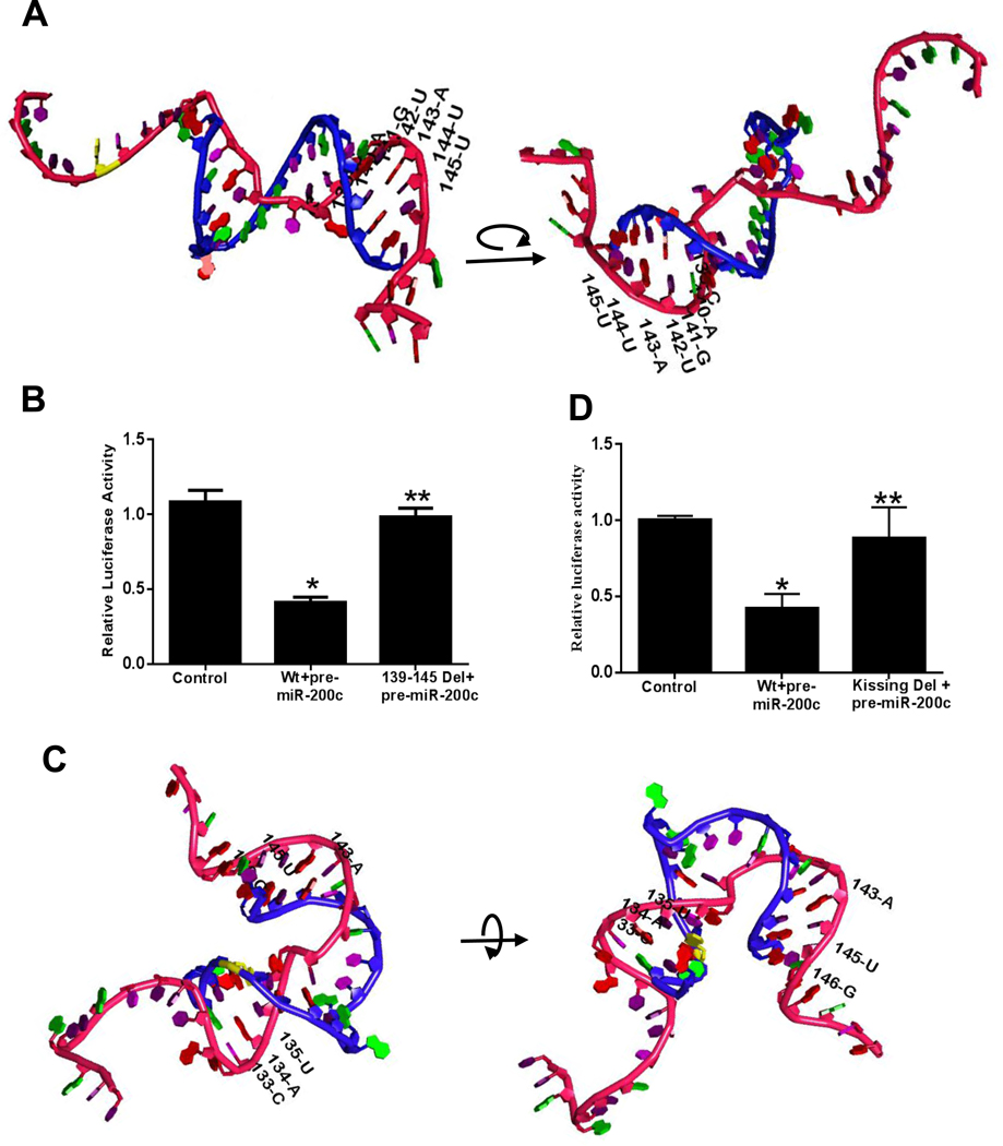
Methods: We used Targetscan to identify microRNAs that would bind the 3' untranslated region (3'UTR) of occludin mRNA; regions that interacted with microRNAs were predicted using the V-fold server and Assemble2, and 3-dimensional models were created using UCSF Chimera linked with Assemble2. Caco-2 cells were transfected with vectors that express microRNAs, analyzed by immunoblots and real-time polymerase chain reaction (PCR), and grown as monolayers; permeability in response to IL1B was assessed with the marker inulin. Male C57BL/6 mice were given intraperitoneal injections of IL1B and intestinal recycling perfusion was measured; some mice were given dextran sodium sulfate to induce colitis and/or gavage with an antagonist to MIR200C-3p (antagomiR-200C) or the nonspecific antagomiR (control). Intestinal tissues were collected from mice and analyzed by histology and real-time PCR; enterocytes were isolated by laser capture microdissection. We also analyzed colon tissues and organoids from patients with and without ulcerative colitis.

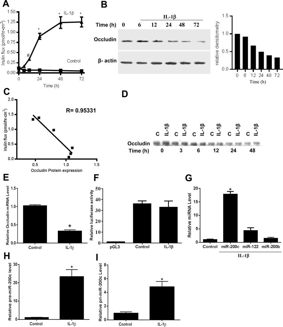
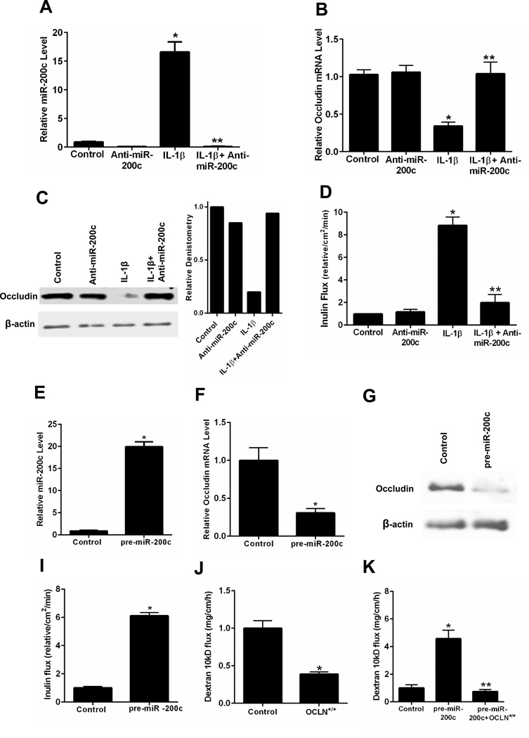
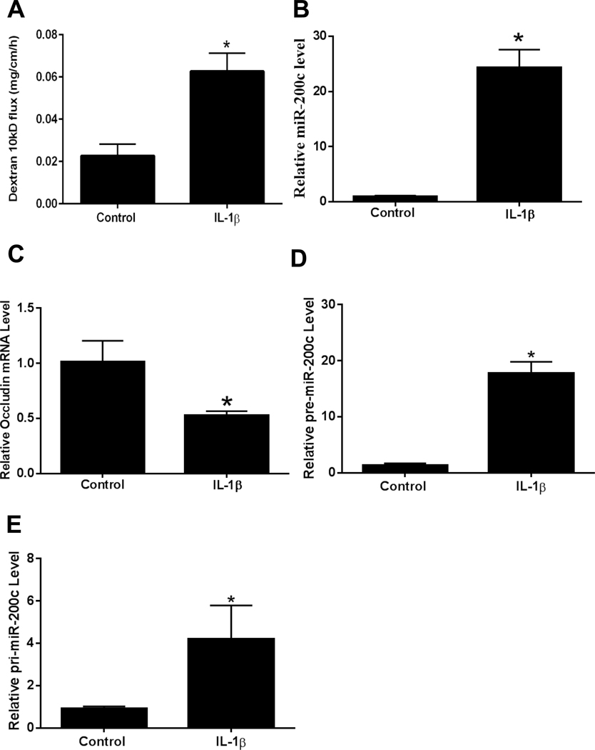
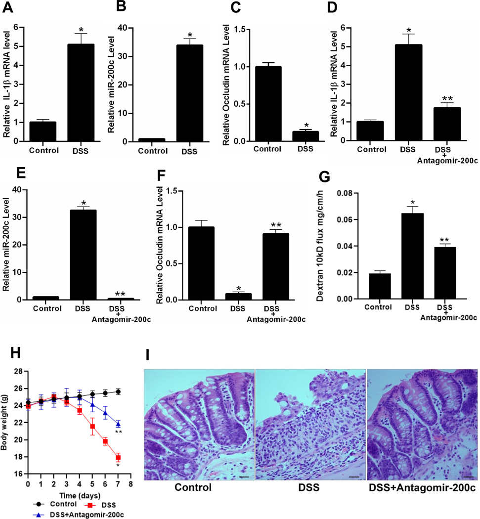
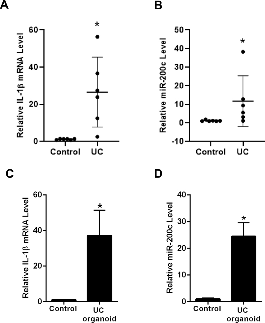
Results: Incubation of Caco-2 monolayers with IL1B increased TJ permeability and reduced levels of occludin protein and mRNA without affecting the expression of other transmembrane TJ proteins. Targetscan identified MIR122, MIR200B-3p, and MIR200C-3p, as miRNAs that might bind to the occludin 3'UTR. MIR200C-3p was rapidly increased in Caco-2 cells incubated with IL1B; the antagomiR-200c prevented the IL1B-induced decrease in occludin mRNA and protein and reduced TJ permeability. Administration of IL1B to mice increased small intestinal TJ permeability, compared with mice given vehicle; enterocytes isolated from mice given IL1B had increased expression of MIR200C-3p and decreased levels of occludin messenger RNA (mRNA) and protein. Intestinal tissues from mice with colitis had increased levels of IL1B mRNA and MIR200C-3p and decreased levels of occludin mRNA; gavage of mice with antagomiR-200C reduced levels of MIR200C-3p and prevented the decrease in occludin mRNA and the increase in colonic permeability. Colon tissues and organoids from patients with ulcerative colitis had increased levels of IL1B mRNA and MIR200C-3p compared with healthy controls. Using 3-dimensional molecular modeling and mutational analyses, we identified the nucleotide bases in the occluding mRNA 3'UTR that interact with MIR200C-3p.

Conclusions: Intestine tissues from patients with ulcerative colitis and mice with colitis have increased levels of IL1B mRNA and MIR200C-3p, which reduces expression of occludin by enterocytes and thereby increases TJ permeability. Three-dimensional modeling of the interaction between MIR200C-3p and the occludin mRNA 3'UTR identified sites of interaction. The antagomiR-200C prevents the decrease in occludin in enterocytes and intestine tissues of mice with colitis, maintaining the TJ barrier.

Keywords: Epigenetic Modification; MicroRNA 200C; RNA Degradation; Translation.


Conflict of interest statement

Conflict of interest

The authors disclose no conflicts.

Figures

Figure 1.
Figure 1.
IL-1β effect on occludin paracellular permeability, protein expression, occludin mRNA transcription, promoter activity and miRNA expression in filter-grown Caco-2 monolayers. (A,B) Time course effect of IL-1β on mucosal-to-serosal flux of inulin and occludin expression (C) Occludin expression vs inulin flux, the correlation coefficient of occludin expression and inulin flux was 0.95. (D) Filter-grown Caco-2 monolayers were treated with IL-1β and the time course of 35S-labelled occludin (pulse-chase) decomposition was determined. (E,F) Effect of IL-1β (10 ng/ml) over 2-hours on occludin mRNA expression and occludin promoter activity. (G) Effect of IL-1β (10 ng/ml) over 2-hour on in silico predicted miRNA expression as determined by real-time PCR. (H,I) Effect of IL-1β on pre-miRNA-200c and pri-miRNA-200c expression. *P < .001 vs control, # P < .005 vs control. pre-miR-200c: precursor microRNA of miR-200c-3p; pri-miR-200c: primary microRNA of miR-200c-3p.
Figure 2.
Figure 2.
Effect of antisense ribonucleotide (ASO) inhibition of miR-200c-3p and miR-200c-3p overexpression on occludin mRNA, protein expression and paracellular permeability in filter-grown Caco-2 monolayers. Effect of anti-miR-200c-3p transfection on IL-1β induced increase on miR-200c-3p expression (A), decrease in occludin mRNA expression (B), decrease in occludin protein expression and relative occludin densitometry (C), and increase in inulin flux (D). Effect of pre-miR-200c-3p transfection on miR-200c-3p expression (E), occludin mRNA expression (F), occludin protein expression (G), and Caco-2 paracellular permeability to inulin (H). Effects of occludin gene transfection on Caco-2 paracellular permeability to dextran 10kD (I), and pre-miR-200c induced increase in dextran 10kD flux (J). * P < 0.01 vs control; * * P < 0.001 vs IL-1β treatment and pre-miR-200c; OCLN+/+: plasmid pCMV3-OCLN-OFPSpark
Figure 3.
Figure 3.
Effect of in vivo intraperitoneal administration of IL-1β (5μg) on mouse intestinal epithelial cell expression of miR-200c-3p and occludin mRNA and mouse intestinal permeability after 24 h treatment period. Effect of intraperitoneal IL-1β on luminal-to-serosal FITC-dextran (MW = 10,000 g/mol) flux (A), mouse enterocyte miR-200c-3p expression (B), mouse enterocyte occludin mRNA expression (C), mouse enterocyte pre-miR-200c expression (D), and mouse enterocyte pri-miR-200c expression (E). Mouse enterocytes were isolated using LCM. * P < 0.01 vs control. pre-miR-200c: precursor microRNA of miR-200c-3p; pri-miR-200c: primary microRNA of miR-200c-3p.
Figure 4.
Figure 4.
The effect of anti-miR-200c-3p transfection and miR-200c-3p overexpression on mouse small intestinal permeability in vivo. Effect of anti-miR-200c-3p transfection on IL-1β induced increase in mouse enterocyte miR-200c-3p expression (A). Mouse intestinal mucosal surface was transfected anti-miR-200c-3p in vivo. Effect of anti-miR-200c-3p transfection on IL-1β induced decrease in mouse enterocyte occludin mRNA expression (B), and increase in mouse intestinal permeability(C). Mouse small intestinal mucosal surface were transfected with pre-miR-200c-3p in vivo. Enterocytes were isolated from the mucosal surface by LCM. Effect of pre-miR-200c-3p transfection on mouse enterocyte miR-200c-3p (D), and occludin mRNA (E) expression. Effect of pre-miR-200c-3p and anti-miR-200c-3p transfection on intestinal tissue occludin protein expression (F), and pre-miR-200c-3p transfection on mouse intestinal permeability in vivo (G). * P < .01 vs control, * * P < .01 vs IL-1β treatment.
Figure 5.
Figure 5.
The effect of murine DSS-induced colitis on cooperation between IL-1β, miR-200c-3p, and mouse intestinal permeability. Effect of DSS oral administration for 7 days on IL-1β mRNA expression (A), miR-200c-3p expression (B) and on occludin mRNA expression (C). The colonic perfusion of DSS mice to analyze the effect of antagomiR-200c on DSS induced increase in IL-1β mRNA expression (D), increase on miR-200c-3p expression (E), decrease in occludin mRNA expression (F), and increase in dextran 10kD flux (G). Effect of antagomiR-200c gavage on body weights of DSS mice (H), Histology of mice colon to see the effect of antagomiR-200c, (I). *P < .01 vs control; * *P < .01 vs DSS; Histology scale-black bar: 20μm.
Figure 6.
Figure 6.
Expression of IL-1β mRNA and miR-200c-3p in human ulcerative colitis (UC) tissue and in human colon organoids. IL-1β mRNA expression (A), and miR-200c-3p expression (B) in human ulcerative colitis (UC) tissue. Control tissues from the six non-IBD/UC individuals and inflamed tissues from gut of the six ulcerative colitis (UC) patients were used. IL-1β mRNA expression (C), and miR-200c-3p expression (D) in ulcerative colitis (UC) vs non-IBD/UC control colon organoids. *P < .05 vs control.
Figure 7.
Figure 7.
The potential molecular interactions of miRNA-200c-3p and occludin mRNA 3’UTR predicted by in silico bioinformatics analysis. Occludin 3’UTR region was cloned into pMIR-REPORT vector in 5’to 3’ direction, and the effect of pre-miR-200c-3p transfection on occludin 3’UTR was determined by luciferase assay. (A) Schematic representation of the predicted 3D final heterodimer formation of miRNA-200c-3p and occludin mRNA 3’UTR as predicted by bioinformatics analyses (rotated for full coverage), Red backbone represents the mature miR-200c-3p, blue backbone represents the occludin 3’UTR fragment (120–151). (B) MiR-200c-3p binding site (139-C, 140-A, 141-G, 142-U, 143-A, 144-U and 145-U) was deleted by site-directed mutagenesis, and effect of pre–miR-200c-3p transfection on deletion (Del) construct was determined. (C) Schematic representation of the initial 3D molecular interactions between miRNA-200c-3p and occludin mRNA 3’UTR (rotated for full coverage), Red backbone represents the mature miR-200c-3p, blue backbone represents the occludin 3’UTR fragment (120–151). (D) Targeted mutation of bases involved in initial kissing interactions (133-C, 134-A, 135-U, 143-A, 145-U and 146-G) prevented pre-miR-200c-3p induced inhibition of occludin mRNA 3’ UTR activity. *P < 0.01 vs control, **P <0.05 vs wild type (Wt) transfected with pre–miR-200c-3p.

References

    1. Konig J, Wells J, Cani PD, et al. Human Intestinal Barrier Function in Health and Disease. Clin Transl Gastroenterol 2016;7:e196. - PMC - PubMed
    1. Ma TY. Intestinal epithelial barrier dysfunction in Crohn’s disease. Proc Soc Exp Biol Med 1997;214:318–27. - PubMed
    1. Madara JL. Loosening tight junctions. Lessons from the intestine. J Clin Invest 1989;83:1089–94. - PMC - PubMed
    1. Turner JR. Intestinal mucosal barrier function in health and disease. Nat Rev Immunol 2009;9:799–809. - PubMed
    1. Arrieta MC, Madsen K, Doyle J, et al. Reducing small intestinal permeability attenuates colitis in the IL10 gene-deficient mouse. Gut 2009;58:41–8. - PMC - PubMed

Publication types

MeSH terms