

The Basis for Natural Multiresistance to Phage in Pseudomonas aeruginosa
- PMID: 32570896
- PMCID: PMC7344871
- DOI: 10.3390/antibiotics9060339
The Basis for Natural Multiresistance to Phage in Pseudomonas aeruginosa
Abstract
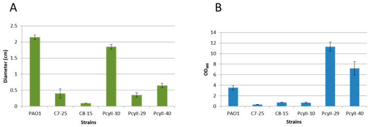
Pseudomonas aeruginosa is responsible for long-term infections and is particularly resistant to treatments when hiding inside the extracellular matrix or biofilms. Phage therapy might represent an alternative to antibiotic treatment, but up to 10% of clinical strains appear to resist multiple phages. We investigated the characteristics of P. aeruginosa clinical strains naturally resistant to phages and compared them to highly susceptible strains. The phage-resistant strains were defective in lipopolysaccharide (LPS) biosynthesis, were nonmotile and displayed an important degree of autolysis, releasing phages and pyocins. Complete genome sequencing of three resistant strains showed the existence of a large accessory genome made of multiple insertion elements, genomic islands, pyocins and prophages, including two phages performing lateral transduction. Mutations were found in genes responsible for the synthesis of LPS and/or type IV pilus, the major receptors for most phages. CRISPR-Cas systems appeared to be absent or inactive in phage-resistant strains, confirming that they do not play a role in the resistance to lytic phages but control the insertion of exogenous sequences. We show that, despite their apparent weakness, the multiphage-resistant strains described in this study displayed selective advantages through the possession of various functions, including weapons to eliminate other strains of the same or closely related species.
Keywords: CRISPR-Cas systems; Pseudomonas aeruginosa; bacteriophage; coevolution; genome sequence; lateral transduction; mechanisms of resistance; mobile genetic elements; phage therapy; prophage induction.
Conflict of interest statement
The authors declare no conflict of interest. The funder did not play any role in the study design, data collection and analysis; decision to publish or preparation of the manuscript.
Figures


References
-
- Larche J., Pouillot F., Essoh C., Libisch B., Straut M., Lee J.C., Soler C., Lamarca R., Gleize E., Gabard J., et al. Rapid identification of international multidrug-resistant Pseudomonas aeruginosa clones by multiple-locus variable number of tandem repeats analysis and investigation of their susceptibility to lytic bacteriophages. Antimicrob. Agents Chemother. 2012;56:6175–6180. doi: 10.1128/AAC.01233-12. - DOI - PMC - PubMed
-
- Croughs P.D., Klaassen C.H.W., van Rosmalen J., Maghdid D.M., Boers S.A., Hays J.P., Goessens W.H.F. Unexpected mechanisms of resistance in Dutch Pseudomonas aeruginosa isolates collected during 14 years of surveillance. Int. J. Antimicrob. Agents. 2018;52:407–410. doi: 10.1016/j.ijantimicag.2018.05.009. - DOI - PubMed
-
- Vu-Thien H., Corbineau G., Hormigos K., Fauroux B., Corvol H., Clement A., Vergnaud G., Pourcel C. Multiple-locus variable-number tandem-repeat analysis for longitudinal survey of sources of Pseudomonas aeruginosa infection in cystic fibrosis patients. J. Clin. Microbiol. 2007;45:3175–3183. doi: 10.1128/JCM.00702-07. - DOI - PMC - PubMed
Grants and funding
LinkOut - more resources
Full Text Sources
