

Clinical and CT findings of COVID-19: differences among three age groups
- PMID: 32571228
- PMCID: PMC7306933
- DOI: 10.1186/s12879-020-05154-9
Clinical and CT findings of COVID-19: differences among three age groups
Abstract
Background: The novel coronavirus pneumonia (coronavirus disease 2019, COVID-19) has spread around the world. We aimed to recapitulate the clinical and CT imaging features of COVID-19 and their differences in three age groups.
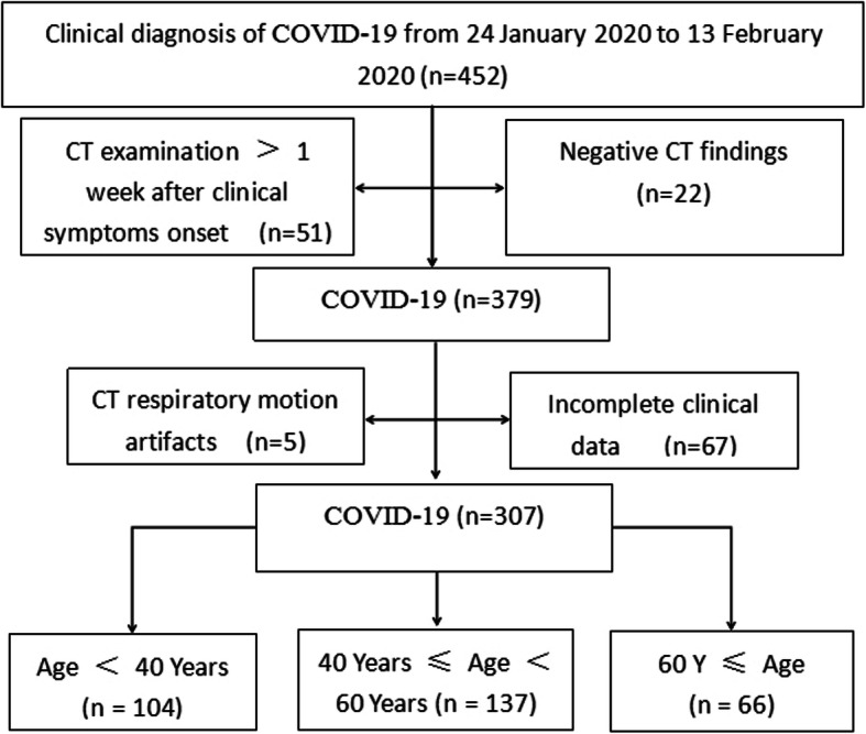
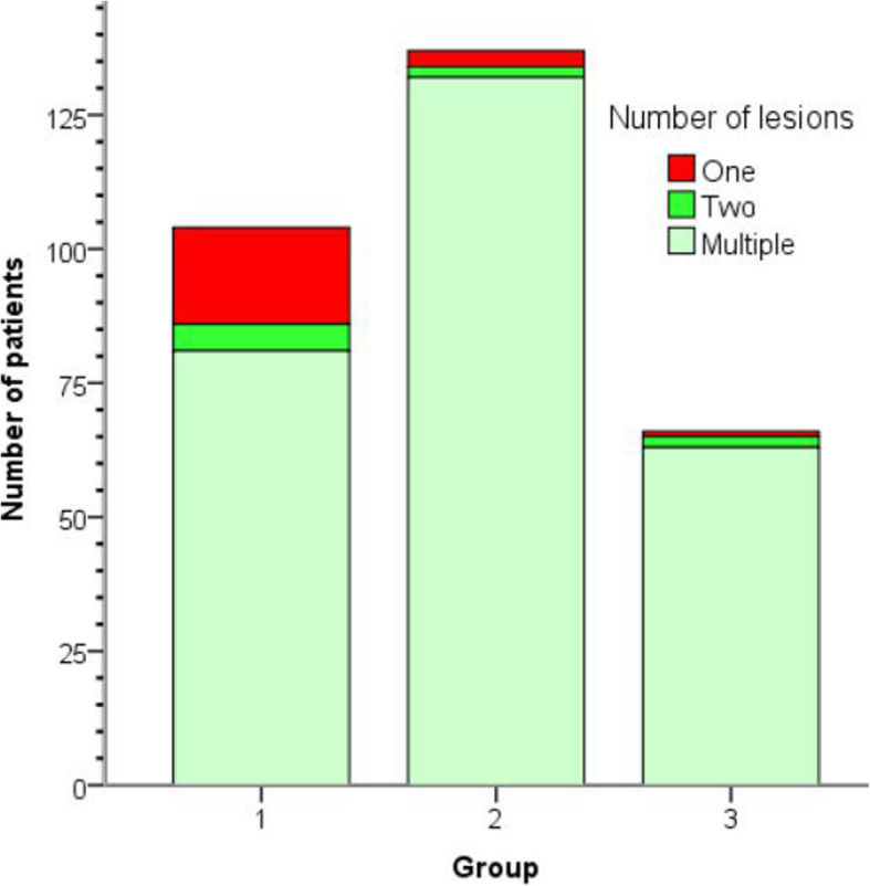
Methods: The clinical and CT data of patients with COVID-19 (n = 307) that had been divided into three groups (Group 1: < 40 years old; Group 2: 40 ≤ age < 60 years old; Group 3: ≥ 60 years old) according to age were analyzed retrospectively.
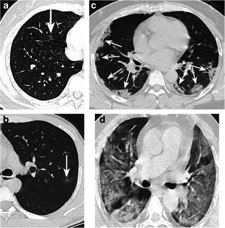
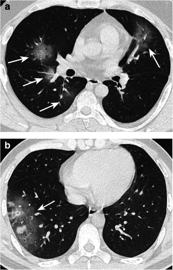
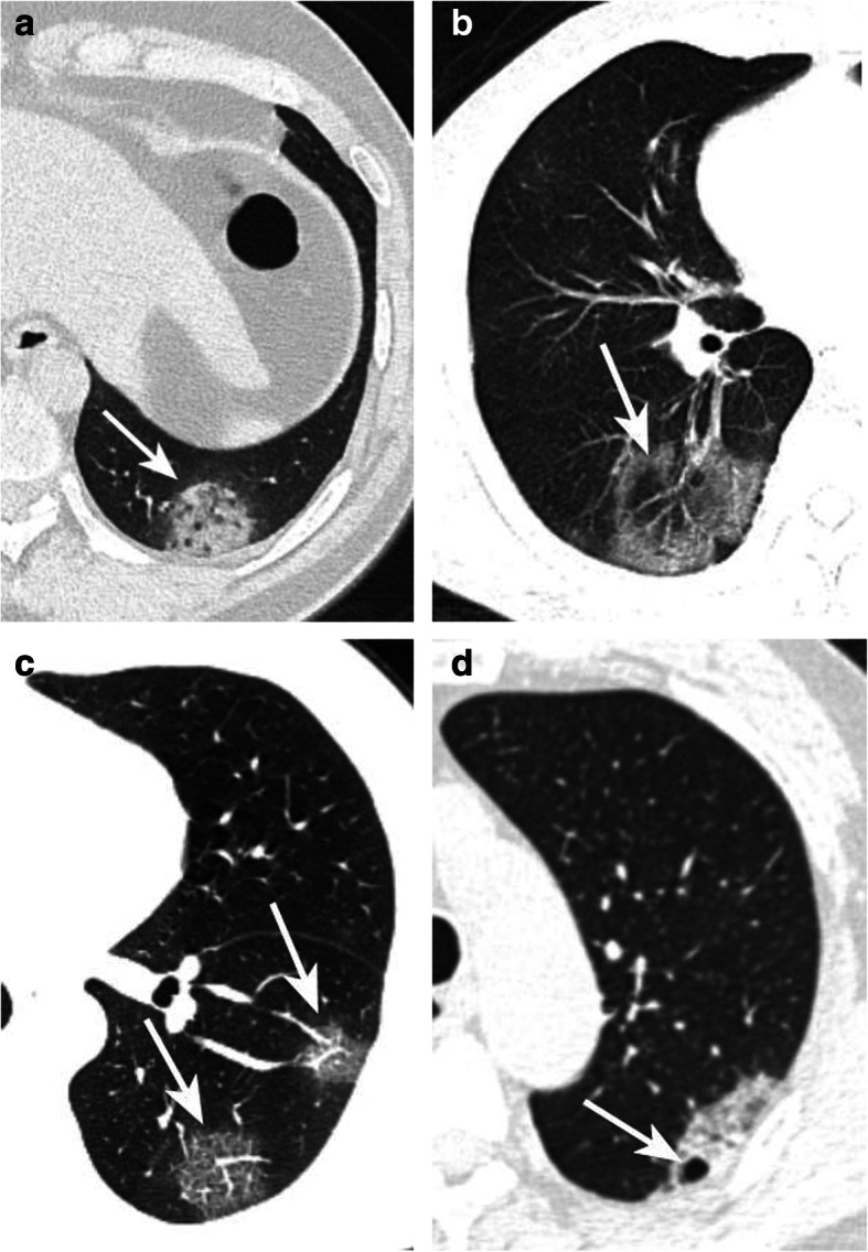
Results: Of all patients, 114 (37.1%) had histories of epidemiological exposure, 48 (15.6%) were severe/critical cases, 31 had hypertension (10.1%), 15 had diabetes mellitus (4.9%), 3 had chronic obstructive pulmonary disease (COPD, 1%). Among the three groups, severe/critical type, hypertension and diabetes occurred more commonly in the elderly group compared with Group 1&2 (P < 0.05, respectively). Cough and chest tightness/pain were more commonly appeared in Group 2&3 compared with Group 1 (P < 0.05, respectively). Compared with Group 1 and 2, there were more abnormal laboratory examination indexes (including CRP increase, abnormal percentage of lymphocytes, neutrophils and monocytes) in Group 3 (P < 0.05, respectively). CT images revealed that more lobes were affected and more subpleural lesions were involved in the elderly group, besides, crazy paving sign, bronchodilatation and pleural thickening were more commonly seen in the elderly group, with significant difference between Group 1&2, Group 2&3 (P < 0.05, respectively).
Conclusions: COVID-19 presented representative clinical manifestations, laboratory examinations and CT findings, but three age groups possessed their own specific characteristics. Grasping the clinical and CT features stratified by age will be helpful for early definite diagnosis of COVID-19.
Keywords: Age; COVID-19; Pulmonary infection; X-ray computed tomography.
Conflict of interest statement
The authors declared that there is no conflict of interest.
Figures
Similar articles
-
A retrospective study of the initial chest CT imaging findings in 50 COVID-19 patients stratified by gender and age.J Xray Sci Technol. 2020;28(5):875-884. doi: 10.3233/XST-200709. J Xray Sci Technol. 2020. PMID: 32804112 Free PMC article.
-
A Comparison of Clinical and Chest CT Findings in Patients With Influenza A (H1N1) Virus Infection and Coronavirus Disease (COVID-19).AJR Am J Roentgenol. 2020 Nov;215(5):1065-1071. doi: 10.2214/AJR.20.23214. Epub 2020 May 26. AJR Am J Roentgenol. 2020. PMID: 32452731
-
[CT imaging analysis of 33 cases with the 2019 novel coronavirus infection].Zhonghua Yi Xue Za Zhi. 2020 Apr 7;100(13):1007-1011. doi: 10.3760/cma.j.cn112137-20200203-00182. Zhonghua Yi Xue Za Zhi. 2020. PMID: 32294858 Chinese.
-
Coronavirus Disease 2019 (COVID-19): A Systematic Review of Imaging Findings in 919 Patients.AJR Am J Roentgenol. 2020 Jul;215(1):87-93. doi: 10.2214/AJR.20.23034. Epub 2020 Mar 14. AJR Am J Roentgenol. 2020. PMID: 32174129
-
Thoracic imaging tests for the diagnosis of COVID-19.Cochrane Database Syst Rev. 2020 Sep 30;9:CD013639. doi: 10.1002/14651858.CD013639.pub2. Cochrane Database Syst Rev. 2020. Update in: Cochrane Database Syst Rev. 2020 Nov 26;11:CD013639. doi: 10.1002/14651858.CD013639.pub3. PMID: 32997361 Updated.
Cited by
-
Evaluation of initial chest computed tomography (CT) findings of COVID-19 pneumonia in 117 deceased patients: a retrospective study.Turk J Med Sci. 2021 Jun 28;51(3):929-938. doi: 10.3906/sag-2009-183. Turk J Med Sci. 2021. PMID: 33315351 Free PMC article.
-
Analysis of Emergency Department Visits and Hospital Activity during Influenza Season, COVID-19 Epidemic, and Lockdown Periods in View of Managing a Future Disaster Risk: A Multicenter Observational Study.Int J Environ Res Public Health. 2020 Nov 10;17(22):8302. doi: 10.3390/ijerph17228302. Int J Environ Res Public Health. 2020. PMID: 33182696 Free PMC article.
-
CT-based radiomic nomogram for predicting the severity of patients with COVID-19.Eur J Med Res. 2022 Jan 25;27(1):13. doi: 10.1186/s40001-022-00634-x. Eur J Med Res. 2022. PMID: 35078525 Free PMC article.
-
Assessment of COVID-19 severity levels and associated factors among patients admitted to the treatment centers in Southern Ethiopia.Front Med (Lausanne). 2024 Oct 31;11:1403615. doi: 10.3389/fmed.2024.1403615. eCollection 2024. Front Med (Lausanne). 2024. PMID: 39544381 Free PMC article.
-
Clinical, biological and radiological features, 4-week outcomes and prognostic factors in COVID-19 elderly inpatients.Infect Dis Now. 2021 Jun;51(4):368-373. doi: 10.1016/j.idnow.2020.12.004. Epub 2021 Jan 18. Infect Dis Now. 2021. PMID: 33495763 Free PMC article.
References
-
- Wu Z, McGoogan JM. Characteristics of and Important Lessons From the Coronavirus Disease 2019(COVID-19) Outbreak in China: Summary of a Report of 72 314 Cases From the Chinese Center for Disease Control and Prevention. JAMA. 2020. 10.1001/jama.2020.2648. - PubMed
-
- World Health Organization. Coronavirus disease 2019 (COVID-19) Situation Report-95. https://www.who.int/docs/default-source/coronaviruse/situation-reports/2....
MeSH terms
LinkOut - more resources
Full Text Sources
Medical
Research Materials
Miscellaneous
