

Incidental diagnosis of Covid-19 pneumonia on chest computed tomography
- PMID: 32571747
- PMCID: PMC7284233
- DOI: 10.1016/j.diii.2020.05.011
Incidental diagnosis of Covid-19 pneumonia on chest computed tomography
Abstract
Purpose: The purpose of this study was to determine the prevalence and imaging characteristics of incidentally diagnosed COVID-19 pneumonia on computed tomography (CT).
Materials and methods: This retrospective study was conducted between March 20th and March 31st, 2020 at Cochin hospital, Paris France. Thoracic CT examinations of all patients referred for another reason than a suspicion of SARS-CoV-2 infection were reviewed. CT images were analyzed by a chest radiologist to confirm the presence of findings consistent with COVID-19 pneumonia and quantify disease extent. Clinical and biological data (C-reactive protein serum level [CRP] and white blood cell count) of patients with CT findings suggestive for COVID-19 pneumonia were retrieved from the electronic medical chart.
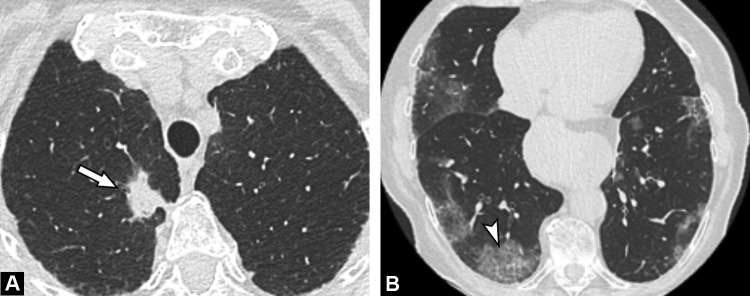
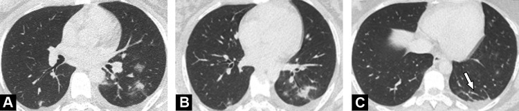
Results: During the study period, among 205 diagnostic CT examinations, six examinations (6/205, 3%) in 6 different patients (4 men, 2 women; median age, 57 years) revealed images highly suggestive of COVID-19 pneumonia. The final diagnosis was confirmed by RT-PCR. Three inpatients were suspected of extra thoracic infection whereas three outpatients were either fully asymptomatic or presented with fatigue only. All had increased CRP serum level and lymphopenia. Disease extent on CT was mild to moderate in 5/6 patients (83%) and severe in 1/6 patient (17%).
Conclusion: Cumulative incidence of fortuitous diagnosis if COVID-19 pneumonia did not exceed 3% during the highest pandemic phase and was predominantly associated with limited lung involvement.
Keywords: COVID-19 pneumonia; Computed tomography; Incidental findings; Multidetector.
Copyright © 2020 Société française de radiologie. Published by Elsevier Masson SAS. All rights reserved.
Figures
Similar articles
-
Thoracic imaging tests for the diagnosis of COVID-19.Cochrane Database Syst Rev. 2020 Sep 30;9:CD013639. doi: 10.1002/14651858.CD013639.pub2. Cochrane Database Syst Rev. 2020. Update in: Cochrane Database Syst Rev. 2020 Nov 26;11:CD013639. doi: 10.1002/14651858.CD013639.pub3. PMID: 32997361 Updated.
-
Imaging and clinical features of patients with 2019 novel coronavirus SARS-CoV-2.Eur J Nucl Med Mol Imaging. 2020 May;47(5):1275-1280. doi: 10.1007/s00259-020-04735-9. Epub 2020 Feb 28. Eur J Nucl Med Mol Imaging. 2020. PMID: 32107577 Free PMC article.
-
Chest CT in patients with a moderate or high pretest probability of COVID-19 and negative swab.Radiol Med. 2020 Dec;125(12):1260-1270. doi: 10.1007/s11547-020-01269-w. Epub 2020 Aug 29. Radiol Med. 2020. PMID: 32862406 Free PMC article.
-
Frequency and Distribution of Chest Radiographic Findings in Patients Positive for COVID-19.Radiology. 2020 Aug;296(2):E72-E78. doi: 10.1148/radiol.2020201160. Epub 2020 Mar 27. Radiology. 2020. PMID: 32216717 Free PMC article.
-
Imaging findings in COVID-19 pneumonia.Clinics (Sao Paulo). 2020 Jun 22;75:e2027. doi: 10.6061/clinics/2020/e2027. eCollection 2020. Clinics (Sao Paulo). 2020. PMID: 32578826 Free PMC article. Review.
Cited by
-
Imaging of COVID-19: An update of current evidences.Diagn Interv Imaging. 2021 Sep;102(9):493-500. doi: 10.1016/j.diii.2021.05.006. Epub 2021 May 25. Diagn Interv Imaging. 2021. PMID: 34088635 Free PMC article. Review.
-
COVID-19 pneumonia: The fight must go on.Diagn Interv Imaging. 2021 Feb;102(2):61-62. doi: 10.1016/j.diii.2021.01.006. Diagn Interv Imaging. 2021. PMID: 33494861 Free PMC article. No abstract available.
-
Rapid quantification of COVID-19 pneumonia burden from computed tomography with convolutional long short-term memory networks.J Med Imaging (Bellingham). 2022 Sep;9(5):054001. doi: 10.1117/1.JMI.9.5.054001. Epub 2022 Sep 6. J Med Imaging (Bellingham). 2022. PMID: 36090960 Free PMC article.
-
rRT-PCR Results of a Covid-19 Diagnosed Geriatric Patient.SN Compr Clin Med. 2020;2(11):2423-2426. doi: 10.1007/s42399-020-00590-9. Epub 2020 Oct 17. SN Compr Clin Med. 2020. PMID: 33103060 Free PMC article.
-
COVID-19 after 18 months: Where do we stand?Diagn Interv Imaging. 2021 Sep;102(9):491-492. doi: 10.1016/j.diii.2021.06.003. Epub 2021 Jun 18. Diagn Interv Imaging. 2021. PMID: 34183299 Free PMC article. No abstract available.
References
MeSH terms
LinkOut - more resources
Full Text Sources
Research Materials
Miscellaneous
