An official website of the United States government
The .gov means it’s official.
Federal government websites often end in .gov or .mil. Before
sharing sensitive information, make sure you’re on a federal
government site.
The site is secure.
The https:// ensures that you are connecting to the
official website and that any information you provide is encrypted
and transmitted securely.
1 CHU Saint-Étienne, Department of Radiology, 42270 Saint-Priest-en-Jarez, France.
2 CHU de Rennes, Department of Radiology, 35033 Rennes, France.
3 CHU de Rennes, Department of Respiratory Medicine, 35033 Rennes, France.
4 CHU de Rennes, Department of Radiology, 35033 Rennes, France; LTSI, INSERM, UMR 1099, Université de Rennes, 35000 Rennes, France. Electronic address: mathieu.lederlin@chu-rennes.fr.
The current COVID-19 pandemic has highlighted the essential role of chest computed tomography (CT) examination in patient triage in the emergency departments, allowing them to be referred to "COVID" or "non-COVID" wards. Initial chest CT examination must be performed without intravenous administration of iodinated contrast material, but contrast material administration is required when pulmonary embolism is suspected, which seems to be frequent in severe forms of the disease. Typical CT features consist of bilateral ground-glass opacities with peripheral, posterior and basal predominance. Lung disease extent on CT correlates with clinical severity. Artificial intelligence could assist radiologists for diagnosis and prognosis evaluation.
64-year-old man with COVID-19 pneumonia. Unenhanced CT images of the chest (lung window:…
Fig. 1
64-year-old man with COVID-19 pneumonia. Unenhanced CT images of the chest (lung window: W1600/L-500 HU) in the axial (A, B) and coronal (C, D) planes reveal bilateral multifocal ground-glass opacities (arrows) predominantly located in the peripheral and posterior part of the lungs.
Fig. 2
Unenhanced CT examination in a…
Fig. 2
Unenhanced CT examination in a 26-year-old woman with COVID-19 pneumonia. Unenhanced CT image…
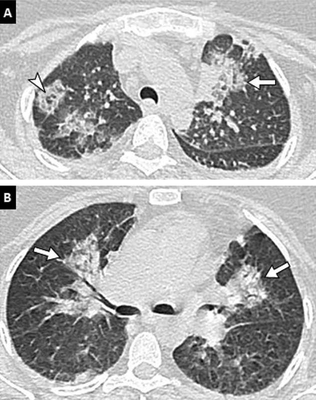
Fig. 2
Unenhanced CT examination in a 26-year-old woman with COVID-19 pneumonia. Unenhanced CT image of the chest (lung window: W1600/L-500 HU) in the axial plane reveals apical and perihilar predominant pulmonary lesions (arrows) with a “reverse halo sign” (arrowhead).
Fig. 3
75-year-old man with history of…
Fig. 3
75-year-old man with history of chronic bronchiolitis recently diagnosed with COVID-19 pneumonia. (A,…
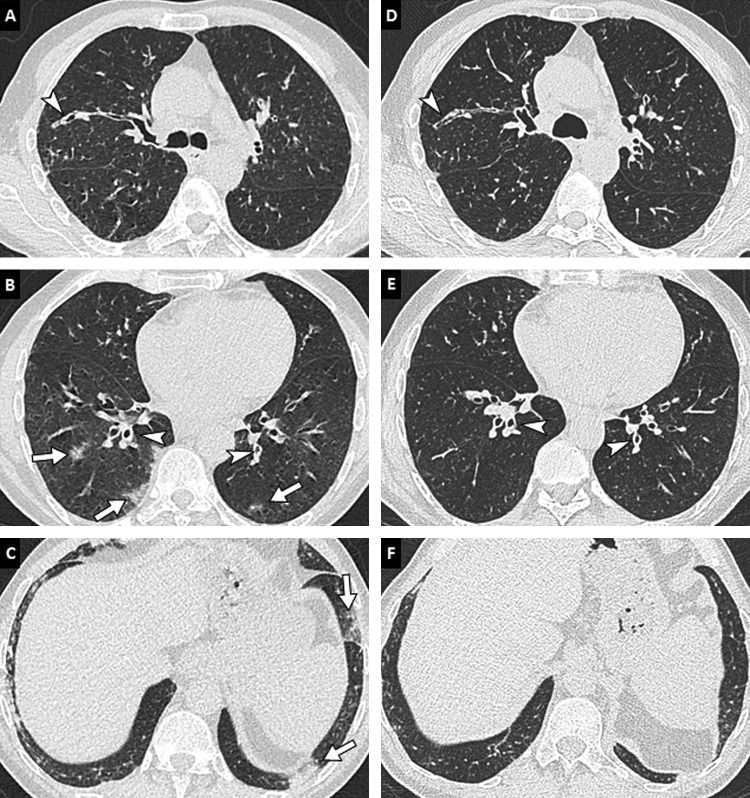
Fig. 3
75-year-old man with history of chronic bronchiolitis recently diagnosed with COVID-19 pneumonia. (A, B, C) Chest CT images in the axial plane show multifocal, patchy, ground-glass opacities (arrows) as well as diffuse thickening of bronchial walls (arrowheads). (D, E, F) The diagnosis of COVID-19 pneumonia was facilitated by the comparison with chest CT images obtained 2 months earlier that already showed bronchitis and bronchiolitis (arrowheads) but no lung opacities.
Fig. 4
78-year-old woman with COVID-19 pneumonia.…
Fig. 4
78-year-old woman with COVID-19 pneumonia. (A, B) Initial unenhanced chest CT image in…
Fig. 4
78-year-old woman with COVID-19 pneumonia. (A, B) Initial unenhanced chest CT image in the axial plane (lung window: W1600/L-500 HU) shows bilateral and peripheral areas of ground-glass and consolidation. (C, D) Follow-up contrast-enhanced CT images performed 13 days later to rule out pulmonary embolism reveal progression in extent and in density of pulmonary lesions with a crazy paving pattern (white arrowheads) and consolidation areas (arrows). (E, F) Contrast-enhanced CT images obtained 28 days after the onset of symptoms show partial regression of consolidation areas but persistence of fibrotic streaks (black arrowheads) with architectural distortion.
Fig. 5
Various degrees of lung involvement…
Fig. 5
Various degrees of lung involvement in COVID-19 pneumonia in four different patients. Unenhanced…
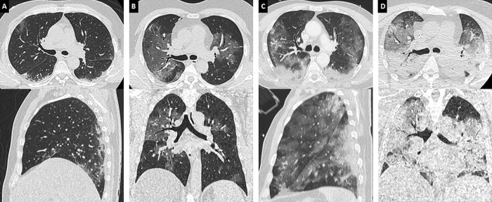
Fig. 5
Various degrees of lung involvement in COVID-19 pneumonia in four different patients. Unenhanced CT images of the chest (lung window: W 1600/L–500 HU) in the axial (up) and coronal (down) planes show typical examples of moderate (< 25%), extensive (25–50%), severe (50–75%) and critical (> 75%) lung involvement (A, B, C, D, respectively). The latter images are (D) characteristic of acute respiratory distress syndrome with a gravitationally dependent gradient.
Fig. 6
59-year-old man with COVID-19 and…
Fig. 6
59-year-old man with COVID-19 and a 3-fold positive endotracheal swab for aspergillus fumigatus.…
Fig. 6
59-year-old man with COVID-19 and a 3-fold positive endotracheal swab for aspergillus fumigatus. Unenhanced CT images of the chest in the axial (A) and coronal (B) planes (lung window: W 1600/L–500 HU) show subpleural ground-glass opacities presumed to correspond to COVID-19 lesions (arrowheads) as well as an extensive apical consolidation area presumed to correspond to invasive aspergillosis (arrow).
Fig. 7
36-year-old woman positive for COVID-19…
Fig. 7
36-year-old woman positive for COVID-19 and pulmonary embolism. CT pulmonary angiography images in…
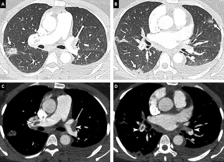
Fig. 7
36-year-old woman positive for COVID-19 and pulmonary embolism. CT pulmonary angiography images in the axial (A, B) and coronal (C, D) planes show typical peripheral ground-glass areas related to COVID-19 pneumonia (arrows) and bilateral proximal pulmonary embolism (arrowheads).
Fig. 8
74-year-old woman with COVID-19 pneumonia.…
Fig. 8
74-year-old woman with COVID-19 pneumonia. (A, B) Baseline CT images obtained after intravenous…
Fig. 8
74-year-old woman with COVID-19 pneumonia. (A, B) Baseline CT images obtained after intravenous administration of contrast material show peripheral ground-glass opacities (black arrowheads), bilateral proximal pulmonary embolism (white arrowheads) and a quadrangular well-demarcated subpleural consolidation containing central lucencies corresponding to a pulmonary infarction (arrow). (C, D) Follow-up CT images obtained 7 days later show progression of COVID-19 pulmonary lesions with reticulations, fibrotic streaks and architectural distortion (black arrowheads) and persisting thrombus (white arrowhead).
Wu Z., McGoogan J.M. Characteristics of and important lessons from the coronavirus disease 2019 (COVID-19) outbreak in China: summary of a report of 72,314 cases from the Chinese Center for Disease Control and Prevention. JAMA. 2020 doi: 10.1001/jama.2020.2648.
-
DOI
-
PubMed
Guan W., Ni Z., Hu Y., Liang W., Ou C., He J. Clinical characteristics of coronavirus disease 2019 in China. N Engl J Med. 2020 doi: 10.1056/NEJMoa2002032.
-
DOI
-
PMC
-
PubMed
Huang C., Wang Y., Li X. Clinical features of patients infected with 2019 novel coronavirus in Wuhan, China. Lancet. 2020;395:497.
-
PMC
-
PubMed
Ai T., Yang Z., Hou H., Zhan C., Chen C., Lv W. Correlation of chest CT and RT-PCR testing in coronavirus disease 2019 (COVID-19) in China: a report of 1014 cases. Radiology. 2020 doi: 10.1148/radiol.2020200432.200642.
-
DOI
-
PMC
-
PubMed