. 2020 Aug 7;295(32):11364-11376.
doi: 10.1074/jbc.RA120.013907. Epub 2020 Jun 22.

The bacterial copper resistance protein CopG contains a cysteine-bridged tetranuclear copper cluster

Affiliations

The bacterial copper resistance protein CopG contains a cysteine-bridged tetranuclear copper cluster

Andrew C Hausrath et al. J Biol Chem. .

Abstract

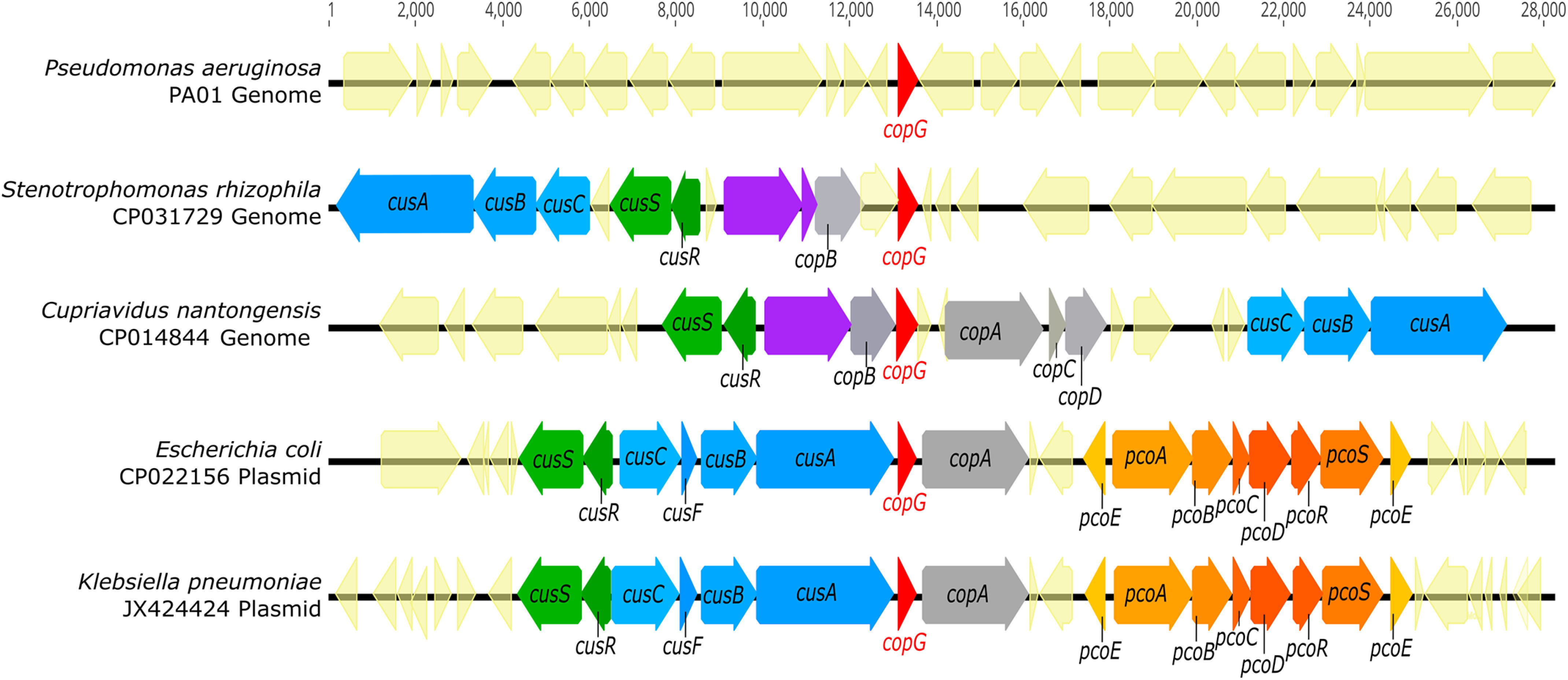
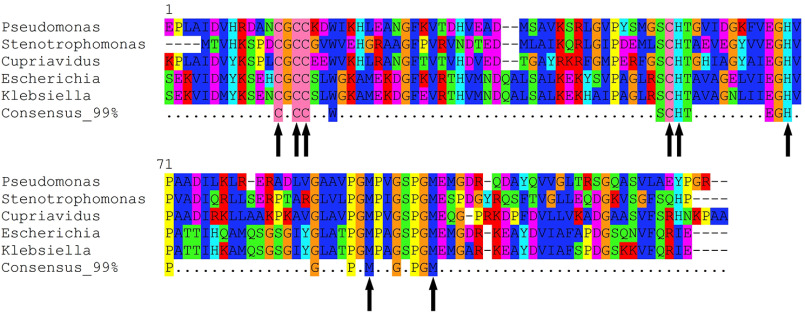
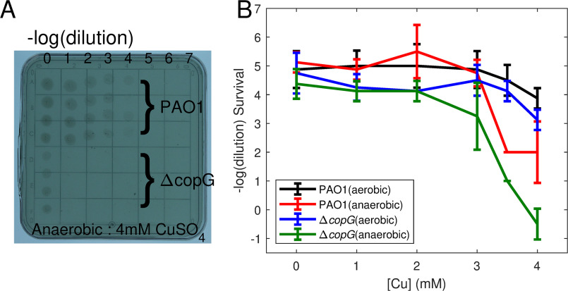
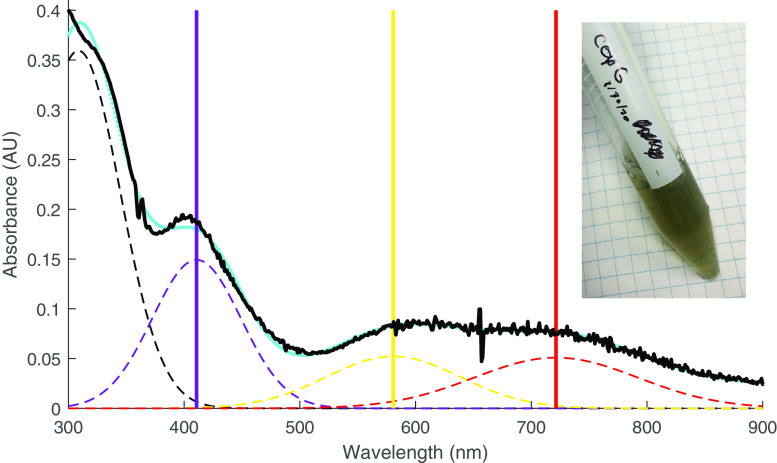
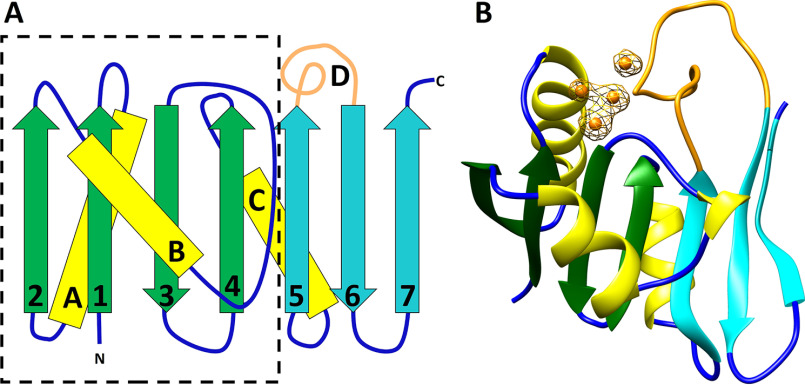
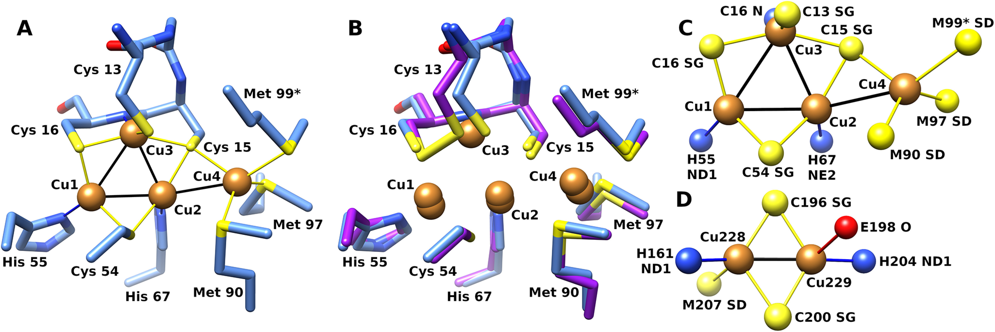
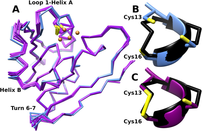
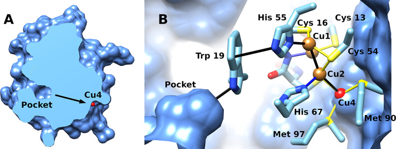
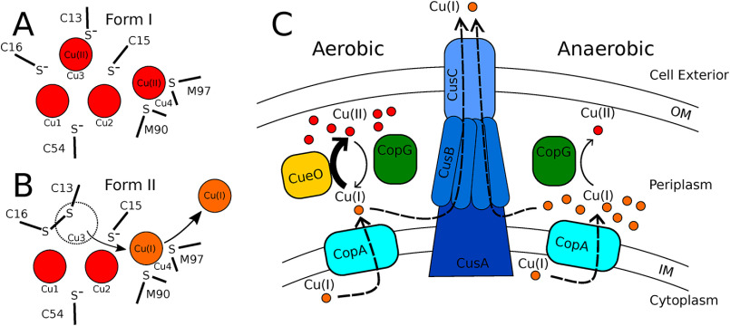
CopG is an uncharacterized protein ubiquitous in Gram-negative bacteria whose gene frequently occurs in clusters of copper resistance genes and can be recognized by the presence of a conserved CxCC motif. To investigate its contribution to copper resistance, here we undertook a structural and biochemical characterization of the CopG protein from Pseudomonas aeruginosa Results from biochemical analyses of CopG purified under aerobic conditions indicate that it is a green copper-binding protein that displays absorbance maxima near 411, 581, and 721 nm and is monomeric in solution. Determination of the three-dimensional structure by X-ray crystallography revealed that CopG consists of a thioredoxin domain with a C-terminal extension that contributes to metal binding. We noted that adjacent to the CxCC motif is a cluster of four copper ions bridged by cysteine sulfur atoms. Structures of CopG in two oxidation states support the assignment of this protein as an oxidoreductase. On the basis of these structural and spectroscopic findings and also genetic evidence, we propose that CopG has a role in interconverting Cu(I) and Cu(II) to minimize toxic effects and facilitate export by the Cus RND transporter efflux system.

Keywords: P. aeruginosa; Pseudomonas aeruginosa; copper; metal homeostasis; metal ion homeostasis; metal resistance; metalloprotein; oxidation-reduction; oxidation-reduction (redox); structure-function; thioredoxin.


Conflict of interest statement

Conflict of interest—The authors declare that they have no conflicts of interest with the contents of this article.

Figures

Figure 1.
Figure 1.
copG is located within metal resistance gene clusters in diverse organisms. Genomic DNA sequences (CP031729 and CP014844) were retrieved from NCBI, and plasmid DNA sequences (CP022156 and JX424424) were retrieved from the PATRIC annotated bacterial plasmid database. Genomes containing CopG homologs were identified via tblastn search using the CopG amino acid sequence from Pseudomonas aeruginosa strain PA01 as a query. All identified sequences displayed at least 40% amino acid sequence identity and at least 90% query coverage to PA01 CopG. Purple gene annotations indicate copper oxidases.
Figure 2.
Figure 2.
Conserved residues in CopG sequences. Comparison of the sequences of CopG homologs from Fig. 1. Alignment of the top-scoring 1,000 homologs to PAO1 CopG was used to generate the consensus sequence at the 99% level. Metal-binding residues within these highly conserved sites in the sequence are marked with arrows.
Figure 3.
Figure 3.
Copper resistance assay. Differences in copper resistance between the PAO1 and ΔcopG Pseudomonas aeruginosa strains were assayed by serial dilution on copper-containing agar plates. A, single plate with 4 replicates of each dilution series. Under anaerobic conditions, the copG knockout shows increased sensitivity to copper. B, comparison of growth on plates containing 0–4 mm copper sulfate for PAO1 and ΔcopG strains under aerobic and anaerobic conditions. The average of the highest dilution allowing growth from 8 replicates (2 plates) is plotted, and error bars indicate 1 standard deviation.
Figure 4.
Figure 4.
Absorption spectrum of CopG. Spectrum of CopG (purified from medium containing supplemental copper) under aerobic conditions. The absorbance data (black) are fitted with a sum of gaussian functions (cyan). The spectrum can be approximated by three main peaks (dashed lines) in the visible range centered at 411 nm (violet), 581 nm (yellow), and 721 nm (red) in addition to the near-UV absorbance from aromatic side chains. For fitting purposes, these maxima are each treated as single peaks (but see the text). The inset shows the visual appearance of the purified CopG protein solution.
Figure 5.
Figure 5.
Structure of CopG. A, secondary structure diagram of CopG, with the dashed line outlining the thioredoxin domain. The C-terminal extension consists of strands 5, 6 and 7 and the extended loop of segment D (cyan and orange). B, ribbon diagram of CopG using the same color scheme. Copper ions are shown in orange. Electron density is from an anomalous difference Fourier map contoured at 4σ. The map was calculated from data collected at 1.2894 Å using phases obtained from the refined model with metal ions omitted.
Figure 6.
Figure 6.
Structure of the CopG copper cluster. A, CopG copper cluster with side chain ligands from crystal form I (blue). B, superposition of corresponding atoms from form II (purple). Met 99 from a symmetry-related molecule is indicated with an asterisk. C, top view of copper cluster with ligand atoms. D, CuA site from cytochrome c oxidase (PDB code 1OCC [25]). Atom color: sulfur, yellow; copper, orange; nitrogen, blue; oxygen, red.
Figure 7.
Figure 7.
Structural consequences of disulfide formation. A, superposition of the Cα traces of the 2 form I models (blue) representing the reduced state and 6 form II models (purple) representing the oxidized state. Disulfide formation between sites 13 and 16 is associated with displacement of helix B and a bending of the loop between strand 1 and helix A relative to the reduced state. The loop between strands 6 and 7 also shows structural variability, but there does not appear to be a discrete difference in this region between the two crystal forms. B and C, detail of residues 13 to 16 showing reduced thiolates on Cys 13 and 16 (form I, chain A) (B) and disulfide (form II, chain A) (C). These are superimposed on the corresponding residues (C32-I38, in black) from the structure of oxidized E. coli thioredoxin (PDB code 2TRX [69]).
Figure 8.
Figure 8.
Proposed electron transfer path. A, section through CopG solvent-accessible surface bisecting pocket at left and showing Cu4 (red) on opposite face of protein. B, protein interior showing potential relay system (black) for reducing equivalents between pocket, disulfide, and copper cluster. Note the visible surface shows the interior face.
Figure 9.
Figure 9.
Models for CopG action. A and B, proposed oxidation states of metal cluster in crystal forms I and II. In crystal form I, the disulfide is reduced and Cu3 and Cu4 are oxidized. In form II, Cu3 is displaced by the disulfide after being reduced, and the Cu4 site remains occupied by Cu(I). C, proposed cellular roles for CopG. Under aerobic conditions, CopG acts as a reductase to facilitate Cu(I) export by the CusCBA efflux pump. Under anaerobic conditions, CopG acts as an oxidase to detoxify Cu(I).

References

    1. Casey A. L., Adams D., Karpanen T. J., Lambert P. A., Cookson B. D., Nightingale P., Miruszenko L., Shillam R., Christian P., and Elliott T. S. J. (2010) Role of copper in reducing hospital environment contamination. J. Hosp. Infect. 74, 72–77 10.1016/j.jhin.2009.08.018 - DOI - PubMed
    1. Lazary A., Weinberg I., Vatine J. J., Jefidoff A., Bardenstein R., Borkow G., and Ohana N. (2014) Reduction of healthcare-associated infections in a long-term care brain injury ward by replacing regular linens with biocidal copper oxide impregnated linens. Int. J. Infect. Dis. 24, 23–29 10.1016/j.ijid.2014.01.022 - DOI - PubMed
    1. White C., Lee J., Kambe T., Fritsche K., and Petris M. J. (2009) A role for the ATP7A copper-transporting ATPase in macrophage bactericidal activity. J. Biol. Chem. 284, 33949–33956 10.1074/jbc.M109.070201 - DOI - PMC - PubMed
    1. Vincent M., Duval R. E., Hartemann P., and Engels-Deutsch M. (2018) Contact killing and antimicrobial properties of copper. J. Appl. Microbiol. 124, 1032–1046 10.1111/jam.13681 - DOI - PubMed
    1. Macomber L., and Imlay J. A. (2009) The iron-sulfur clusters of dehydratases are primary intracellular targets of copper toxicity. Proc. Natl. Acad. Sci. U S A 106, 8344–8349 10.1073/pnas.0812808106 - DOI - PMC - PubMed

Publication types