. 2020 Aug 14;295(33):11656-11668.
doi: 10.1074/jbc.RA120.013695. Epub 2020 Jun 22.

Crystal structures of human PAICS reveal substrate and product binding of an emerging cancer target

Affiliations

Crystal structures of human PAICS reveal substrate and product binding of an emerging cancer target

Jana Škerlová et al. J Biol Chem. .

Abstract

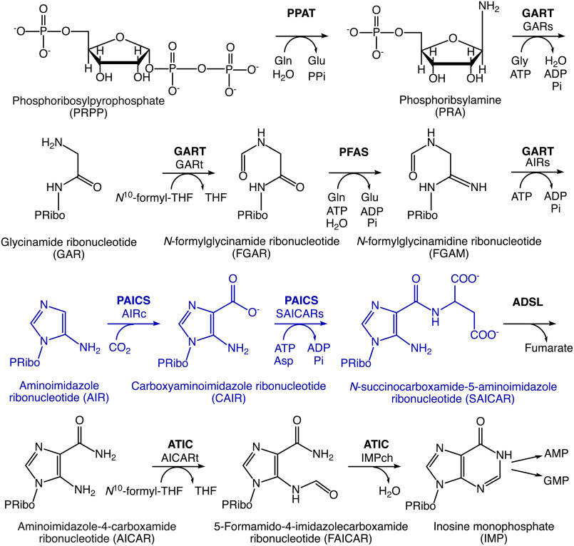
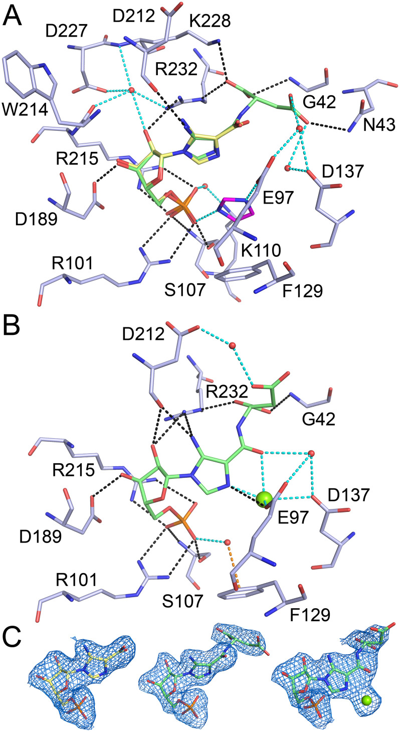
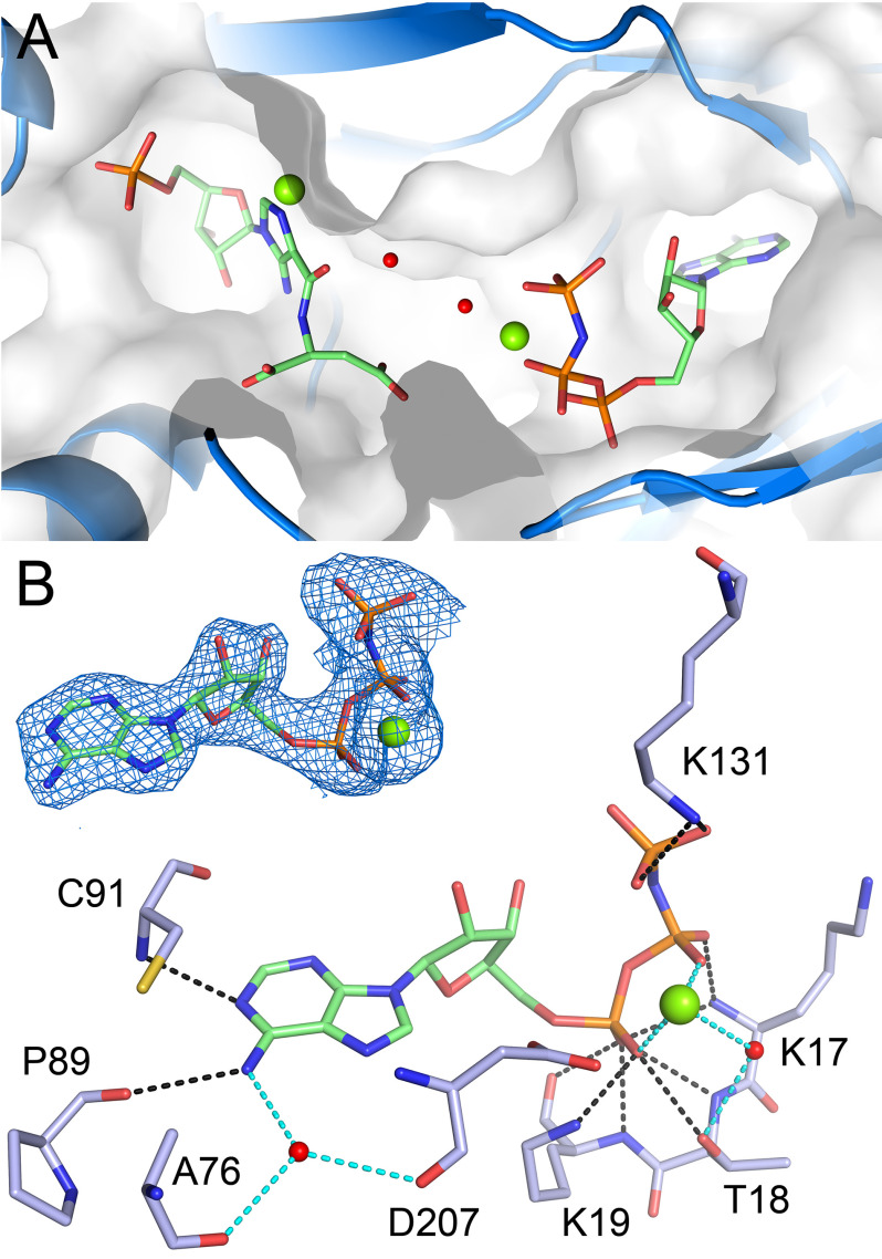
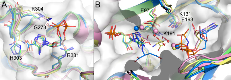
The bifunctional human enzyme phosphoribosylaminoimidazole carboxylase and phosphoribosylaminoimidazolesuccinocarboxamide synthetase (PAICS) catalyzes two essential steps in the de novo purine biosynthesis pathway. PAICS is overexpressed in many cancers and could be a promising target for the development of cancer therapeutics. Here, using gene knockdowns and clonogenic survival and cell viability assays, we demonstrate that PAICS is required for growth and survival of prostate cancer cells. PAICS catalyzes the carboxylation of aminoimidazole ribonucleotide (AIR) and the subsequent conversion of carboxyaminoimidazole ribonucleotide (CAIR) and l-aspartate to N-succinylcarboxamide-5-aminoimidazole ribonucleotide (SAICAR). Of note, we present the first structures of human octameric PAICS in complexes with native ligands. In particular, we report the structure of PAICS with CAIR bound in the active sites of both domains and SAICAR bound in one of the SAICAR synthetase domains. Moreover, we report the PAICS structure with SAICAR and an ATP analog occupying the SAICAR synthetase active site. These structures provide insight into substrate and product binding and the architecture of the active sites, disclosing important structural information for rational design of PAICS inhibitors as potential anticancer drugs.

Keywords: N-succinylcarboxamide-5-aminoimidazole ribonucleotide (SAICAR); cancer target; cancer therapy; carboxyaminoimidazole ribonucleotide (CAIR); de novo purine biosynthesis; drug design; nucleoside/nucleotide biosynthesis; nucleotide metabolism; phosphoribosylaminoimidazole carboxylase and phosphoribosylaminoimidazolesuccinocarboxamide synthetase (PAICS); prostate cancer; purine; rational drug design; structural biology.


Conflict of interest statement

Conflict of interest—The authors declare that they have no conflicts of interest with the contents of this article.

Figures

Figure 1.
Figure 1.
Scheme of the de novo purine biosynthesis pathway in humans. The reactions catalyzed by PAICS are highlighted in blue. The following abbreviations are used: ADSL, adenylosuccinate lyase; AICARt, aminoimidazole-4-carboxamide ribonucleotide transformylase; ATIC, aminoimidazole-4-carboxamide ribonucleotide transformylase/inosine monophosphate cyclohydrolase; GARs, glycinamide ribonucleotide synthetase; GARt, glycinamide ribonucleotide transformylase; GART, glycinamide ribonucleotide synthetase/glycinamide ribonucleotide transformylase/aminoimidazole ribonucleotide synthetase; IMPch, inosine monophosphate cyclohydrolase; PFAS, N-formylglycinamidine ribonucleotide synthase; PPAT, phosphoribosylpyrophosphate amidotransferase; THF, tetrahydrofolate.
Figure 2.
Figure 2.
The effect of PAICS knockdown on proliferation and clonogenic survival of prostate cancer cell lines. A, Western blots showing knockdown of PAICS after doxycycline induction of prostate cancer cell lines stably transfected with two different shRNAs against PAICS. B, Resazurin assay measuring short-time effects of PAICS depletion on proliferation of prostate cancer cell lines DU-145 (left panel) and PC3 (right panel). Graphs shows survival (%) normalized to shRNA NT + Dox (n = 2). C, representative plates from colony formation assay of DU-145 (left panel) and PC3 (right panel) with and without doxycycline-induced shRNA-mediated PAICS depletion. D, quantification of colony formation assay shown in C. Graphs display survival (%) of minus doxycycline control (n = 3). Statistical significance was analyzed using two-way ANOVA or ordinary one-way ANOVA in GraphPad Prism.
Figure 3.
Figure 3.
Overall structure of the PAICS octamer. PAICS octamer is displayed in a view along the 4-fold rotation axis (top) and rotated by 90° along the x axis (bottom). Each chain is a different color in panel A. In panel B one monomer is highlighted in blue and the ligands CAIR in the AIRc active site and SAICAR and AMP-PNP in the SAICARs active site are shown as pink, yellow, and green spheres, respectively. The CAIR originates from the PAICS-CAIR structure and the SAICAR and AMP-PNP were superposed here from the structure of the PAICS-SAICAR-Mg2+-AMP-PNP-Mg2+ complex to demonstrate the relative location of the individual binding sites in the PAICS octamer.
Figure 4.
Figure 4.
CAIR bound in the AIRc active site. The CAIR molecule is shown as pink sticks and the active site residues involved in direct (black dashed lines) or water-mediated (cyan dashed lines) hydrogen bonds are shown as pale blue sticks. Residue Ser-370 from another PAICS monomer in the octamer that participates in ligand binding through hydrogen bonds (yellow dashed lines) is shown as yellow sticks. Refined 2FoFc electron density map for CAIR contoured at 1σ and mFo – DFc Polder omit map for CAIR contoured at 4 σ are shown in the upper right corner in blue and yellow, respectively.
Figure 5.
Figure 5.
CAIR, SAICAR, and SAICAR-Mg2+ bound in the SAICARs active site. A, the active site hydrogen bond network is shown here for SAICAR (green sticks), and the CAIR molecule (yellow sticks) from the second monomer is shown in superposition. The imidazole molecule is shown as magenta sticks. B, the active site hydrogen bond network is shown here for SAICAR-Mg2+ (green sticks and green sphere). The active site residues involved in direct (black dashed lines) or water-mediated, imidazole-mediated, and magnesium-mediated (cyan dashed lines) hydrogen bonds are shown as pale blue sticks. Residue Phe-129 that stacks with the imidazole molecule or forms a π-stacking interaction (orange dashed line) with a ligand-bound water molecule is also shown. C, refined 2FoFc electron density maps for CAIR, SAICAR, and SAICAR-Mg2+ contoured at 1σ are shown left to right, respectively.
Figure 6.
Figure 6.
SAICARs active site with SAICAR-Mg2+ and AMP-PNP-Mg2+. A, the SAICAR-Mg2+ (left) and AMP-PNP-Mg2+ (right) molecules (green sticks and green spheres) and water molecules bridging the two ligands (red spheres) are shown in the SAICARs active pocket (blue cartoon and semi-transparent surface). B, the ATP-binding site hydrogen bond network is shown for AMP-PNP-Mg2+ (green sticks and green sphere). Active site residues involved in direct (black dashed lines) or water-mediated and magnesium-mediated (cyan dashed lines) hydrogen bonds are shown as pale blue sticks. Refined 2FoFc electron density map for AMP-PNP-Mg2+ contoured at 1σ is shown in the upper left corner.
Figure 7.
Figure 7.
Intermediate tunnel system in PAICS. A, the tunnel connecting the AIRc (labeled with asterisks) and SAICARs (labeled with circles) active sites of 4 different PAICS monomers (differentiated by colors) in the octameric assembly is depicted in yellow, as identified by the program Caver (36). The CAIR and SAICAR molecules in the active sites are shown as spheres. Details of the AIRc site tunnel entrance and SAICARs site tunnel exit are shown in panels B and C, respectively. Key residues facilitating the entry/exit of the intermediate are shown as magenta sticks with polar interactions highlighted as black dotted lines, and the possible intermediate trajectory is shown as red dotted arrows.
Figure 8.
Figure 8.
Ligand-induced structural changes in PAICS active sites. Cartoon representations of PAICS in complex with CAIR, SAICAR-Mg2+, and AMP-PNP-Mg2+, and without any ligand (PDB code 2h31 (29)) are overlaid in gray, orange, and cyan, respectively. Side chains of residues located in the positions structurally affected by ligand binding are shown in sticks with residue numbers indicated. A, AIRc site is shown with CAIR in pink stick representation. B, SAICARs site is shown with CAIR and imidazole molecules in gray sticks and with SAICAR-Mg2+ and AMP-PNP-Mg2+ in yellow sticks and green spheres. The portion of the protein that is unstructured in the apo structure and folds in the presence of CAIR is highlighted in pink in the otherwise gray cartoon of the PAICS-CAIR complex.
Figure 9.
Figure 9.
Ligand binding in PAICS compared with homologues. A, AIRc site of PAICS in complex with CAIR (blue) is shown in superposition with prokaryotic PurE II from T. denticola in complex with AIR (green, PDB code 3rgg (23)), with PurE I from A. aceti in complex with AIR and CO2 (pink, H59N catalytically inactive mutant, PDB code 5clj), and with PurE I from E. coli in complex with CAIR (yellow, H45N catalytically inactive mutant, PDB code 2nsl (21)). Differentially conserved, class-specific residues involved in CAIR binding, His-303, and Gly-273 (and equivalent residues) are shown as sticks. B, SAICARs site of PAICS in complex with SAICAR-Mg2+ and AMP-PNP-Mg2+ (blue) and in complex with CAIR (green) is shown in superposition with SAICARs (PurC) from E. coli in complex with CAIR, ADP, formate, and magnesium cations (pink, PDB code 2gqs (24)) and with SAICARs (PurC) from S. pneumoniae in complex with AIR, ADP, aspartate, and magnesium cations (yellow, PDB code 4fe2 (25)). Side chains of suggested catalytic residues are shown as sticks. Semitransparent surface representation of PAICS is also shown in both panels.

References

    1. Hartman S. C., and Buchanan J. M. (1959) Biosynthesis of the purines: XXVI. the identification of the formyl donors of the transformylation reactions. J. Biol. Chem. 234, 1812–1816 - PubMed
    1. Lukens L. N., and Buchanan J. M. (1959) Biosynthesis of the purines: XXIV. the enzymatic synthesis of 5-amino-1-ribosyl-4-imidazolecarboxylic acid 5'-phosphate from 5-amino-1-ribosylimidazole 5'-phosphate and carbon dioxide. J. Biol. Chem. 234, 1799–1805 - PubMed
    1. Yin J., Ren W., Huang X., Deng J., Li T., and Yin Y. (2018) Potential mechanisms connecting purine metabolism and cancer therapy. Front. Immunol. 9, 1697 10.3389/fimmu.2018.01697 - DOI - PMC - PubMed
    1. Meng M., Chen Y., Jia J., Li L., and Yang S. (2018) Knockdown of PAICS inhibits malignant proliferation of human breast cancer cell lines. Biol Res 51, 24 10.1186/s40659-018-0172-9 - DOI - PMC - PubMed
    1. Barfeld S. J., Fazli L., Persson M., Marjavaara L., Urbanucci A., Kaukoniemi K. M., Rennie P. S., Ceder Y., Chabes A., Visakorpi T., and Mills I. G. (2015) Myc-dependent purine biosynthesis affects nucleolar stress and therapy response in prostate cancer. Oncotarget 6, 12587–12602 10.18632/oncotarget.3494 - DOI - PMC - PubMed

Publication types

MeSH terms

Substances