. 2020 Jul 31;295(31):10741-10748.
doi: 10.1074/jbc.AC120.013449. Epub 2020 Jun 22.

Structural and functional conservation of the programmed -1 ribosomal frameshift signal of SARS coronavirus 2 (SARS-CoV-2)

Affiliations

Structural and functional conservation of the programmed -1 ribosomal frameshift signal of SARS coronavirus 2 (SARS-CoV-2)

Jamie A Kelly et al. J Biol Chem. .

Abstract

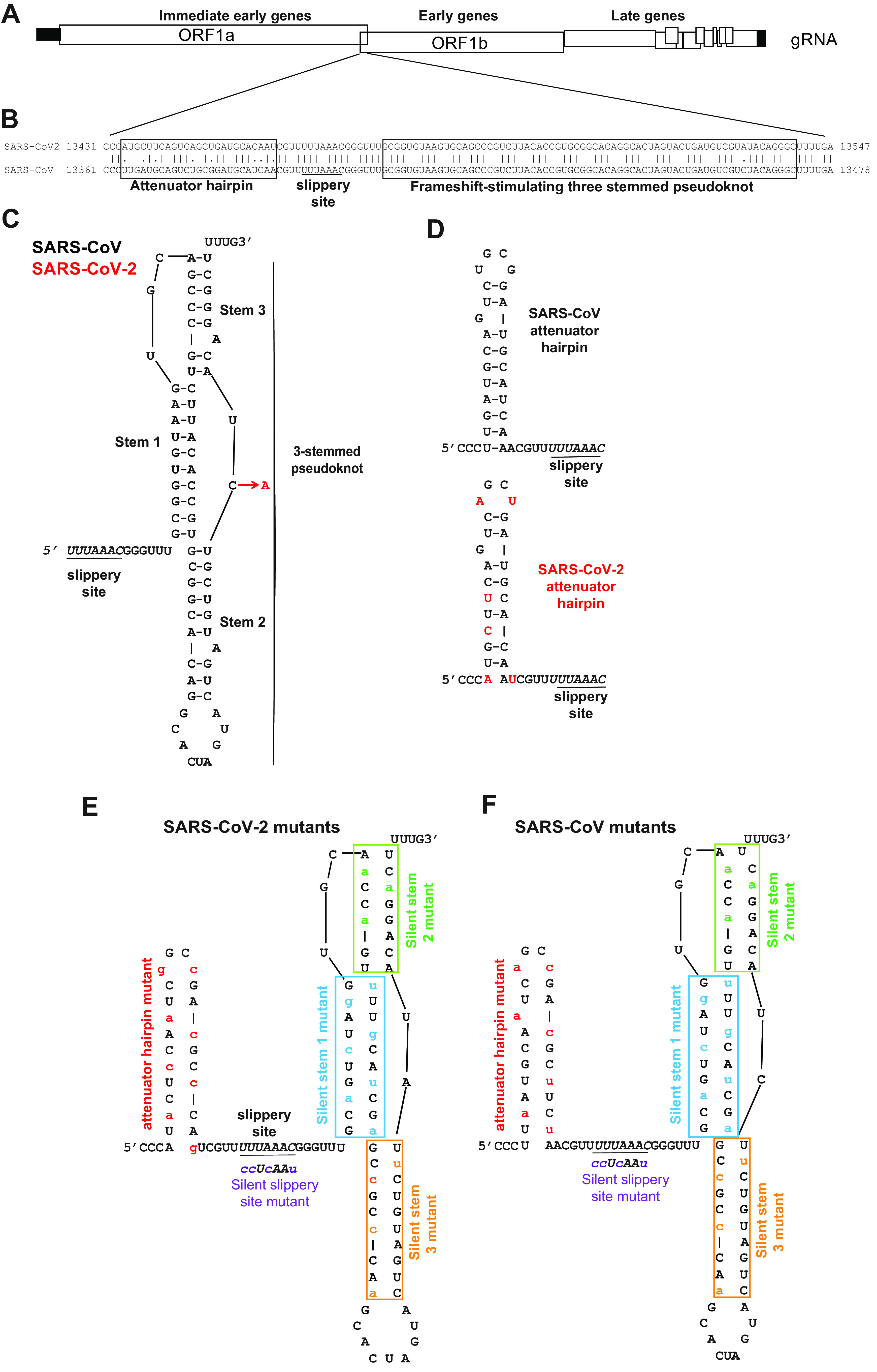
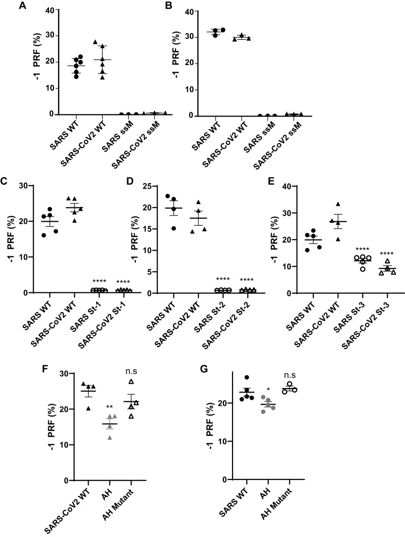
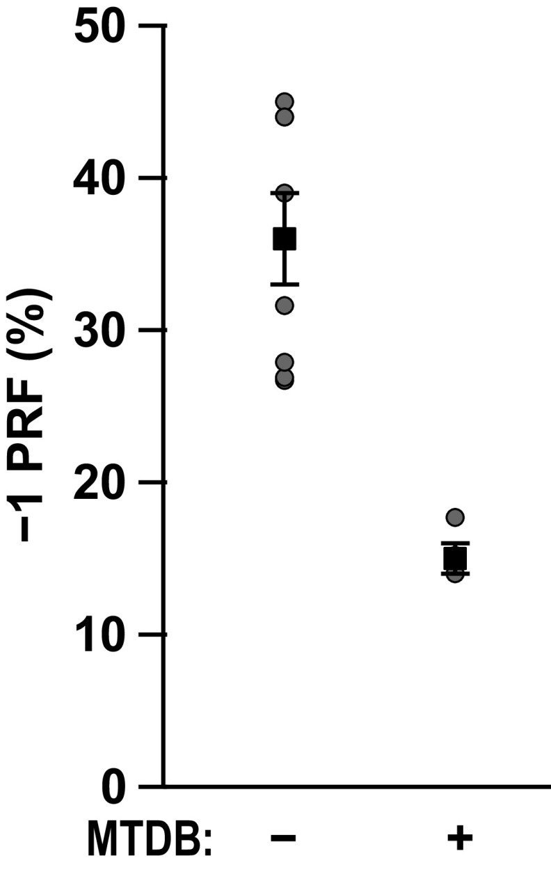
Approximately 17 years after the severe acute respiratory syndrome coronavirus (SARS-CoV) epidemic, the world is currently facing the COVID-19 pandemic caused by SARS corona virus 2 (SARS-CoV-2). According to the most optimistic projections, it will take more than a year to develop a vaccine, so the best short-term strategy may lie in identifying virus-specific targets for small molecule-based interventions. All coronaviruses utilize a molecular mechanism called programmed -1 ribosomal frameshift (-1 PRF) to control the relative expression of their proteins. Previous analyses of SARS-CoV have revealed that it employs a structurally unique three-stemmed mRNA pseudoknot that stimulates high -1 PRF rates and that it also harbors a -1 PRF attenuation element. Altering -1 PRF activity impairs virus replication, suggesting that this activity may be therapeutically targeted. Here, we comparatively analyzed the SARS-CoV and SARS-CoV-2 frameshift signals. Structural and functional analyses revealed that both elements promote similar -1 PRF rates and that silent coding mutations in the slippery sites and in all three stems of the pseudoknot strongly ablate -1 PRF activity. We noted that the upstream attenuator hairpin activity is also functionally retained in both viruses, despite differences in the primary sequence in this region. Small-angle X-ray scattering analyses indicated that the pseudoknots in SARS-CoV and SARS-CoV-2 have the same conformation. Finally, a small molecule previously shown to bind the SARS-CoV pseudoknot and inhibit -1 PRF was similarly effective against -1 PRF in SARS-CoV-2, suggesting that such frameshift inhibitors may be promising lead compounds to combat the current COVID-19 pandemic.

Keywords: (+) ssRNA; RNA; RNA structure; coronavirus; coronavirus disease 2019 (COVID-19); inhibitor; mRNA pseudoknot; programmed −1 ribosomal frameshifting (−1 PRF); small molecule inhibitor; translation; virus.


Conflict of interest statement

Conflict of interest—The authors declare that they have no conflicts of interest with the contents of this article.

Figures

Figure 1.
Figure 1.
Structural comparison of the SARS-CoV and SARS-CoV-2 −1 PRF signals. A, cartoon depicting SARS-CoV and SARS-CoV-2 genome organization including a −1 PRF between ORF1a and ORF1b. B, pairwise analysis of the two −1 PRF signals. The attenuator elements and three-stemmed pseudoknot sequences are boxed as indicated. The U UUA AAC slippery site is underlined. C, structure of the SARS-CoV −1 PRF signal (11) is composed of the 5′ slippery site, a 6-nt spacer, and the three-stemmed pseudoknot stimulatory element. The single-base difference in SARS-CoV-2 (red) maps to the short loop linking stems 2 and 3. D, comparison of the SARS-CoV and SARS-CoV-2 −1 PRF attenuator elements. SARS-CoV-2–specific bases are indicated in red. E and F, silent coding mutations designed to disrupt the attenuators, slippery sites, and stems 1, 2, and 3 in the SARS-CoV-2 (E) and SARS-CoV (F) −1 PRF signals. gRNA, genomic RNA.
Figure 2.
Figure 2.
Functional characterization of the SARS-CoV and SARS-CoV-2 −1 PRF signals. A and B, analyses of silent slippery site mutants. The efficiencies of −1 PRF promoted by the WT (U UUA AAC) and silent slippery site mutant (C CUC AAC) −1 PRF signals were assayed in HEK (A) and HeLA (B). ssM denotes silent slippery site mutant. C–E, analyses of the importance of the three stems in the −1 PRF stimulating RNA pseudoknot. Silent stem 1 (St-1, C), stem 2 (St-2, D), and stem 3 (St-3, E) mutants were assayed in HEK cells. F and G, analyses of the attenuator hairpins. AH denotes constructs that included attenuator hairpin sequences. AH mutant denotes mutants harboring the silent coding attenuator hairpin sequences shown in Fig. 1 (E and F). Assays were performed using Dual-Luciferase assays as previously described (15, 16). Each data point represents a single biological replicate comprised of three technical replicates. Error bars denote S.E. n.s, not significant.
Figure 3.
Figure 3.
Small-molecule ligand MTDB inhibits −1 PRF stimulation by SARS-CoV-2 pseudoknot. −1 PRF efficiency was reduced almost 60% in the presence of 5 μm MTDB (right), compared with −1 PRF efficiency in the absence of MTDB (left).
Figure 4.
Figure 4.
SAXS analyses. A, scattering profiles from laboratory-purified SAXS samples containing pseudoknots from SARS-CoV (blue) and SARS-CoV-2 (red). Inset, scattering profiles from inline SEC-SAXS measurements, containing purely monomeric pseudoknots. B and C, difference between the scattering profiles for SARS-CoV and SARS-CoV-2 pseudoknots obtained from lab-purified SAXS (B) and inline SEC-SAXS (C) samples. Arb., arbitrary.

Update of

References

    1. Coronaviridae Study Group of the International Committee on Taxonomy of Viruses (2020) The species severe acute respiratory syndrome–related coronavirus: classifying 2019-nCoV and naming it SARS-CoV-2. Nat. Microbiol. 5, 536–544 10.1038/s41564-020-0695-z - DOI - PMC - PubMed
    1. Brian D. A., and Baric R. S. (2005) Coronavirus genome structure and replication. In Coronavirus Replication and Reverse Genetics, pp. 1–30, Springer-Verlag, Berlin/Heidelberg - PMC - PubMed
    1. Dinman J. D. (2012) Mechanisms and implications of programmed translational frameshifting. Wiley Interdiscip. Rev. RNA 3, 661–673 10.1002/wrna.1126 - DOI - PMC - PubMed
    1. Atkins J. F., Loughran G., Bhatt P. R., Firth A. E., and Baranov P. V. (2016) Ribosomal frameshifting and transcriptional slippage: From genetic steganography and cryptography to adventitious use. Nucleic Acids Res. 44, 7007–7078 10.1093/nar/gkw530 - DOI - PMC - PubMed
    1. Jacks T., and Varmus H. E. (1985) Expression of the Rous sarcoma virus pol gene by ribosomal frameshifting. Science 230, 1237–1242 10.1126/science.2416054 - DOI - PubMed

Publication types