. 2020 Aug 10;202(17):e00124-20.
doi: 10.1128/JB.00124-20. Print 2020 Aug 10.

Programmed Proteolysis of Chemotaxis Proteins in Sinorhizobium meliloti: Features in the C-Terminal Region Control McpU Degradation

Affiliations

Programmed Proteolysis of Chemotaxis Proteins in Sinorhizobium meliloti: Features in the C-Terminal Region Control McpU Degradation

Timofey D Arapov et al. J Bacteriol. .

Abstract

Chemotaxis and motility are important traits that support bacterial survival in various ecological niches and in pathogenic and symbiotic host interaction. Chemotactic stimuli are sensed by chemoreceptors or methyl-accepting chemotaxis proteins (MCPs), which direct the swimming behavior of the bacterial cell. In this study, we present evidence that the cellular abundance of chemoreceptors in the plant symbiont Sinorhizobium meliloti can be altered by the addition of several to as few as one amino acid residues and by including common epitope tags such as 3×FLAG and 6×His at their C termini. To further dissect this phenomenon and its underlying molecular mechanism, we focused on a detailed analysis of the amino acid sensor McpU. Controlled proteolysis is important for the maintenance of an appropriate stoichiometry of chemoreceptors and between chemoreceptors and chemotactic signaling proteins, which is essential for an optimal chemotactic response. We hypothesized that enhanced stability is due to interference with protease binding, thus affecting proteolytic efficacy. Location of the protease recognition site was defined through McpU stability measurements in a series of deletion and amino acid substitution mutants. Deletions in the putative protease recognition site had similar effects on McpU abundance, as did extensions at the C terminus. Our results provide evidence that the programmed proteolysis of chemotaxis proteins in S. meliloti is cell cycle regulated. This posttranslational control, together with regulatory pathways on the transcriptional level, limits the chemotaxis machinery to the early exponential growth phase. Our study identified parallels to cell cycle-dependent processes during asymmetric cell division in Caulobacter crescentusIMPORTANCE The symbiotic bacterium Sinorhizobium meliloti contributes greatly to growth of the agriculturally valuable host plant alfalfa by fixing atmospheric nitrogen. Chemotaxis of S. meliloti cells toward alfalfa roots mediates this symbiosis. The present study establishes programmed proteolysis as a factor in the maintenance of the S. meliloti chemotaxis system. Knowledge about cell cycle-dependent, targeted, and selective proteolysis in S. meliloti is important to understand the molecular mechanisms of maintaining a suitable chemotaxis response. While the role of regulated protein turnover in the cell cycle progression of Caulobacter crescentus is well understood, these pathways are just beginning to be characterized in S. meliloti In addition, our study should alert about the cautionary use of epitope tags for protein quantification.

Keywords: ClpXP protease; alphaproteobacteria; cell cycle; chemotaxis; epitope tags.


Figures

FIG 1
FIG 1
Mutational analysis of the C-terminal region of McpU and its effect on stability. Depicted is a diagram of the C-terminal region of McpU (amino acids [aa] 666 to 707) and the extensions, deletions, and substitutions made in its native chromosomal locus (Table 1). Boxes in the wild-type sequence and the respective mutants indicate specific motifs and their substitutions, respectively. Black bars represent extent of deletions. The column on the right symbolizes McpU abundance relative to that in the wild type; +, increase, −, decrease; +/−, no change.
FIG 2
FIG 2
Representative immunoblots for direct comparison of McpU abundance in cell lysates of wild type compared to that in mutant strains. Each lane contains cell lysates from 1 ml of culture at an optical density at 600 nm (OD600) of 0.25. Each panel is the result of different film exposures; therefore, comparisons can be made only within a panel.
FIG 3
FIG 3
Representative immunoblots used to quantify the relative abundance of McpU in cell lysates of the wild type compared to mutant strains. Lanes 1 to 3 contain RU11/001 lysates (WT) from 1 ml of culture at an OD600 of 0.25. Lanes denoted by a wedge shape contain a mix of each mutant strain and RU11/828 (ΔmcpU) at ratios of 1:1, 3:1, 5.7:1, 7:1, and 9:1, yielding a total volume of 1 ml of culture at an OD600 of 0.25. The last lane in each panel contains lysates from 1 ml of culture of the respective mutant strain at an OD600 of 0.25.
FIG 4
FIG 4
Relative abundance of McpU in mutant strains compared to that in the wild type. Asterisks indicate quantifications obtained through direct comparison (Fig. 2); all other values were obtained through generation of a standard curve (Fig. 3). The dashed line represents wild-type abundance of McpU. Values and error bars are the means and standard deviations from three biological replicates. Statistical significance was determined by a two-tailed Student t test (P < 0.05). All values but those marked by an octothorpe denote statistically significant differences from the wild type.
FIG 5
FIG 5
Representative immunoblots used to quantify chemotaxis proteins with C-terminal 3×FLAG fusions. (A) Lanes 1 to 3 contain BS193 (McpY-3×FLAG expressing strain) cell lysate from 1 ml of culture at an OD600 of 0.25, and wild type (WT) + McpV-3×FLAG lanes contain RU11/001 (WT) cell lysate from 1 ml of culture at an OD600 of 0.25 with McpV-LBD-3×FLAG (0.4, 0.3, 0.2, 0.01, 0.05, and 0 ng). (B) Lanes 1 to 3 contain BS194 (McpT-3×FLAG-expressing strain) cell lysate from 1 ml of culture at an OD600 of 0.25, and WT + McpV-3×FLAG lanes contain RU11/001 (WT) cell lysate from 1 ml of culture at an OD600 of 0.25 with McpV-LBD-3×FLAG (1.8, 1.5, 1, 0.5, 0.3, and 0 ng). (C) Lanes 1 to 3 contain BS201 (CheW1-3×FLAG-expressing strain) cell lysate from 1 ml of culture at an OD600 of 0.25, and WT + McpV-3×FLAG lanes contain RU11/001 (WT) cell lysate from 1 ml of culture at an OD600 of 0.25 with McpV-LBD-3×FLAG (8, 6.5, 5.2, 4, 2.6, and 0 ng). (D) Lanes 1 to 3 contain BS203 (IcpA-3×FLAG-expressing strain) cell lysate from 1 ml of culture at an OD600 of 0.25, and WT + McpV-3×FLAG lanes contain RU11/001 (WT) cell lysate from 1 ml of culture at an OD600 of 0.25 with McpV-LBD-3×FLAG (10, 8, 6, 4, 2, and 0 ng). (E) Direct comparison of McpV abundance in cell lysates of the wild type compared to those of BS221. Each lane contains cell lysates from 1 ml of culture at an OD600 of 0.25.
FIG 6
FIG 6
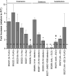
Relative swim ring diameter of S. meliloti mutant strains in Bromfield and in Rhizobium basal medium containing 10−4 M lysine. Plates containing 0.3% agar were inoculated with 3 μl stationary-phase TYC culture and incubated for 3 and 5 days, respectively, at 30°C. Dark and light gray bars represent results obtained in Bromfield and Rhizobium basal medium with 10−4 M lysine, respectively. Values are the means, and error bars reflect the standard deviation from five replicates. Statistical significance was determined by a two-tailed Student t test (P < 0.05). Asterisks denote statistically significant differences from the wild type.
FIG 7
FIG 7
Comparison of the region comprising 47 amino acid residues proximal to the carboxy terminus of C. crescentus McpA and McpB, five S. meliloti MCPs, and CheW1. Residues critical for correct proteolysis are indicated by shaded boxes according to Tsai et al. (28), Potocka et al. (27), and this work. Putative residues important for proteolysis are marked by unshaded boxes. Numbers at the end of each sequence reflect overall protein length.

Similar articles

References

    1. Pratt LA, Kolter R. 1998. Genetic analysis of Escherichia coli biofilm formation: roles of flagella, motility, chemotaxis and type I pili. Mol Microbiol 30:285–293. doi:10.1046/j.1365-2958.1998.01061.x. - DOI - PubMed
    1. Locke J. 2013. How bacteria choose a lifestyle. Nature 503:476–477. doi:10.1038/nature12837. - DOI - PubMed
    1. Davey ME, O'Toole GA. 2000. Microbial biofilms: from ecology to molecular genetics. Microbiol Mol Biol Rev 64:847–867. doi:10.1128/mmbr.64.4.847-867.2000. - DOI - PMC - PubMed
    1. Raina J-B, Fernandez V, Lambert B, Stocker R, Seymour JR. 2019. The role of microbial motility and chemotaxis in symbiosis. Nat Rev Microbiol 17:284–294. doi:10.1038/s41579-019-0182-9. - DOI - PubMed
    1. Oliveira NM, Foster KR, Durham WM. 2016. Single-cell twitching chemotaxis in developing biofilms. Proc Natl Acad Sci U S A 113:6532–6537. doi:10.1073/pnas.1600760113. - DOI - PMC - PubMed

Publication types

Substances

LinkOut - more resources