. 2020 Oct;18(10):1560-1573.
doi: 10.1158/1541-7786.MCR-20-0291. Epub 2020 Jun 22.

SPANX Control of Lamin A/C Modulates Nuclear Architecture and Promotes Melanoma Growth

Affiliations

SPANX Control of Lamin A/C Modulates Nuclear Architecture and Promotes Melanoma Growth

Ikrame Lazar et al. Mol Cancer Res. 2020 Oct.

Abstract

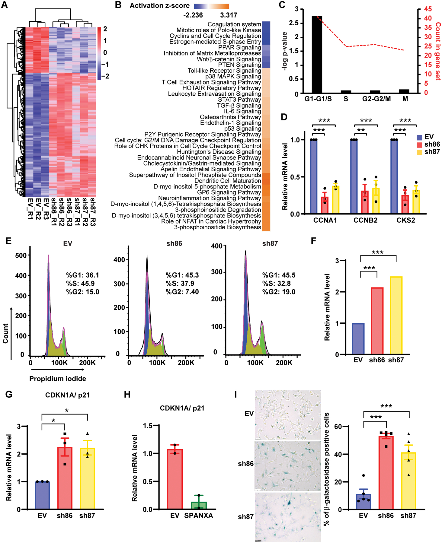
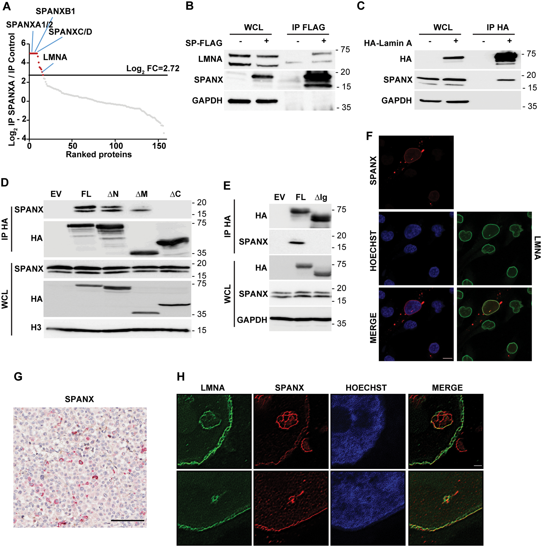
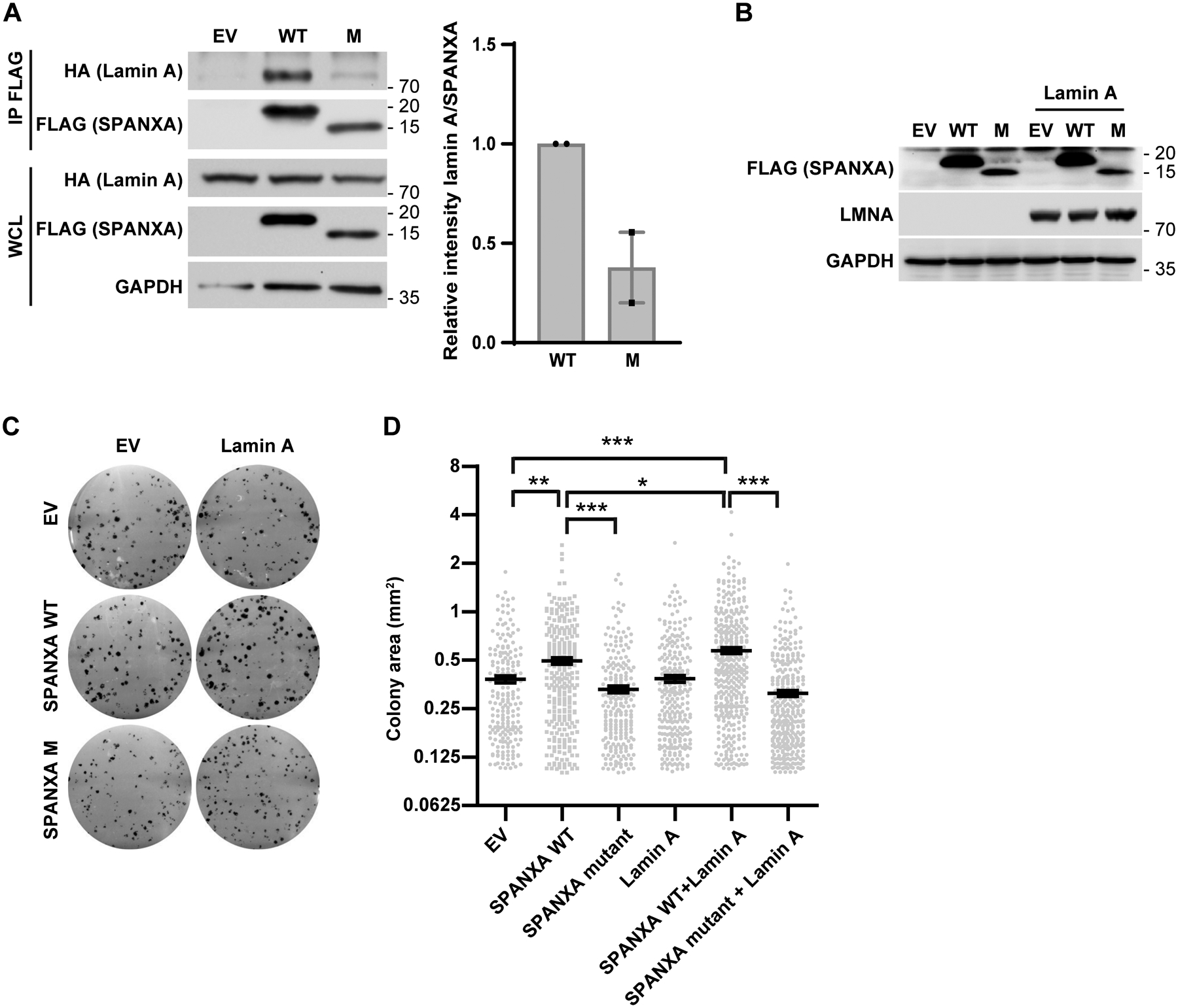
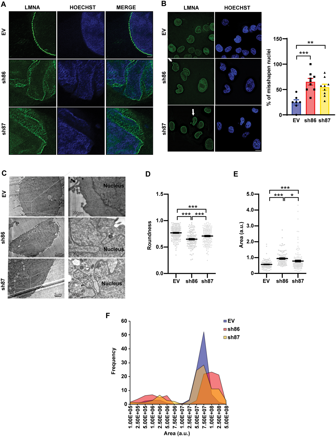
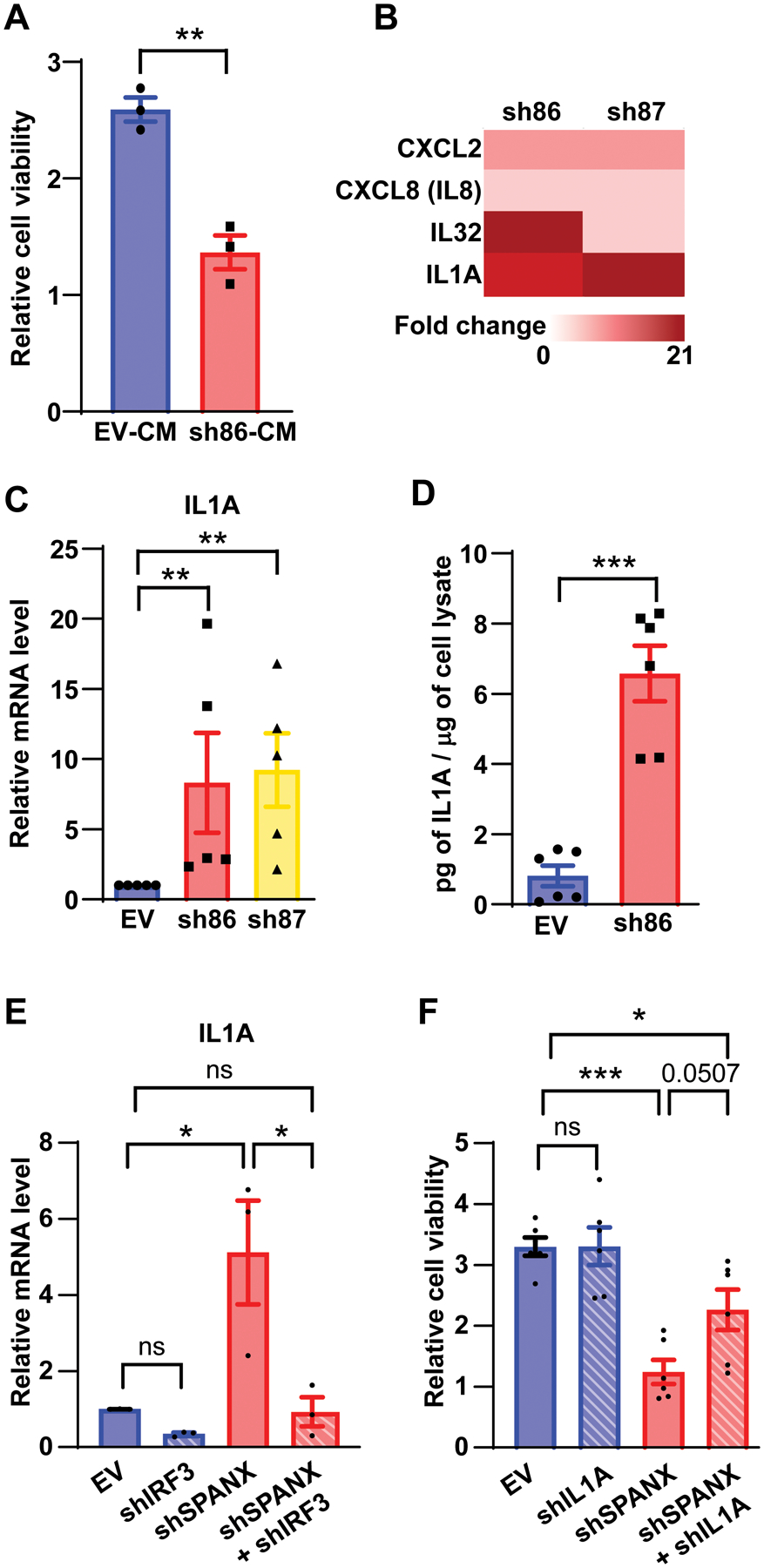
Mechanisms regulating nuclear organization control fundamental cellular processes, including the cell and chromatin organization. Their disorganization, including aberrant nuclear architecture, has been often implicated in cellular transformation. Here, we identify Lamin A, among proteins essential for nuclear architecture, as SPANX (sperm protein associated with the nucleus on the X chromosome), a cancer testis antigen previously linked to invasive tumor phenotypes, interacting protein in melanoma. SPANX interaction with Lamin A was mapped to the immunoglobulin fold-like domain, a region critical for Lamin A function, which is often mutated in laminopathies. SPANX downregulation in melanoma cell lines perturbed nuclear organization, decreased cell viability, and promoted senescence-associated phenotypes. Moreover, SPANX knockdown (KD) in melanoma cells promoted proliferation arrest, a phenotype mediated in part by IRF3/IL1A signaling. SPANX KD in melanoma cells also prompted the secretion of IL1A, which attenuated the proliferation of naïve melanoma cells. Identification of SPANX as a nuclear architecture complex component provides an unexpected insight into the regulation of Lamin A and its importance in melanoma. IMPLICATIONS: SPANX, a testis protein, interacts with LMNA and controls nuclear architecture and melanoma growth.


Conflict of interest statement

Competing Interests: ZR is a co-founder and serves as scientific advisor to Pangea Therapeutics. All other authors declare no competing interests.

Figures

Figure 1:
Figure 1:. SPANX expression promotes tumor cell growth
A. RT-qPCR analysis of SPANX expression in melanoma patient-derived xenografts (PDX). B. RT-qPCR and western blot analysis of SPANX expression in A375 cells transduced with empty vector (EV) or SPANX-targeting shRNA (n=3). C. A375 cells transduced with EV or SPANX-targeting shRNA were monitored for altered viability using a Cell Titer Glo luminescence-based kit (n=4). Statistical significance was assessed by two-way ANOVA with post hoc Tukey’s test, ***p-value < 0.001. D. Colony formation assay (left) and quantification (right) of A375 colony number following transduction with SPANX shRNA or EV control (n=3). E. Viability assay based on crystal violet staining of MM150922 cells transfected with control (ctrl) or SPANX-targeting siRNA (left) and quantification (right, upper) (n=3). Statistical significance was assessed by Student’s t-test, *p-value < 0.05. (right, lower) Western blot analysis of SPANX expression in MM150922 cells transfected with ctrl or SPANX-targeting si-RNA. F. Western blot analysis of A375 cells transduced with EV (pLVX) or vector expressing SPANXA (A) or SPANXC (C). SPANX expression was evaluated 4 days after induction with doxycycline. G. Sphere formation assay of A375 cells transduced as in (F) and pre-treated for 3 days with doxycycline. (left) Representative pictures of the obtained spheroids. Scale bar: 100μm. (right) Calculation of sphere area 8 days after cell seeding (n=3, 6 spheres per independent experiment). Error bars represent means ± SEM. Statistical significance was assessed by one-way ANOVA with post hoc Tukey’s test, **p-value < 0.01, ***p-value < 0.001 in panels B, D and G.
Figure 2:
Figure 2:. SPANX depletion induces G1/S arrest
A. Heat map representing RNAseq data in A375 cells transduced with EV or SPANX-targeting shRNA (log2 fold-change >0.4 and p-value <0.01false discovery rate [FDR] adjusted p-value < 0.05 by Benjamini-Hochberg method. R: replicate. B. Ingenuity Pathway Analysis of canonical pathways associated wih deregulated genes identified by RNAseq in A375 cells subjected to SPANX KD. C. Analysis using the Reactome database of cell cycle-related genes identified as downregulated by RNAseq in SPANX KD A375 cells. D. RT-qPCR confirmation of selected genes identified by RNAseq in A375 cells transduced with EV or SPANX-targeting shRNAs. E. Cell cycle analysis of A375 cells transduced with EV or SPANX-targeting shRNAs. F. Relative CDKN1A/p21 expression as determined by RNAseq in A375 cells transduced with EV or SPANX-targeting shRNAs. Statistical significance was assessed by Benjamini-Hochberg method, ***p-value < 0.001. G. RT-qPCR analysis of CDKN1A/p21 expression in A375 cells transduced with EV or SPANX-targeting shRNAs. H. RT-qPCR analysis of CDKN1A/p21 expression in A375 cells transduced with EV or an inducible vector encoding SPANXA grown in 3D, 3 days after doxycycline treatment (n=2). I. (left) β-galactosidase staining in A375 cells transduced with EV or SPANX-targeting shRNAs. Scale bars: 50μm. (right) Quantification of β-galactosidase-positive cells (n=5). Error bars represent means ± SEM. Statistical significance was assessed by one-way ANOVA with post hoc Tukey’s test, *p-value < 0.05, ***p-value < 0.001 in panels D, G and I.
Figure 3:
Figure 3:. SPANX interacts with A type lamins
A. A375 cells transduced with EV (pLVX) or vector expressing SPANXA-FLAG were treated for 3 days with doxycycline before seeding, lysis, immunoprecipitation (IP) using anti-FLAG beads. Precipitates were analyzed by nano LC-MS/MS. Graph shows log2 ratio of SPANXA-FLAG IP over IP control as measured by nano LC-MS/MS. Proteins are ranked from highest to lowest ratio. The log2 fold-change (FC) threshold of 2.72 was determined as described in Materials and Methods. B. IP with anti-FLAG beads of A375 cells expressing FLAG-tagged SPANXA (SP) followed by immunoblot with indicated antibodies. WCL, whole cell lysate. C. IP with anti-HA beads of A375 cells expressing HA-tagged lamin A followed by immunoblot with indicated antibodies. D. IP of lamin A mutants (described in Fig S3D) followed by immunoblot for indicated proteins. WCL, whole cell lysate; FL, full length; ΔN, deletion of lamin A head; ΔM, deletion of lamin A central rod domain; ΔC, deletion of lamin A tail. E. IP of either FL lamin A or lamin A deleted of the Ig-like domain (ΔIg), as described in Fig S3E, followed by immunoblot for indicated proteins. F. Immunofluorescence staining of endogenous SPANX and LMNA in A375 cells, as detected by confocal microscopy. Scale bar: 10μm. G. Immunohistochemical staining of SPANX in a tumor derived from a patient with melanoma. Sample is counterstained with hematoxylin. Scale bar: 100μm. H. Super-Resolution Radial Fluctuations (SRRF) microscopy analysis of endogenous SPANX and LMNA in A375 cells. Scale bar: 1μm.
Figure 4:
Figure 4:. SPANX effects on cell proliferation are, in part, lamin A-dependent
A. (left) HEK293T cells were transfected with wild type (WT) or mutant (M) FLAG-tagged SPANXA, plus or minus HA-lamin A. Immunoprecipitation (IP) of WT or M FLAG-tagged SPANXA was performed using anti-FLAG beads followed by immunoblot. WCL: whole cell lysate. (right) Quantification of results (n=2). B. Western blot analysis of indicated proteins in HEK293T cells transfected with WT or M FLAG-tagged SPANXA, plus or minus HA-lamin A. C. Colony formation assay in HEK293T cells transfected with WT or M FLAG-tagged SPANXA, plus or minus HA-lamin A. D. Measurement of colony area in colony formation assays shown in C (n=3). Statistical significance was assessed by one-way ANOVA with post hoc Tukey’s test, *p-value < 0.05, **p-value < 0.01, ***p-value < 0.001. Error bars represent means ± SEM.
Figure 5:
Figure 5:. SPANX loss alters LMNA organization and nuclear architecture
A. SRRF microscopy analysis of LMNA in A375 cells transduced with EV or shRNAs targeting SPANX. Scale bar: 1μm. B. (left) Immunostaining of LMNA followed by fluorescence microscopy of A375 cells transduced as in (A). Arrows indicate aberrant nuclear structures. Scale bar: 10μm. (right) Quantification of disorganized nuclei observed in these cells (n=3). C. Electron micrographs of A375 cells transduced as in (A). D. Analysis of degree of nuclear roundness using Image J software in A375 cells transduced as in (A) (n=3). E. Nuclear area measurement using Image J software in A375 cells transduced as in (A) (n=3). a.u., arbitrary units. F. Distribution of size of the nuclear area of A375 cells transduced as in (A). Error bars represent means ± SEM. Statistical significance was assessed by one-way ANOVA with post hoc Tukey’s test in panels B, D and E, *p-value < 0.05, **p-value < 0.01, ***p-value < 0.001.
Figure 6:
Figure 6:. SPANX loss-induced growth arrest is, in part, IRF3-dependent
A. Immunostaining for LMNA followed by fluorescence microscopy analysis in A375 cells transduced with EV or shRNAs targeting SPANX. Arrows indicate micronuclei. Scale bar: 10μm. B. Quantification of findings shown in (A) (n=4). C. Electron micrographs of A375 cells transduced as in (A). D. Immunostaining of IRF3 followed by fluorescence microscopy analysis in A375 cells transduced as in (A). Scale bar: 10μm. E. Quantification of findings shown in (D) (n=4). F. Viability assay of A375 cell following transduction with EV or with SPANX- or IRF3-targeting shRNA, or a combination of both (n=3). Statistical significance was assessed by one-way ANOVA with post hoc Tukey’s test, *p-value < 0.05, ***p-value < 0.001. Error bars represent means ± SEM. Statistical significance was assessed by Student’s t-test, *p-value < 0.05 in panels B and E.
Figure 7:
Figure 7:. Growth arrest of A375 cells following SPANX loss is in part IRF3/IL1A-dependent
A. Viability assay of A375 cells incubated 48h with conditioned medium from A375 cells transduced with EV or SPANX-targeting shRNA (n=3), CM: conditioned media B. Heat map representing fold-change as determined from RNAseq data of indicated genes in A375 cells transduced with SPANX-targeting shRNA versus control cells, p-value < 0.01false discovery rate [FDR] adjusted p-value < 0.05 by Benjamini-Hochberg method. C. RT-qPCR analysis of IL1A expression in A375 cells transduced with EV or SPANX-targeting shRNA (n=3). D. Measurement by ELISA of IL1A protein levels in conditioned medium from A375 cells transduced with EV or SPANX-targeting shRNA (n=6). E. RT-qPCR analysis of IL1A expression in A375 cells transduced with the EV or with SPANX- or IRF3-targeting shRNA, or a combination of both (n=3). F. Viability assay of A375 cells transduced with EV or with SPANX-targeting or IL1A-targeting shRNA, or a combination of both (n=3). Error bars represent means ± SEM. Statistical significance was assessed by Student’s t-test in panels A,C and D and by one-way ANOVA with post hoc Tukey’s test in panels E and F, ns: not significant, *p-value < 0.05, **p-value < 0.01, ***p-value < 0.001

Similar articles

Cited by

References

    1. Miller AJ, Mihm MC Jr. Melanoma. N Engl J Med 2006;355(1):51–65 doi 10.1056/NEJMra052166. - DOI - PubMed
    1. Jain RK, Martin JD, Stylianopoulos T. The role of mechanical forces in tumor growth and therapy. Annu Rev Biomed Eng 2014;16:321–46 doi 10.1146/annurev-bioeng-071813-105259. - DOI - PMC - PubMed
    1. Dechat T, Adam SA, Taimen P, Shimi T, Goldman RD. Nuclear lamins. Cold Spring Harb Perspect Biol 2010;2(11):a000547 doi 10.1101/cshperspect.a000547. - DOI - PMC - PubMed
    1. Gruenbaum Y, Foisner R. Lamins: nuclear intermediate filament proteins with fundamental functions in nuclear mechanics and genome regulation. Annu Rev Biochem 2015;84:131–64 doi 10.1146/annurev-biochem-060614-034115. - DOI - PubMed
    1. Davidson PM, Lammerding J. Broken nuclei--lamins, nuclear mechanics, and disease. Trends Cell Biol 2014;24(4):247–56 doi 10.1016/j.tcb.2013.11.004. - DOI - PMC - PubMed

Publication types