. 2020 Jun 22;10(1):10069.
doi: 10.1038/s41598-020-66917-7.

A 2,000-year-old specimen with intraerythrocytic Bartonella quintana

Affiliations

A 2,000-year-old specimen with intraerythrocytic Bartonella quintana

R Barbieri et al. Sci Rep. .

Abstract

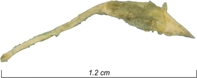
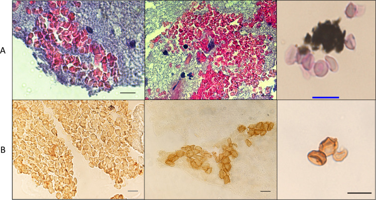
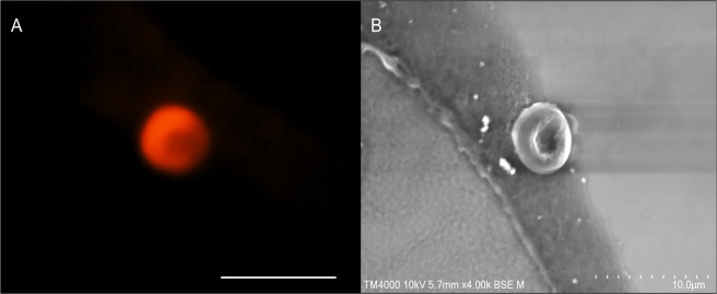
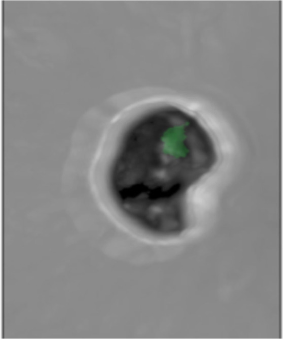
Photogrammetry and cascading microscopy investigations of dental pulp specimens collected from 2,000-year-old individuals buried in a Roman necropolis in Besançon, France, revealed unprecedented preserved tissular and cellular morphology. Photogrammetry yielded 3-D images of the smallest archaeological human remains ever recovered. Optical microscopy examinations after standard haematoxylin-phloxine-saffron staining and anti-glycophorin A immunohistochemistry exposed dental pulp cells, in addition erythrocytes were visualised by electron microscopy, which indicated the ancient dental pulp trapped a blood drop. Fluorescence in situ hybridisation applied on red blood cells revealed the louse-borne pathogen Bartonella quintana, a finding confirmed by polymerase chain reaction assays. Through paleohistology and paleocytology, we demonstrate that the ancient dental pulp preserved intact blood cells at the time of the individual's death, offering an unprecedented opportunity to engage in direct and indirect tests to diagnose pathogens in ancient buried individuals.


Conflict of interest statement

The authors declare no competing interests.

Figures

Figure 1
Figure 1
Photogrammetry of a 2,000-year-old intact dental pulp recovered from Ind. 17. qPCR-based detection of B. quintana.
Figure 2
Figure 2
Identification of ancient erythrocytes in individual 33 (B. quintana-negative) and individual 35 (B. quintana-positive) (A) HPS staining (10-µm scale) and (B) anti-glycophorin A staining (10-µm scale).
Figure 3
Figure 3
(A) Fluorescence microscopy of an erythrocyte in Ind. 35 using wavelength 555 nm. (B) The same erythrocyte was observed by scanning electron microscopy (10-µm scale).
Figure 4
Figure 4
FISH revealed B. quintana-infected erythrocytes from individual 35, with a B. quintana-negative autofluorescent erythrocyte from individual 33 used as a negative control. (A–F): Optical microscopy; (B–G): confocal microscopy with DAPI staining. (C–H): confocal microscopy with EUB probe. (D–I): Confocal microscopy with a B. quintana- specific probe. (E–J): Confocal microscopy with a nonEUB probe.
Figure 5
Figure 5
3D-FISH revealed B. quintana inside individual 35 erythrocytes under a green channel-specific probe.

References

    1. Hublin JJ, et al. New fossils from Jebel Irhoud, Morocco and the pan-African origin of Homo sapiens. Nature. 2017;546:289–292. doi: 10.1038/nature22336. - DOI - PubMed
    1. Moncel MH, Condemi S. Découverte de dents humaines dans le site Paléolithique moyen de Payre (Ardèche, France) Comptes rendus de l’Académie des sciences. Série 2. Sciences de la terre et des planètes. 1996;322:251–257.
    1. Higgins D, Austin JJ. Teeth as a source of DNA for forensic identification of human remains: a review. Science & Justice. 2013;53:433–441. doi: 10.1016/j.scijus.2013.06.001. - DOI - PubMed
    1. Drancourt M, Raoult D. Palaeomicrobiology: current issues and perspectives. Nature Reviews Microbiology. 2005;3:23–35. doi: 10.1038/nrmicro1063. - DOI - PubMed
    1. Lindahl T. Instability and decay of the primary structure of DNA. nature. 1993;362:709–715. doi: 10.1038/362709a0. - DOI - PubMed