

Validation of a digital pathology system including remote review during the COVID-19 pandemic
- PMID: 32572154
- PMCID: PMC7306935
- DOI: 10.1038/s41379-020-0601-5
Validation of a digital pathology system including remote review during the COVID-19 pandemic
Abstract
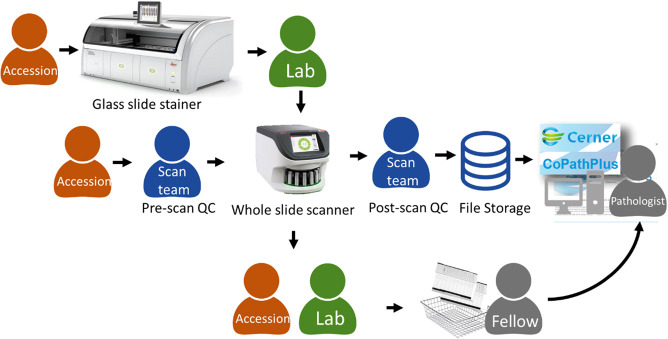
Remote digital pathology allows healthcare systems to maintain pathology operations during public health emergencies. Existing Clinical Laboratory Improvement Amendments regulations require pathologists to electronically verify patient reports from a certified facility. During the 2019 pandemic of COVID-19 disease, caused by the SAR-CoV-2 virus, this requirement potentially exposes pathologists, their colleagues, and household members to the risk of becoming infected. Relaxation of government enforcement of this regulation allows pathologists to review and report pathology specimens from a remote, non-CLIA certified facility. The availability of digital pathology systems can facilitate remote microscopic diagnosis, although formal comprehensive (case-based) validation of remote digital diagnosis has not been reported. All glass slides representing routine clinical signout workload in surgical pathology subspecialties at Memorial Sloan Kettering Cancer Center were scanned on an Aperio GT450 at ×40 equivalent resolution (0.26 µm/pixel). Twelve pathologists from nine surgical pathology subspecialties remotely reviewed and reported complete pathology cases using a digital pathology system from a non-CLIA certified facility through a secure connection. Whole slide images were integrated to and launched within the laboratory information system to a custom vendor-agnostic, whole slide image viewer. Remote signouts utilized consumer-grade computers and monitors (monitor size, 13.3-42 in.; resolution, 1280 × 800-3840 × 2160 pixels) connecting to an institution clinical workstation via secure virtual private network. Pathologists subsequently reviewed all corresponding glass slides using a light microscope within the CLIA-certified department. Intraobserver concordance metrics included reporting elements of top-line diagnosis, margin status, lymphovascular and/or perineural invasion, pathology stage, and ancillary testing. The median whole slide image file size was 1.3 GB; scan time/slide averaged 90 s; and scanned tissue area averaged 612 mm2. Signout sessions included a total of 108 cases, comprised of 254 individual parts and 1196 slides. Major diagnostic equivalency was 100% between digital and glass slide diagnoses; and overall concordance was 98.8% (251/254). This study reports validation of primary diagnostic review and reporting of complete pathology cases from a remote site during a public health emergency. Our experience shows high (100%) intraobserver digital to glass slide major diagnostic concordance when reporting from a remote site. This randomized, prospective study successfully validated remote use of a digital pathology system including operational feasibility supporting remote review and reporting of pathology specimens, and evaluation of remote access performance and usability for remote signout.
Figures


References
-
- Borowsky AD, Glassy EF, Wallace WD, Kallichanda NS, Behling CA, et al. Digital whole slide imaging compared with light microscopy for primary diagnosis in surgical pathology: a multicenter, double-blinded, randomized study of 2045 cases. Arch Pathol Lab Med. 2020. 10.5858/arpa.2019-0569-OA. Online ahead of print. - DOI - PubMed
-
- Mukhopadhyay S, Feldman MD, Abels E, Raheela Ashfaq, Beltaifa S, Cacciabeve NG, et al. Whole slide imaging versus microscopy for primary diagnosis in surgical pathology: a multicenter blinded randomized noninferiority study of 1992 cases (pivotal study) Am J Surg Pathol. 2018;42:39–52. doi: 10.1097/PAS.0000000000000948. - DOI - PMC - PubMed
Publication types
MeSH terms
Grants and funding
LinkOut - more resources
Full Text Sources
Other Literature Sources
Miscellaneous
