. 2020 Nov;33(11):2128-2138.
doi: 10.1038/s41379-020-0603-3. Epub 2020 Jun 22.

A systematic review of pathological findings in COVID-19: a pathophysiological timeline and possible mechanisms of disease progression

Affiliations

A systematic review of pathological findings in COVID-19: a pathophysiological timeline and possible mechanisms of disease progression

Samuel B Polak et al. Mod Pathol. 2020 Nov.

Abstract

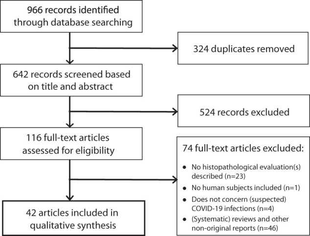
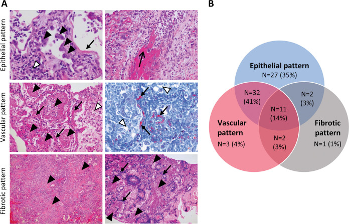
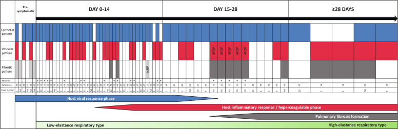
Since the outbreak of the COVID-19 pandemic, much has been learned regarding its clinical course, prognostic inflammatory markers, disease complications, and mechanical ventilation strategy. Clinically, three stages have been identified based on viral infection, pulmonary involvement with inflammation, and fibrosis. Moreover, low and high elastance phenotypes can be distinguished in mechanically ventilated patients, based on lung mechanics, ventilation-to-perfusion ratio, and CT scans; these two phenotypes have presumed differences in their underlying pathophysiology. Although essential for therapeutic guidance, the pathophysiology of COVID-19 is poorly understood. Here, we systematically reviewed published case reports and case series in order to increase our understanding of COVID-19 pathophysiology by constructing a timeline and correlating histopathological findings with clinical stages of COVID-19. Using PRISMA-IPD guidelines, 42 articles reporting 198 individual cases were included in our analysis. In lung samples (n = 131 cases), we identified three main histological patterns: epithelial (n = 110, 85%), with reactive epithelial changes and DAD; vascular (n = 76, 59%) with microvascular damage, (micro)thrombi, and acute fibrinous and organizing pneumonia; and fibrotic (n = 28, 22%) with interstitial fibrosis. The epithelial and vascular patterns can present in all stages of symptomatic COVID-19, whereas the fibrotic pattern presents starting at ~3 weeks. Moreover, patients can present with more than one pattern, either simultaneously or consecutively. These findings are consistent with knowledge regarding clinical patterns of viral infection, development of hyperinflammation and hypercoagulability, and fibrosis. Close collaboration among medical staff is necessary in order to translate this knowledge and classification of pathophysiological mechanisms into clinical stages of disease in individual patients. Moreover, further research, including histopathological studies, is warranted in order to develop reliable, clinically relevant biomarkers by correlating these pathological findings with laboratory results and radiological findings, thus, increasing our understanding of COVID-19 and facilitating the move to precision medicine for treating patients.


Figures

Fig. 1
Fig. 1
Flow chart depicting the search strategy.
Fig. 2
Fig. 2
Summary of cases with the epithelial, vascular, and/or fibrotic pattern of lung injury. a Example images of lung sections showing the epithelial (top; hematoxylin and eosin stains), vascular (middle; left hematoxylin and eosin stain, right fibrin-Lendrum (MSB) stain), and fibrotic (bottom; left hematoxylin and eosin stain, right Verhoeff-van Gieson stain) pattern of lung injury in COVID-19. In the top panels, atypia and detachment of type II pneumocytes (closed arrowheads), hyaline membrane formation (closed arrow), an interstitial inflammatory response (open arrowhead), and denudation of bronchiolar epithelium (open arrow) are indicated. In the middle panels, intracapillary hyaline thrombi (arrows), acute fibrinous and organizing pneumonia (closed arrowheads), and edema (open arrowhead) are indicated. In the bottom panels, notable intra-alveolar fibroelastosis (closed arrowheads) with pre-existing alveolar septal elastin (arrows) are indicated, possibly representing fibrosing organizing pneumonia. Images were obtained from autopsies of COVID-19 patients performed at the Erasmus Medical Center. For low power images, see Supplementary Figures S1–S3. b Venn diagram summarizing the histological patterns of lung injury in 78 COVID-19 patients.
Fig. 3
Fig. 3
Timeline correlating the epithelial, vascular, and fibrotic patterns of lung injury with the duration of COVID-19 symptoms. Shown are the identified histological pulmonary patterns (epithelial, vascular, and fibrotic) for 65 individual cases; each case is shown in a single column. Cases are organized chronologically in four time periods: the number of days since the onset of symptoms of COVID-19 symptoms is indicated. For the fibrotic pattern, cases shaded in light gray are cases in which fibrotic changes were likely pre-existing and were not associated with COVID-19, and cases shaded in dark gray indicate fibrotic changes. For cases indicated with an asterisk (*), the duration of the clinical course was estimated from a report with aggregated pathological findings. Shown below the timeline is the approximate occurrence of COVID-19 clinical phases, color-coded based on the three patterns of histological pulmonary changes, and the transition from L- to H-respiratory phenotypes [5]. In pre-symptomatic patients, tissue was obtained antemortem before the onset of COVID-19 symptoms. Other antemortem samples are: reference [18, 22] (cases 1 and 2), and 33; the other samples are obtained at autopsy. AFOP acute fibrinous and organizing pneumonia, NSIP nonspecific interstitial pneumonia, NA not applicable, NS not specified.

Comment in

References

    1. Wu F, Zhao S, Yu B, Chen YM, Wang W, Song ZG, et al. A new coronavirus associated with human respiratory disease in China. Nature. 2020;579:265–269. PMCID 7094943; PubMed PMID: 32015508. - PMC - PubMed
    1. WHO Director-General's remarks at the media briefing on 2019-nCoV on 11 February 2020 2020 [Internet] [cited April 22, 2020]: https://www.who.int/dg/speeches/detail/who-director-general-s-remarks-at....
    1. Wu C, Chen X, Cai Y, Xia J, Zhou X, Xu S, et al. Risk factors associated with acute respiratory distress syndrome and death in patients with coronavirus disease 2019 pneumonia in Wuhan, China. JAMA Intern Med. 2020;e200994. - PMC - PubMed
    1. Zhou F, Yu T, Du R, Fan G, Liu Y, Liu Z, et al. Clinical course and risk factors for mortality of adult inpatients with COVID-19 in Wuhan, China: a retrospective cohort study. Lancet. 2020;395:1054–1062. PMCID 7270627; PubMed PMID: 32171076. - PMC - PubMed
    1. Gattinoni L, Chiumello D, Caironi P, Busana M, Romitti F, Brazzi L, et al. COVID-19 pneumonia: different respiratory treatments for different phenotypes? Intensive Care Med. 2020; 10.1007/s00134-020-06033-2 - PMC - PubMed

Publication types

LinkOut - more resources