Multicenter Study
. 2021 Jan;34(1):233-248.
doi: 10.1038/s41379-020-0593-1. Epub 2020 Jun 22.

The histologic presentation of hepatitis E reflects patients' immune status and pre-existing liver condition

Affiliations
Multicenter Study

The histologic presentation of hepatitis E reflects patients' immune status and pre-existing liver condition

Daniela Lenggenhager et al. Mod Pathol. 2021 Jan.

Abstract

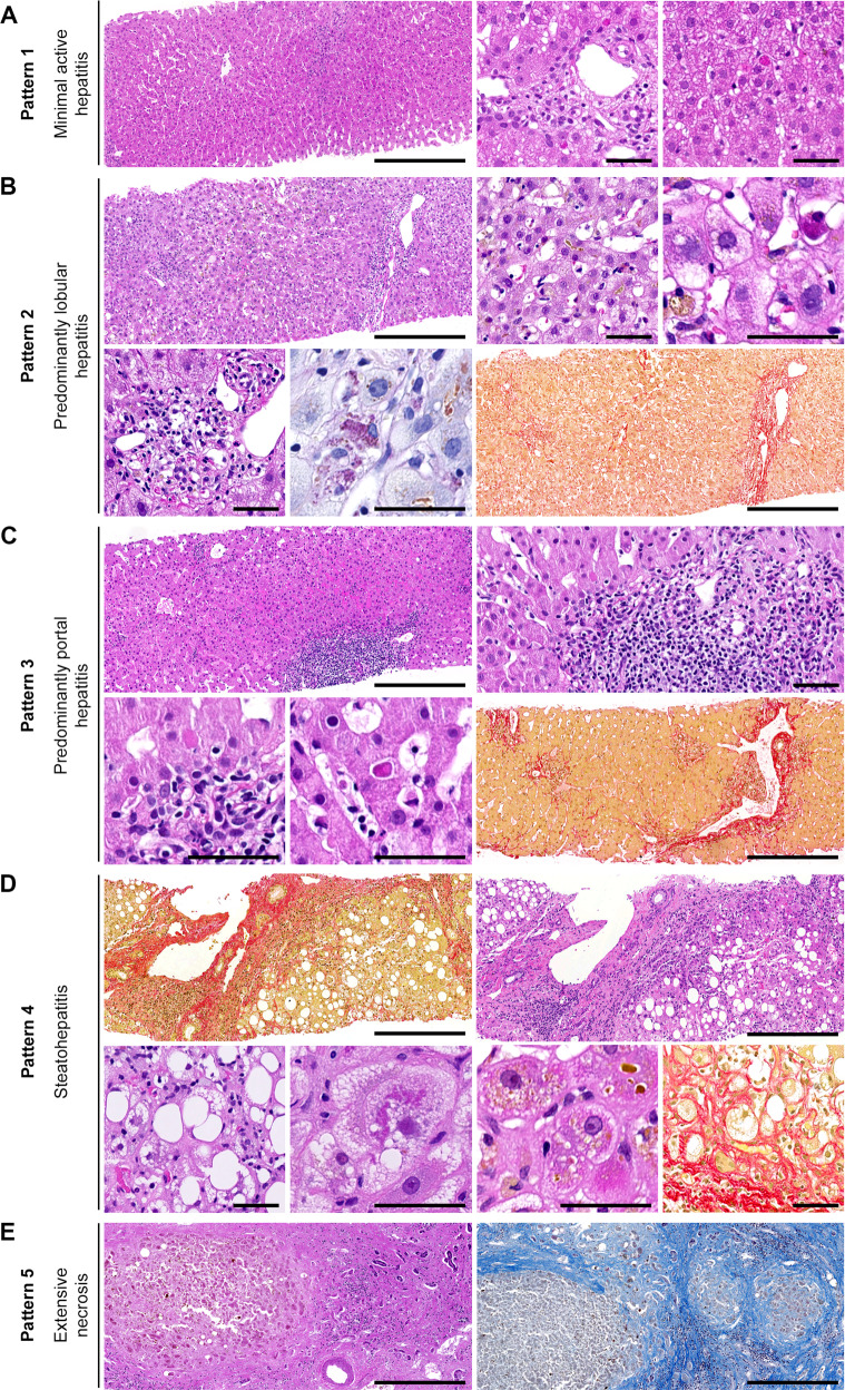
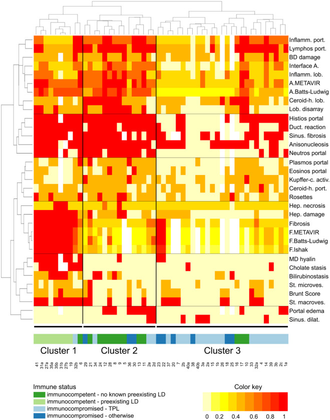
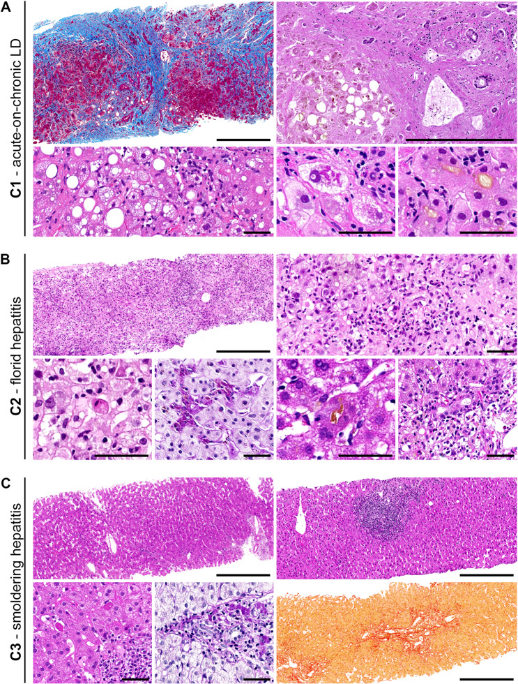
Infection with the hepatitis E virus (HEV) is one of the main causes of acute hepatitis worldwide. Given that, the histopathology of hepatitis E is relatively poorly characterized, and it is unclear what exactly determines its remarkable variability. The aim of our study was a systematic analysis of hepatitis E histology, especially with regard to the clinical setting. Fifty-two liver samples (48 biopsies, 1 liver explant, 3 autopsy livers) from 41 patients with molecularly proven hepatitis E (28 HEV genotype (gt) 3, three gt 1, one gt 4 and 9 undetermined gt) were systematically evaluated for 33 histopathologic features. Following one approach, the biopsies were assigned to one of five generic histologic patterns. In another approach, they were subjected to hierarchical clustering. We found that 23/41 (56%) patients were immunocompromised, whereas 18 (44%) had no known immunosuppression. Five patients (12%) had pre-existing liver disease (LD). The histopathologic spectrum ranged from almost normal to acute, chronic, and steato-hepatitis to subtotal necrosis, and was thus distributed across all five generic patterns. Hierarchical clustering, however, identified three histopathologic clusters (C1-C3), which segregated along the immune status and pre-existing LD: C1 comprised mostly patients with pre-existing LD; histology mainly reflected the respective LD without pointing to the additional hepatitis E. C2 comprised mostly immunocompetent patients; histology mainly displayed florid hepatitis. C3 comprised mostly immunocompromised patients; histology mainly displayed smoldering hepatitis. Accordingly, C1-C3 differed markedly with respect to their clinical and histopathologic differential diagnoses. Hierarchical clustering suggests three groups with distinct histopathologies, indicating biologically different manifestations of hepatitis E. The association of histopathologic changes with the patient's immune status and pre-existing LD plausibly explains the diversity of hepatitis E histopathology, and suggests that these factors are the crucial underlying determinants. We expect our results to improve patient management by guiding the clinico-pathologic diagnosis of hepatitis E.


Conflict of interest statement

The authors declare that they have no conflict of interest.

Figures

Fig. 1
Fig. 1. Histopathologic findings observed in hepatitis E represented in five generic histologic patterns.
a Mimimal active hepatitis with normal architecture, minimal portal, and lobular inflammation and only single scattered hepatocyte apoptosis. b Predominantly lobular hepatitis with prominent lobular disarray, rosettes formation, bilirubinostasis, hepatocyte damage, necrotic hepatocytes, bile duct damage, signs of beginning removal with lobular ceroid-laden histiocytes (PAS stain) and some parenchymal collapse (Sirius red stain). c Predominantly portal inflammation with preserved lobular architecture and some interface and lobular inflammatory activity with few hepatocyte apoptoses. Developing fibrosis possible (Sirius red stain). d Cirrhotic parenchyma (Sirius red stain) with steatosis, necroinflammation, hepatocyte ballooning, Mallory-Denk hyaline, bilirubinostasis and pronounced pericellular fibrosis (Sirius red stain). e Subtotal necrosis of liver parenchyma shown here in a cirrhotic liver (CAB stain). H&E stain, if not otherwise specified. Scale bars overviews 400 μm, scale bars details 50 μm.
Fig. 2
Fig. 2. Hierarchical clustering of histopathologic findings.
Hierarchical clustering of the 52 liver specimens (x axis) with respect to the 33 histopathologic features (y axis) represented in a heatmap: Identification of three clusters (C1–C3) that segregate along the patient’s immune status and pre-existing LD. The 33 histopathologic features clustered mainly according to the different pathologies they reflect (i.e., from top to bottom: features of acute inflammatory activity, features of early subacute inflammatory activity, features of late subacute inflammatory activity, features of chronicity and features of cellular damage). Only two features (portal edema and sinusoidal dilatation) did not cluster in any of the above mentioned groups. A activity, BD bile duct, Ceroid-h. Ceroid-histiocytes, Duct. ductular, F fibrosis, Hep. hepatocyte, Inflamm. inflammation, Kupffer-c. activ. Kupffer cell activation, LD liver disease, lob. lobular, MD Mallory-Denk, port. portal, Sinus. sinusoidal, St. steatosis, TPL transplantation.
Fig. 3
Fig. 3. Histopathologic findings observed in hepatitis E represented in three histopathologic clusters.
a Samples in C1 showed mainly features of acute-on-chronic LD, such as cirrhosis (Masson trichrome stain), necrosis, steatosis, steatohepatitis, hepatocyte damage, Mallory-Denk hyaline and bilirubinostasis (from upper left to lower right). b Samples in C2 displayed mainly features of florid hepatitis, such as lobular inflammation and parenchymal disarray, foci of hepatocyte necrosis and many scattered apoptotic hepatocytes as well as signs of beginning removal with lobular ceroid-laden histiocytes (PAS-D stain), cholestasis and bile duct damage (from upper left to lower right). c Samples in C3 were mainly characterized by features of smoldering hepatitis, ranging from minimal histologic changes over predominantly portal based inflammation with mild interface and lobular activity with only rare apoptotic hepatocytes as well as signs of previous damage such as ceroid-laden histiocytes (PAS-D stain) in portal tracts and in some cases with fibrosis (Sirius red stain) (from upper left to lower right). H&E stain, if not otherwise specified. Scale bars overviews 400 μm, scale bars details 50 μm. C cluster, LD liver disease.
Fig. 4
Fig. 4. Association of the three clusters with the clinical setting and the five histologic patterns.
Clinico-pathologic settings of the three clusters including their most relevant differential diagnoses. The thickness of the lines between the patterns and the clusters represent the relative association of both. ASH alcoholic steatohepatitis, C cluster, DILI drug-induced liver injury, GVHD graft-versus-host disease, LD liver disease, NASH nonalcoholic steatohepatitis, TPL transplantation.

References

    1. Debing Y, Moradpour D, Neyts J, Gouttenoire J. Update on hepatitis E virology: implications for clinical practice. J Hepatol. 2016;65:200–12. doi: 10.1016/j.jhep.2016.02.045. - DOI - PubMed
    1. Nimgaonkar I, Ding Q, Schwartz RE, Ploss A. Hepatitis E virus: advances and challenges. Nat Rev Gastroenterol Hepatol. 2018;15:96–110. doi: 10.1038/nrgastro.2017.150. - DOI - PMC - PubMed
    1. Kamar N, Izopet J, Pavio N, Aggarwal R, Labrique A, Wedemeyer H, et al. Hepatitis E virus infection. Nat Rev Dis Prim. 2017;3:17086. doi: 10.1038/nrdp.2017.86. - DOI - PubMed
    1. European Association for the Study of the Liver. EASL clinical practice guidelines on hepatitis E virus infection. J Hepatol. 2018;68:1256–71. doi: 10.1016/j.jhep.2018.03.005. - DOI - PubMed
    1. Dalton HR, Izopet J. Transmission and epidemiology of hepatitis E virus genotype 3 and 4 infections. Cold Spring Harb Perspect Med. 2018;8:a032144. doi: 10.1101/cshperspect.a032144. - DOI - PMC - PubMed

Publication types

MeSH terms