. 2020 Sep;16(9):1019-1025.
doi: 10.1038/s41589-020-0575-0. Epub 2020 Jun 22.

Structural insight into the formation of lipoprotein-β-barrel complexes

Affiliations

Structural insight into the formation of lipoprotein-β-barrel complexes

Raquel Rodríguez-Alonso et al. Nat Chem Biol. 2020 Sep.

Abstract

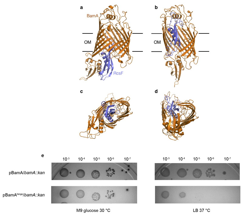
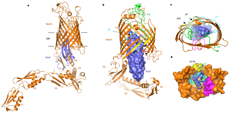
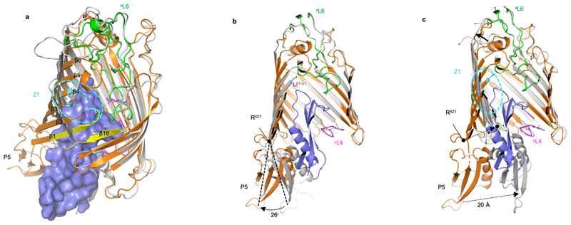
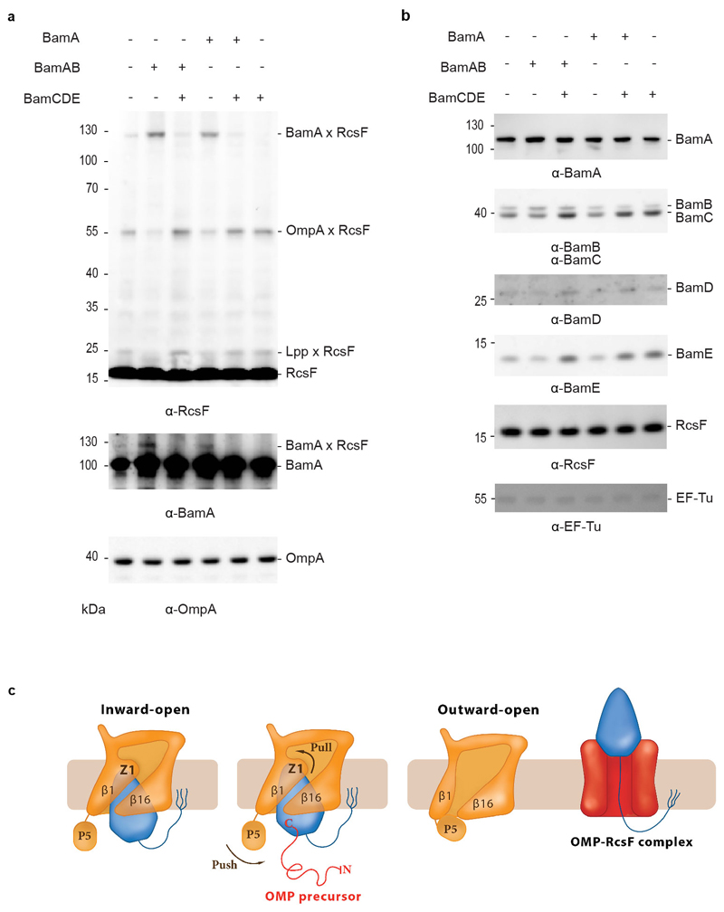
The β-barrel assembly machinery (BAM) inserts outer membrane β-barrel proteins (OMPs) in the outer membrane of Gram-negative bacteria. In Enterobacteriacea, BAM also mediates export of the stress sensor lipoprotein RcsF to the cell surface by assembling RcsF-OMP complexes. Here, we report the crystal structure of the key BAM component BamA in complex with RcsF. BamA adopts an inward-open conformation, with the lateral gate to the membrane closed. RcsF is lodged deep within the lumen of the BamA barrel, binding regions proposed to undergo outward and lateral opening during OMP insertion. On the basis of our structural and biochemical data, we propose a push-and-pull model for RcsF export following conformational cycling of BamA, and provide a mechanistic explanation for how RcsF uses its interaction with BamA to detect envelope stress. Our data also suggest that the flux of incoming OMP substrates is involved in the control of BAM activity.


Conflict of interest statement

Author Information

The authors declare no competing financial interests.

Figures

Extended Data Fig. 1
Extended Data Fig. 1. RcsF can be co-purified with the BAM complex
(a,b,c) Gel filtration profiles of the affinity-purified BamAB-RcsF, BAM-RcsF and BAM complexes. The size exclusion chromatography was performed using a HiLoad 10/300 Superdex 200pg. The input and peak fractions were collected and the samples were analyzed by blue native electrophoresis with Coomassie staining. The migration pattern of BamABCDE-RcsF (b) was modified compared to BamABCDE (c) upon size exclusion chromatography (band 8 increases), reflecting the higher instability of the BamABCDE-RcsF complex. n= 4 biologically independent experiments.
Extended Data Fig. 2
Extended Data Fig. 2. Crystal structure of the BamA-RcsF complex
(a, b) Final 2Fo-Fc electron map of the BamA-RcsF complex, shown with a map contour level of 0.08 e-/Å3 (root mean square deviation 1.02 Å). The asymmetric unit of the crystals holds two BamA-RcsF copies, one revealing interpretable electron density for the full BamA sequence (a), and a second revealing unambiguous density for POTRA domain 4 only (b). In the second copy (b), the electron density corresponding to POTRA domains 1, 2, 3, and 5 is too weak to allow unambiguous rigid body placement of the domains. All descriptions and images in the main text are based on the first copy (a). (c) Overlay of two BamA-RcsF complexes in the asymmetric unit. The first complex depicts BamA in gold and RcsF in blue, while these molecules are yellow and light blue, respectively, in the second complex. In both copies, RcsF makes an average displacement of 4 Å relative to the BamA β-barrel. (d) Crystal packing of the BamA-RcsF complex viewed along the a- (left) and c-axis (right). For the first copy of the BamA-RcsF complex in the asymmetric unit (orange-slate) the conformation of the POTRA domains is stabilized by the packing along the b-axis, whilst for the second copy (cyan-slate) only POTRA domain 4 is involved in crystal contacts. In the latter, POTRA 5, 3, 2 and 1 are not in contact with neighboring molecules and show weak electron density only due to the lack of conformational stabilization.
Extended Data Fig. 3
Extended Data Fig. 3. Structural dynamics of the BamA POTRA domains
(a, b) Superimposition of BamA-RcsF (gold and blue, respectively) with the POTRA domains in the inward-open BamABCDE complex (PDB: 5D0O; light blue) or the outward-open BamACDE complex (PDB: 5EKQ; green). Complexes are superimposed based on 400 equivalent Cα atoms in the BamA β-barrel, and shown in side (a) or periplasmic (b) view. For 5d0o and 5ekq, the accessory Bam subunits and the BamA β-barrel are omitted for clarity. (c) Periplasmic view of the inward-open BamABCDE complex, showing binding of the BAM accessory proteins BamB (magenta), BamC (red), BamD (blue), and BamE (yellow). Pulldown experiments showed that RcsF binds the BamABCDE complex (Fig. 1). In agreement with this observation, structural comparisons reveal that RcsF binding would not result in direct steric clashes with any BAM accessory protein. However, the positions of the POTRA domains in the BamA-RcsF and BamABCDE complexes are markedly different. In the BamA-RcsF complex, POTRA5 makes a 26 outward rotation to accommodate RcsF (see also Fig. 3), and a reorganization in the joint between POTRA domains 3 and 2 results in a more extended conformation of the POTRA “arm” and the projection of POTRA domains 2 and 1 further from the BamA β-barrel, a conformation not previously reported in available BamA structures. In the BamABCDE complex, BamD contacts both POTRA5 and the joint of POTRA domains 1 and 2. In the BamA-RcsF complex, POTRA5 and POTRA domains 2 and 1 are too distant to be bridged by BamD; binding of BamD to BamA-RcsF therefore requires a conformational change in the POTRA arm or the dissociation of BamD at either of these two contact points.
Extended Data Fig. 4
Extended Data Fig. 4. Validation of the BamA-RcsF structure
(a) RcsF aminoacid sequence. The sequence coverage of the XL-MS experiment was about 60% as highlighted in violet (b) Ribbon diagram of the BamA-RcsF structure. Highlighted residues show sites mutated to amber for incorporation of the photoreactive lysine analog DiZPK. Sites that crosslink to RcsF are green, sites that show no crosslinking are magenta. Mutation of extracellular loop 1 (eL1; red) leads to loss of RcsF binding (see panel g). BamA sidechains found to crosslink with RcsF by means of the homobifunctional amine-reactive crosslinker disuccinimidyl dibutyric urea (DSBU) are shown as sticks and colored cyan. Residue K61 from RcsF, which was found to crosslink to BamA using DSBU, is shown as a stick and colored orange. The other two RcsF residues (K42 and K134) that could be crosslinked to BamA are not visible in this structural model. (c) In vivo photocrosslinking experiment in which cells expressing the BamA mutants containing DiZPK at the indicated positions were treated (+) or not (-) with ultraviolet light. Proteins samples were analyzed via SDS-PAGE and immunoblotted with anti-RcsF or anti-BamA antibodies, showing that the photo-crosslinked complexes contain BamA and RcsF. WT, wild type. (d,e) Sensorgrams from biolayer interferometry (left) and corresponding equilibrium binding plots (right) of immobilized RcsF titrated with BamA (d) or immobilized BamA titrated with RcsF (e), n=1 biologically independent experiment. (f) The levels of major OMPs are slightly decreased in cells expressing BamAΔloop1. WT cells harboring the empty plasmid (pAM238) were used as control and EF-Tu expression levels were analyzed as loading control. n= 3 biologically independent experiments. (g) Deletion of loop 1 in BamA prevents RcsF from being pulled down with BamA. WT cells harboring the empty plasmid (pAM238) were used as control. n= 3 biologically independent experiments. (h) Overexpression of pBamAΔLoop1 in a bamA deletion strain activates the Rcs system compared to WT. A chromosomal rprA::lacZ fusion was used to monitor Rcs activity, and specific β-galactosidase activity was measured from cells at mid-log phase (OD600=0.5). Boxplot with whiskers from minimum to maximum. All values were normalized to the average activity obtained for WT cells harbouring the empty plasmid (pET3a) obtained from N=8 biologically independent experiments. Mean is showed as +. WT, wild-type; Kan, kanamycin.
Extended Data Fig. 5
Extended Data Fig. 5. RcsF binds the inward-open conformation of BamA
Models for the BamAG393C/G584C 5 and BamAG433C/N805C 26 double cysteine mutants, which are locked in the outward-open or inward-open conformation, respectively, when oxidized. Mutated cysteines are shown as atom spheres. (b) BamA barrel locking and RcsF binding. Overexpression of double cysteine mutants pBamAG393C/G584C-B and BamAG433C/N805C-B in a wild-type strain. RcsF can be co-purified with the BamA β-barrel locked in the inward-open conformation (BamAG433C/N805C) by a disulfide bond (ox) but not in the outward-open conformation (BamAG393C/G584C). BamA mutants become reduced (red) following treatment with tris(2-carboxyethyl) phosphine (TCEP) and migrate similarly. The oxidized form of BamAG393C/G584C migrates more slowly than wild-type BamA. As a result, two bands are visible for BamA in the input of BamAG393C/G584C, the lower migrating band corresponding to wild-type BamA expressed from the chromosome. n= 3 biologically independent experiments. (c) Sensorgram from biolayer interferometry of immobilized RcsF titrated with BamAG393C/G584C, without (oxidized; - DTT) or with dithiothreitol (reduced; + DTT). When the β-barrel is locked in the outward-open conformation (- DTT), RcsF is unable to bind BamA. When reduced, BamAG393C/G584C regains binding, demonstrating that BamA reverts to the inward-open conformation in which it can bind RcsF.
Extended Data Fig. 6
Extended Data Fig. 6. The movement of POTRA5 towards the periplasmic exit of the lumen of the BamA barrel could push RcsF upwards
(a,b) Lateral view of the initial and final conformations, respectively, of the BamA-RcsF complex during the dynamic importance sampling simulation (DIMS) of the BamA-RcsF complex. (c,d) Bottom view (from the periplasm) of the above conformations. BamA and RcsF are colored in orange and blue, respectively. The initial conformation of the system (BamA and RcsF) corresponds to the structure determined in this work (PDB code 6T1W) with the POTRA1-4 domains removed. The final conformation of BamA is similar to the outward-open structure (PDB code 5D0Q). The explicit outer membrane and solvent are not shown for clarity. (e) Expression from BamAhinge from a plasmid in ΔbamA cells leads to a severe growth defect when cells are grown at 37°C in rich media, but not when they are grown in minimal media at 30°C. Cells were grown in M9 minimal glucose medium at 30°C until they reached OD600 =1. Tenfold serial dilutions were made in M9 minimal glucose, plated onto M9 minimal glucose or LB agar, and incubated at 30°C or 37°C. Plates were supplemented with ampicillin (200μg/ml). n=3 biologically independent experiments.
Figure 1
Figure 1. RcsF forms a complex with BamAB and BamABCDE.
(a, b) SDS-PAGE (a) and blue native (b) analysis of purified BAM, BAM-RcsF and BamAB-RcsF complexes obtained via BamA-affinity chromatography. The bands analyzed in (c) are labelled 1 to 8. (c) SDS-PAGE analysis of the complexes shown in panel b (bands 1 to 8). The BAM complex expressed from pRRA1 is a mixture of BamABCDE and BamABDE. n= 4 biologically independent experiments.
Figure 2
Figure 2. Structure of the BamA-RcsF complex.
(a) Ribbon diagram of the BamA-RcsF complex in side view. BamA, gold; RcsF, blue. (b, c) Front (b) and extracellular (c) views of BamA-RcsF, with RcsF shown as a solvent-accessible surface. POTRA domains 1 and 2 have been omitted for clarity. BamA eL6, green; pL4, magenta. Putative RcsF-interacting residues in contact zones Z1 and Z2 of the BamA β-barrel are colored cyan and magenta, respectively, and shown as sticks. Strands β1 and β16, which form the proposed “lateral gate” of the BamA β-barrel, are yellow. (d) Periplasmic view of the BamA-RcsF complex, with the BamA β-barrel shown as a solvent-accessible surface and RcsF as a ribbon. Colors are as in panels b and c. POTRA domains were omitted for clarity.
Figure 3
Figure 3. Conformational characteristics of the BamA-RcsF complex.
(a, c) Tilted top view and slabbed side view of the overlay of the BamA-RcsF complex and BamA in the outward-open conformation (grey, taken from BamACDE complex PDB:5EKQ4). The BamA β-barrel undergoes a ~45 outward rotation at strands β1-β6, and a 20 Å inward displacement of POTRA5 compared to the structure of BamA-RcsF presented here. (b) Slabbed side view of the overlay of BamA-RcsF and BamA in the inward-open conformation (grey, taken from BamABCDE complex PDB:5D0O 5). In the structure of BamA-RcsF presented here, POTRA5 makes a 26 outward rotation relative to R421, where it connects to the BamA β-barrel. (a-c) Color scheme for BamA-RcsF is as in Fig. 2b. RcsF is shown as a solvent-accessible surface (a) or a ribbon (b, c). Panels (b, c) show side views, slabbed down to view the interior of the complex. For 5EKQ and 5D0O, the BAM accessory proteins BamB, C, D, and E were omitted for clarity, as were POTRA domains 1-4 in all shown BamA structures.
Figure 4
Figure 4. BamA-RcsF is a proxy for the inward-open conformation of BamA.
(a) In vivo chemical crosslinking of RcsF with BamA and OmpA. The BamA-RcsF complex accumulates when either BamA alone or BamAB together are moderately over-expressed from a plasmid in cells also expressing BAM at physiological levels from the chromosome. The copies of BamA and BamAB in excess are not functional (BamCDE is required for BAM activity) and do not funnel RcsF to its OMP partners. As a result, RcsF accumulates on BamA and OmpA-RcsF does not form. Over-expression of BamCDE (also from a plasmid) in these cells restores the stoichiometry between the BAM components: BamA-RcsF does not accumulate and the formation of OmpA-RcsF is restored. As shown previously, levels of OmpA-RcsF are inversely correlated with BamA-RcsF. Overexpression of the BamCDE sub-complex alone does not impact the activity of the BAM complex expressed from the chromosome: wild-type BamA-RcsF and OmpA-RcsF levels are observed. Wild-type BamA-RcsF and OmpA-RcsF levels are also detected when BamA and BamCDE are overexpressed together, as expected given that BamB is not essential. RcsF also forms a complex with the abundant lipoprotein Lpp (Lpp-RcsF), as in. Protein expression levels of OmpA were analyzed by immunoblot in the non-crosslinked samples, showing no differences. The additional bands that are detected in the lanes where BamA-RcsF is not observed likely correspond to poorly abundant complexes between RcsF and unknown proteins. n= 3 biologically independent experiments. (b) Protein expression levels of BamB, BamC, BamD, BamE and RcsF from no-crosslinked samples overexpressing BamA (pBamA), BamAB (pBamA-B) and BamCDE (pSC263) were analyzed by western blot. EF-Tu expression levels were analyzed as loading control. n= 3 biologically independent experiments. (c) Model proposing that BamA conformational cycling is triggered by incoming OMP substrates on the BAM holocomplex. A BamA inward-to-outward open transition could result in an upward displacement of RcsF via a push-and-pull mechanism, resulting in an OMP-RcsF complex. The push-and-pull mechanism involves BamA POTRA5 (P5) and Z1. The topology of the OMP-RcsF complex remains to be established. For clarity, POTRA1-4 and the BAM lipoproteins have been ommitted.

References

    1. Noinaj N, Gumbart JC, Buchanan SK. The beta-barrel assembly machinery in motion. Nat Rev Microbiol. 2017;15:197–204. doi: 10.1038/nrmicro.2016.191. - DOI - PMC - PubMed
    1. Hagan CL, Silhavy TJ, Kahne D. beta-Barrel membrane protein assembly by the Bam complex. Annu Rev Biochem. 2011;80:189–210. doi: 10.1146/annurev-biochem-061408-144611. - DOI - PubMed
    1. Iadanza MG, et al. Lateral opening in the intact beta-barrel assembly machinery captured by cryo-EM. Nat Commun. 2016;7 doi: 10.1038/ncomms12865. 12865. - DOI - PMC - PubMed
    1. Bakelar J, Buchanan SK, Noinaj N. The structure of the beta-barrel assembly machinery complex. Science. 2016;351:180–186. doi: 10.1126/science.aad3460. - DOI - PMC - PubMed
    1. Gu Y, et al. Structural basis of outer membrane protein insertion by the BAM complex. Nature. 2016;531:64–69. doi: 10.1038/nature17199. - DOI - PubMed

Publication types

MeSH terms