. 2020 Jun 23:9:e55279.
doi: 10.7554/eLife.55279.

Skd3 (human ClpB) is a potent mitochondrial protein disaggregase that is inactivated by 3-methylglutaconic aciduria-linked mutations

Affiliations

Skd3 (human ClpB) is a potent mitochondrial protein disaggregase that is inactivated by 3-methylglutaconic aciduria-linked mutations

Ryan R Cupo et al. Elife. .

Abstract

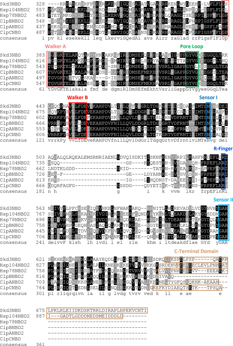
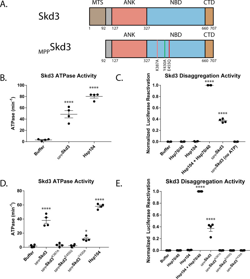
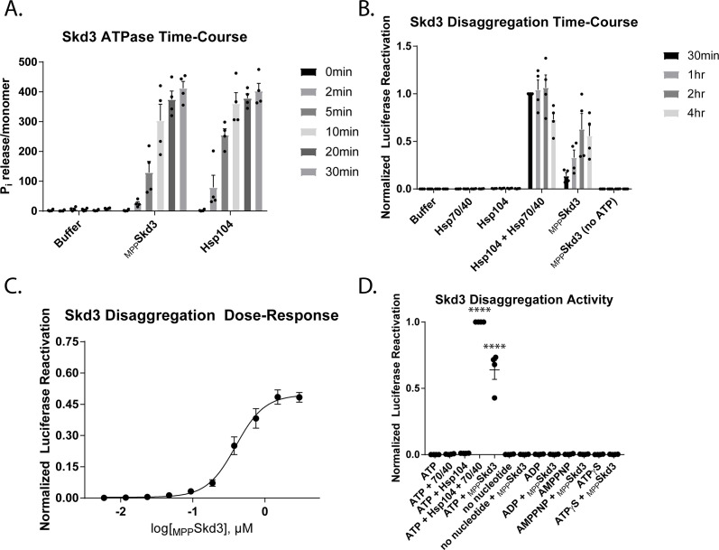
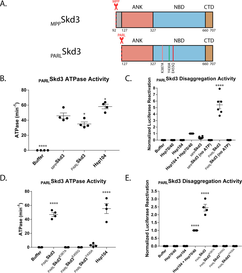
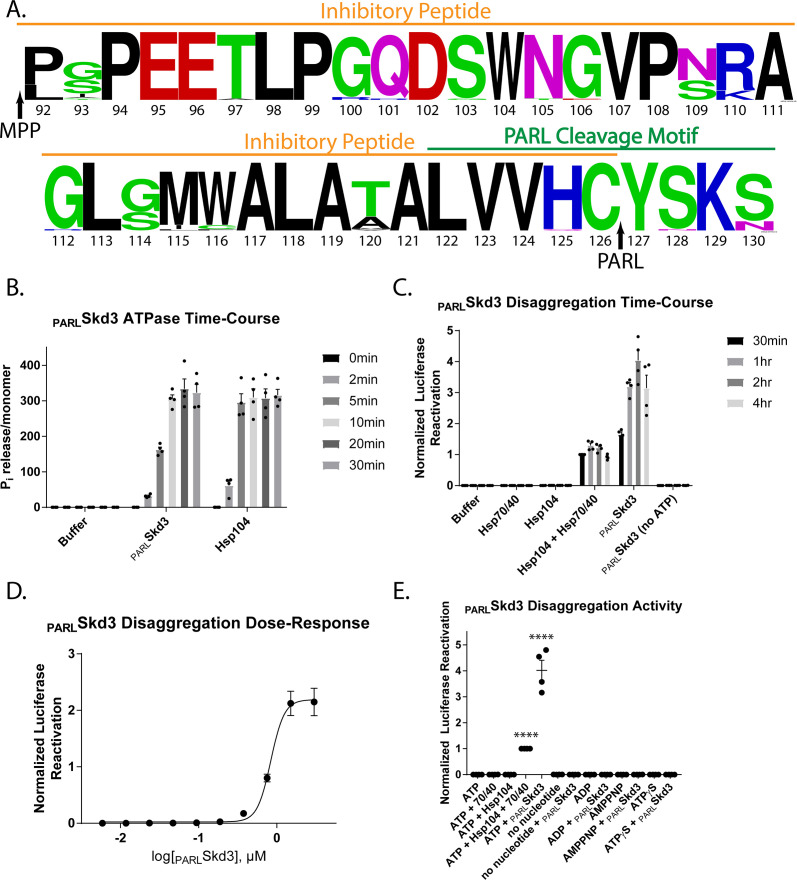
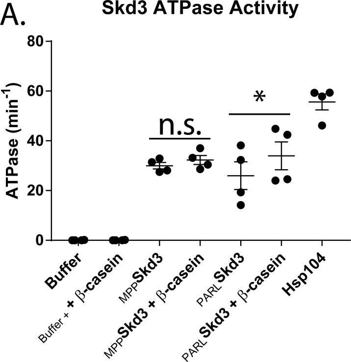
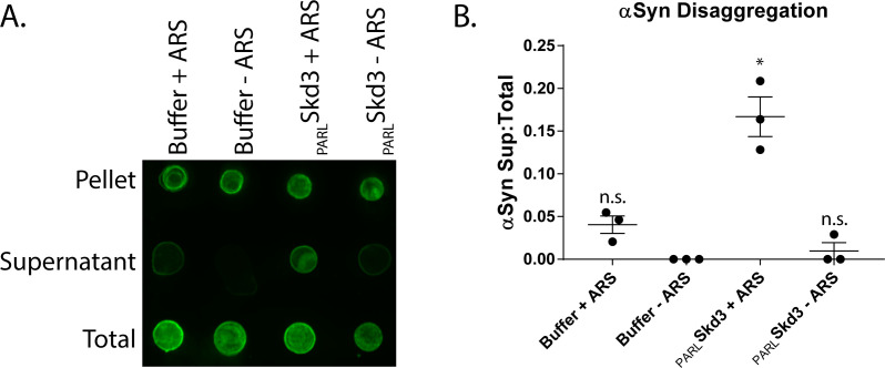
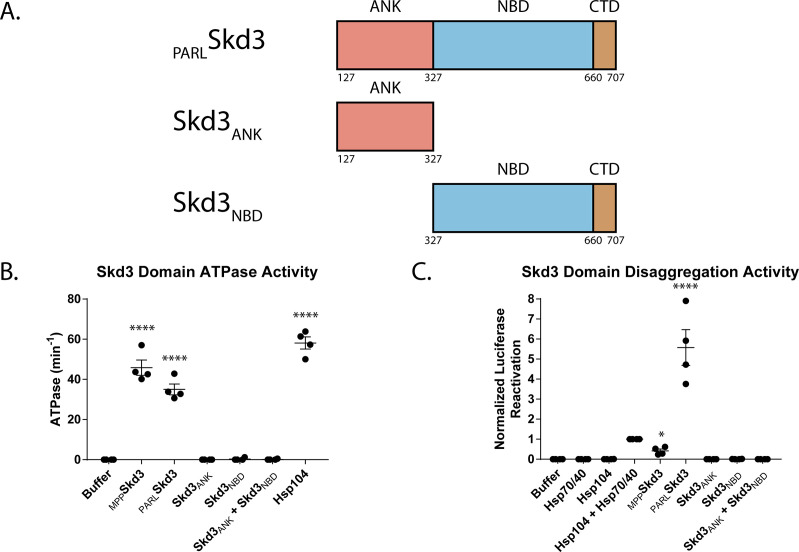
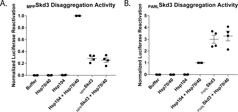
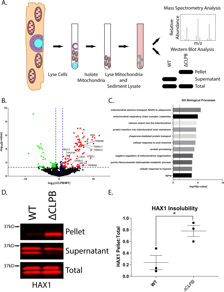
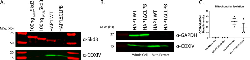
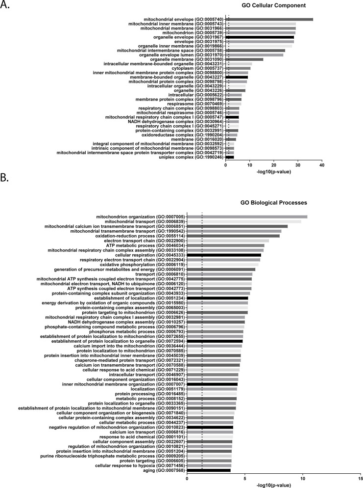
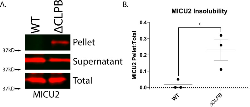
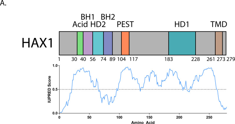
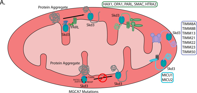
Cells have evolved specialized protein disaggregases to reverse toxic protein aggregation and restore protein functionality. In nonmetazoan eukaryotes, the AAA+ disaggregase Hsp78 resolubilizes and reactivates proteins in mitochondria. Curiously, metazoa lack Hsp78. Hence, whether metazoan mitochondria reactivate aggregated proteins is unknown. Here, we establish that a mitochondrial AAA+ protein, Skd3 (human ClpB), couples ATP hydrolysis to protein disaggregation and reactivation. The Skd3 ankyrin-repeat domain combines with conserved AAA+ elements to enable stand-alone disaggregase activity. A mitochondrial inner-membrane protease, PARL, removes an autoinhibitory peptide from Skd3 to greatly enhance disaggregase activity. Indeed, PARL-activated Skd3 solubilizes α-synuclein fibrils connected to Parkinson's disease. Human cells lacking Skd3 exhibit reduced solubility of various mitochondrial proteins, including anti-apoptotic Hax1. Importantly, Skd3 variants linked to 3-methylglutaconic aciduria, a severe mitochondrial disorder, display diminished disaggregase activity (but not always reduced ATPase activity), which predicts disease severity. Thus, Skd3 is a potent protein disaggregase critical for human health.

Keywords: AAA+ protein; Hsp104; Hsp78; Skd3; biochemistry; chemical biology; disaggregase; human; protein misfolding.


Conflict of interest statement

RC, JS No competing interests declared

Figures

Figure 1.
Figure 1.. Skd3 is homologous to Hsp104 and Hsp78 and is conserved across diverse metazoan lineages.
(A) Domain map depicting S. cerevisiae Hsp104, S. cerevisiae Hsp78, and H. sapiens Skd3. Hsp104 is composed of a N-terminal domain (NTD), nucleotide-binding domain 1 (NBD1), middle domain (MD), nucleotide-binding domain 2 (NBD2), and C-terminal domain (CTD). Hsp78 is composed of a mitochondrial-targeting signal (MTS), NBD1, MD, NBD2, and CTD. Skd3 is composed of a MTS, a short hydrophobic stretch of unknown function, an ankyrin-repeat domain (ANK) containing four ankyrin repeats, an NBD that is homologous to Hsp104 and Hsp78 NBD2, and a CTD. (B) Phylogenetic tree depicting a Clustal Omega alignment of Skd3 sequences from divergent metazoan lineages and the protozoan Monosiga brevicollis. The alignment shows conservation of Skd3 across diverse species and shows high similarity between mammalian Skd3 proteins.
Figure 1—figure supplement 1.
Figure 1—figure supplement 1.. Skd3 NBD alignment to other AAA+ proteins reveals high similarity to Hsp104 and Hsp78.
Alignment of the NBD from H. sapiens Skd3 and NBD2 from S. cerevisiae Hsp104, S. cerevisiae Hsp78, E. coli ClpB, E. coli ClpA, and S. aureus ClpC. Alignments were constructed using Clustal Omega. Bottom row shows consensus sequence of alignment. Highlighted in red are the Walker A and Walker B motifs. Highlighted in green are the Pore-Loop motifs. Highlighted in blue are the Sensor I, Sensor II, and Arginine-Finger motifs.
Figure 2.
Figure 2.. Skd3 is a protein disaggregase.
(A) Domain map depicting the Mitochondrial-processing peptidase (MPP) cleavage site and mature-length Skd3 (MPPSkd3). The MTS was predicted using MitoProt in agreement with previous work, as outlined in the Materials and methods. The positions of the Walker A mutation (K387A) predicted to disrupt ATP binding and hydrolysis, pore-loop tyrosine mutation (Y430A) predicted to disrupt substrate binding, and Walker B mutation (E455Q) predicted to disrupt ATP hydrolysis are shown. (B) MPPSkd3 is an ATPase. ATPase assay comparing MPPSkd3 and Hsp104. MPPSkd3 and Hsp104 ATPase were compared to buffer using one-way ANOVA and a Dunnett’s multiple comparisons test (N = 4, individual data points shown as dots, bars show mean ± SEM, ****p<0.0001). (C) Luciferase disaggregation/reactivation assay showing that MPPSkd3 has disaggregase activity in the presence but not absence of ATP. Luciferase activity was buffer subtracted and normalized to Hsp104 + Hsp70/Hsp40. Luciferase activity was compared to buffer using one-way ANOVA and a Dunnett’s multiple comparisons test (N = 4, individual data points shown as dots, bars show mean ± SEM, ****p<0.0001). (D) ATPase assay comparing MPPSkd3, MPPSkd3K387A (Walker A mutant), MPPSkd3E455Q (Walker B mutant), and MPPSkd3Y430A (Pore-Loop mutant), showing that both Walker A and Walker B mutations abolish Skd3 ATPase activity, whereas the Pore Loop mutation reduces ATPase activity. ATPase activity was compared to buffer using one-way ANOVA and a Dunnett’s multiple comparisons test (N = 4, individual data points shown as dots, bars show mean ± SEM, *p<0.05, ****p<0.0001). (E) Luciferase disaggregation/reactivation assay comparing MPPSkd3 to Walker A, Walker B, and Pore-Loop variants demonstrating that ATP binding, ATP hydrolysis, and pore-loop contacts are essential for Skd3 disaggregase activity. Luciferase activity was buffer subtracted and normalized to Hsp104 + Hsp70/Hsp40. Luciferase activity was compared to buffer using one-way ANOVA and a Dunnett’s multiple comparisons test (N = 4, individual data points shown as dots, bars show mean ± SEM, ****p<0.0001).
Figure 2—figure supplement 1.
Figure 2—figure supplement 1.. Recombinant Skd3 is highly pure and immunoreactive with several commercially available antibodies.
(A) Representative gel of MPPSkd3 showing high purity via Coomassie Brilliant Blue stain. (B) Western blot with Skd3 antibody (Abcam ab76179) showing immunoreactivity of a singular band of purified MPPSkd3. (C) Western blot with Skd3 antibody (Abcam ab87253) showing immunoreactivity of a singular band of purified MPPSkd3. (D) Western blot with Skd3 antibody (Proteintech #15743–1-AP) showing immunoreactivity of a singular band of purified MPPSkd3.
Figure 2—figure supplement 2.
Figure 2—figure supplement 2.. Skd3 is a protein disaggregase.
(A) ATPase assay time course showing that MPPSkd3 ATPase activity is approximately linear over the first five minutes of the assay. (N = 4, bars show mean ± SEM). (B) Luciferase disaggregation/reactivation activity time course showing that MPPSkd3 disaggregates more luciferase over time. Luciferase activity was buffer subtracted and normalized to Hsp104 plus Hsp70 and Hsp40 (N = 4, bars show mean ± SEM). (C) Luciferase disaggregation/reactivation assay showing dose-response relationship between MPPSkd3 concentration and luciferase reactivation. Luciferase activity was buffer subtracted and normalized to Hsp104 plus Hsp70 and Hsp40. (N = 4, dots show mean ± SEM, EC50 = 0.394 μM). (D) Luciferase disaggregation/reactivation assay showing MPPSkd3 disaggregase activity in the presence of different nucleotides. Results show that MPPSkd3 can disaggregate luciferase in the presence of ATP, but not in the absence of ATP, in the presence of ADP, or in the presence of ATP analogues ATPγS (slowly hydrolyzable) or AMP-PNP (non-hydrolyzable). Luciferase assay incubated for 30 min and no ATP regeneration system was used. Luciferase activity was buffer subtracted and normalized to Hsp104 plus Hsp70 and Hsp40 (N = 4, individual data points shown as dots, bars show mean ± SEM, ****p<0.0001).
Figure 3.
Figure 3.. PARL cleavage enhances Skd3 disaggregase activity.
(A) Domain map depicting MPPSkd3 and the PARL cleavage site and corresponding PARL-cleaved Skd3 (PARLSkd3). The positions of the Walker A mutation (K387A) predicted to disrupt ATP binding and hydrolysis, pore-loop tyrosine mutation (Y430A) predicted to disrupt substrate binding, and Walker B mutation (E455Q) predicted to disrupt ATP hydrolysis are shown. (B) ATPase assay comparing MPPSkd3 and PARLSkd3. PARLSkd3 is catalytically active, but is slightly less active than MPPSkd3. PARLSkd3 and Hsp104 ATPase were compared to MPPSkd3 ATPase using one-way ANOVA and a Dunnett’s multiple comparisons test (N = 6, individual data points shown as dots, bars show mean ± SEM, *p<0.05, ****p<0.0001). (C) Luciferase disaggregation/reactivation assay comparing MPPSkd3 disaggregase activity to PARLSkd3. PARLSkd3 was over 10-fold more active than MPPSkd3. Luciferase activity was buffer subtracted and normalized to Hsp104 + Hsp70/Hsp40. Luciferase activity was compared to MPPSkd3 using one-way ANOVA and a Dunnett’s multiple comparisons test (N = 6, individual data points shown as dots, bars show mean ± SEM, ****p<0.0001). (D) ATPase assay comparing PARLSkd3, PARLSkd3K387A (Walker A), PARLSkd3E455Q (Walker B), and PARLSkd3Y430A (Pore Loop), showing that both Walker A and Walker B mutations abolish Skd3 ATPase activity, whereas the Pore-Loop mutation reduces ATPase activity. ATPase activity was compared to buffer using one-way ANOVA and a Dunnett’s multiple comparisons test (N = 4, individual data points shown as dots, bars show mean ± SEM, ****p<0.0001). (E) Luciferase disaggregation/reactivation assay comparing PARLSkd3 to Walker A, Walker B, and Pore-Loop variants showing that ATP binding, ATP hydrolysis, and pore-loop contacts are essential for PARLSkd3 disaggregase activity. Luciferase activity was buffer subtracted and normalized to Hsp104 + Hsp70/Hsp40. Luciferase activity was compared to buffer using one-way ANOVA and a Dunnett’s multiple comparisons test (N = 4, individual data points shown as dots, bars show mean ± SEM, ****p<0.0001).
Figure 3—figure supplement 1.
Figure 3—figure supplement 1.. The auto-inhibitory peptide of Skd3 is hydrophobic.
(A) Kyte-Doolittle hydrophobicity score was calculated for Skd3 using the ExPASy web server (Kyte and Doolittle, 1982; Wilkins et al., 1999). A positive hydrophobicity score indicates highly hydrophobic regions. Analysis shows a spike in hydrophobicity corresponding to the C-terminal portion of the Skd3 auto-inhibitory peptide (92-126), which is removed by PARL cleavage.
Figure 3—figure supplement 2.
Figure 3—figure supplement 2.. PARL cleavage of Skd3 enhances Skd3 disaggregase activity.
(A) Sequence logo depicting the conservation of the auto-inhibitory peptide (orange) and PARL-cleavage motif (green) of Skd3. Arrows indicate MPP and PARL cleavage sites. Logo shows a high level of homology suggesting conserved importance. Sequence Logo was built with WebLogo using Skd3 protein sequence from 42 different mammalian species. (B) ATPase assay time course showing that PARLSkd3 ATPase activity is approximately linear over the first five minutes of the assay (N = 4, bars show mean ± SEM). (C) Luciferase disaggregation/reactivation activity time course showing that PARLSkd3 disaggregates more luciferase over time. Luciferase activity was buffer subtracted and normalized to Hsp104 plus Hsp70 and Hsp40 (N = 4, bars show mean ± SEM). (D) Luciferase disaggregation/reactivation assay showing dose-response relationship between PARLSkd3 concentration and luciferase reactivation. Luciferase activity was buffer subtracted and normalized to Hsp104 plus Hsp70 and Hsp40 (N = 4, dots show mean ± SEM, EC50 = 0.836 μM). (E) Luciferase disaggregation/reactivation assay showing PARLSkd3 disaggregase activity in the presence of different nucleotides. Results show that PARLSkd3 can disaggregate luciferase in the presence of ATP, but not in the absence of ATP, in the presence of ADP, or in the presence of ATP analogues ATPγS (slowly hydrolyzable) or AMP-PNP (non-hydrolyzable). Luciferase assay incubated for 30 min and no ATP regeneration system was used. Luciferase activity was buffer subtracted and normalized to Hsp104 plus Hsp70 and Hsp40 (N = 4, individual data points shown as dots, bars show mean ± SEM, ****p<0.0001).
Figure 3—figure supplement 3.
Figure 3—figure supplement 3.. PARLSkd3, but not MPPSkd3, ATPase activity is stimulated by a model substrate.
(A) ATPase assay showing that PARLSkd3 but not MPPSkd3 is stimulated by the model substrate β-casein. ATPase activity with substrate was compared to controls without substrate using a two-tailed, unpaired t-test. (N = 4, individual data points shown as dots, bars show mean ± SEM, *p<0.05).
Figure 4.
Figure 4.. Skd3 disaggregates α-synuclein fibrils.
(A) Representative dot blot of α-synuclein disaggregation assay. Blot shows solubilization of α-synuclein fibrils by PARLSkd3 in the presence of an ATP regeneration system (ARS), but not in the presence of PARLSkd3 or ARS alone. (N = 3). (B) Quantification of α-synuclein disaggregation assay showing that PARLSkd3 in the presence of an ARS disaggregates α-synuclein fibrils. Results are normalized as fraction in the supernatant relative to the fraction in the supernatant and the pellet. The fraction of α-synuclein in the supernatant was compared to buffer using a repeated measure one-way ANOVA and a Dunnett’s multiple comparisons test (N = 3, individual data points shown as dots, bars show mean ± SEM, *p<0.05).
Figure 5.
Figure 5.. The ankyrin-repeat domain and NBD are required for Skd3 disaggregase activity.
(A) Domain maps showing the Skd3ANK and Skd3NBD constructs. (B) ATPase assay comparing Skd3ANK and Skd3NBD ATPase activity. Results show that Skd3ANK, Skd3NBD, and Skd3ANK + Skd3NBD do not have ATPase activity. Data are from the same experiments as Figure 3B. ATPase activity was compared to buffer using one-way ANOVA and a Dunnett’s multiple comparisons test (N = 4, individual data points shown as dots, bars show mean ± SEM, ****p<0.0001). (C) Luciferase disaggregation/reactivation assay comparing Skd3ANK, Skd3NBD, and Skd3ANK + Skd3NBD d activity. Results show that Skd3ANK, Skd3NBD, or Skd3ANK + Skd3NBD are inactive disaggregases. Data are from same experiments as Figure 3C. Luciferase activity was buffer subtracted and normalized to Hsp104 plus Hsp70 and Hsp40. Luciferase disaggregase activity was compared to buffer using one-way ANOVA and a Dunnett’s multiple comparisons test (N = 4, individual data points shown as dots, bars show mean ± SEM, ****p<0.0001).
Figure 6.
Figure 6.. Skd3 does not collaborate with Hsp70 and Hsp40 in protein disaggregation.
(A) Luciferase disaggregation/reactivation comparing MPPSkd3 disaggregase activity in the presence or absence of Hsp70 (Hsc70) and Hsp40 (Hdj1). Luciferase activity was buffer subtracted and normalized to Hsp104 plus Hsp70 and Hsp40. Results show a stimulation of Hsp104 disaggregase activity by Hsp70 and Hsp40, but no stimulation of disaggregase activity for MPPSkd3. MPPSkd3 plus Hsp70 and Hsp40 was compared to MPPSkd3 using a two-tailed, unpaired t-test. Test found no significant difference in disaggregation activity. (N = 4, individual data points shown as dots, bars show mean ± SEM). (B) Luciferase disaggregation/reactivation comparing PARLSkd3 disaggregase activity in the presence or absence of Hsp70 and Hsp40. Luciferase activity was buffer subtracted and normalized to Hsp104 plus Hsp70 and Hsp40. Results show no stimulation of disaggregase activity for PARLSkd3 by Hsp70 and Hsp40. PARLSkd3 plus Hsp70 and Hsp40 was compared to PARLSkd3 using a two-tailed, unpaired t-test. Test found no significant difference in disaggregation activity. (N = 4, individual data points shown as dots, bars show mean ± SEM).
Figure 7.
Figure 7.. Skd3 maintains the solubility of key mitochondrial proteins in human cells.
(A) Schematic showing sedimentation assay design. HAP1 cells were lysed and the mitochondrial fraction was separated from the cytosolic fraction. The mitochondrial fraction was then lysed, and the soluble fraction was separated from the insoluble fraction via sedimentation. The samples were then either analyzed via mass-spectrometry or western blotting. (B) Volcano plot showing the log2 fold change of protein in the Skd3 (CLPB) knockout pellet compared to the wild-type (WT) pellet. The 99 proteins that were enriched in the Skd3 pellet are highlighted in red. The 53 proteins that were enriched in the wild-type (WT) pellet are highlighted in green. Significance cutoffs were set as fold change >2.0 and p<0.05, indicated with blue dashed lines (N = 3, p<0.05). (C) Select statistically significant terms for GO biological processes from the enriched proteins in the Skd3 knockout pellet. Dashed line shows p=0.05 (p<0.05). For full list see Figure 7—figure supplement 2b. (D) Representative western blot of sedimentation assay showing relative solubility of HAX1 protein in wild-type (WT) and Skd3 (CLPB) knockout cells. Results show a marked decrease in HAX1 solubility when Skd3 is knocked out. (N = 3). (E) Quantification of HAX1 sedimentation assay shows an overall increase in the insoluble HAX1 relative to the total protein in the Skd3 (CLPB) knockout cell line. Quantification is normalized as signal in the pellet divided by the sum of the signal in the pellet and supernatant. The fraction in the pellet for the Skd3 knockout was compared to the wild-type cells using a two-way, unpaired, t-test. (N = 3, individual data points shown as dots, bars show mean ± SEM, *p<0.05).
Figure 7—figure supplement 1.
Figure 7—figure supplement 1.. Verification of Skd3 knockout and mitochondria isolation in HAP1 cells.
(A) Representative western blot of HAP1 cells showing knockout of Skd3. First and second lane show 100 ng load of recombinant MPPSkd3 and PARLSkd3. Anti-Skd3 (Abcam CAT# ab235349) and anti-COXIV (Abcam CAT# ab14744) antibodies were used. (B) Representative western blot of HAP1 whole cell lysate and mitochondria extract showing enrichment of the mitochondrial protein COXIV and depletion of the cytoplasmic protein GAPDH from whole cell lysates to mitochondrial extract (N = 3). (C) Quantification of the western blots from (B) showing the relative enrichment of COXIV:GAPDH in the purified mitochondria.
Figure 7—figure supplement 2.
Figure 7—figure supplement 2.. Skd3 deletion increases insolubility of mitochondrial inner membrane and intermembrane space proteins.
(A) Terms for GO cellular component associated with the enriched proteins in the Skd3 knockout pellet. Dashed line shows p=0.05 (p<0.05). (B) Full list of terms for GO biological processes associated with the enriched proteins in the Skd3 knockout pellet. Dashed line shows p=0.05 (p<0.05).
Figure 7—figure supplement 3.
Figure 7—figure supplement 3.. Skd3 maintains the solubility of MICU2 in human cells.
(A) Representative western blot of sedimentation assay showing relative solubility of MICU2 protein in wild-type and Skd3 (CLPB) knockout cells. Results show a decrease in MICU2 solubility when Skd3 is knocked out. (N = 3). (B) Quantification of MICU2 sedimentation assay shows an overall increase in the insoluble MICU2 relative to the total protein in the Skd3 (CLPB) knockout cell line. Quantification is normalized as signal in the pellet divided by the sum of the signal in the pellet and supernatant. The fraction in the pellet for the Skd3 knockout was compared to the wild-type cells using a two-way, unpaired, t-test. (N = 3, individual data points shown as dots, bars show mean ± SEM, *p<0.05).
Figure 7—figure supplement 4.
Figure 7—figure supplement 4.. HAX1 is a highly disordered protein.
(A) Domain map of HAX1 with IUPRED disorder prediction score plotted underneath (Mészáros et al., 2018; Mészáros et al., 2009). IUPRED scores higher than 0.5 predict disorder. Analysis suggests that HAX1 is a highly disordered protein. Acidic domain labeled in green, BH1 and BH2 domains labeled in purple, HD1 and HD2 domains labeled in blue, PEST domain labeled in orange, and transmembrane domain (TMD) labeled in tan.
Figure 8.
Figure 8.. Skd3 disaggregase activity predicts the clinical severity of MGCA7-associated mutations.
(A) Domain map depicting all published mutations in Skd3 that have been associated with MGCA7. Mutants in red are studied further here. (B) ATPase assay showing the effect of four homozygous MGCA7 mutations on Skd3 activity. PARLSkd3T268M has increased ATPase activity, PARLSkd3R475Q and PARLSkd3A591V have decreased ATPase activity, and PARLSkd3R650P has unchanged ATPase activity compared to wild type. PARLSkd3 MGCA7 mutants ATPase activities were compared to PARLSkd3 wild-type using one-way ANOVA and a Dunnett’s multiple comparisons test (N = 3, individual data points shown as dots, bars show mean ± SEM, ****p<0.0001). (C) Luciferase disaggregation/reactivation assay showing the effect of the same four homozygous MGCA7 mutations on Skd3 activity. PARLSkd3T268M had reduced disaggregase activity, whereas PARLSkd3R475Q, PARLSkd3A591V, and PARLSkd3R650Pwere almost completely inactive compared to wild type. Luciferase activity was buffer subtracted and normalized to Hsp104 plus Hsp70 and Hsp40. Luciferase disaggregase activity was compared to PARLSkd3 wild type using one-way ANOVA and a Dunnett’s multiple comparisons test (N = 3, individual data points shown as dots, bars show mean ± SEM, ****p<0.0001). (D) Table summarizing the clinical severity of each MGCA7 mutation as well as the ATPase activity and luciferase disaggregase activity. The table shows a relationship between luciferase disaggregase activity and clinical severity, but no relationship between either the ATPase activity and clinical severity or ATPase and luciferase disaggregase activity. Values represent ATPase activity and luciferase disaggregase activity normalized to wild-type PARLSkd3 activity. Values show mean ± SEM (N = 3).
Figure 9.
Figure 9.. Skd3 is a protein disaggregase that is activated by PARL and inactivated by MGCA7-linked mutations.
(A) Schematic illustrating (i) that Skd3 is a protein disaggregase that is activated by PARL cleavage of a hydrophobic auto-inhibitory peptide, (ii) that Skd3 works to solubilize key substrates in the mitochondrial intermembrane space and inner membrane that are involved in apoptosis, protein import, calcium handling, and respiration, and (iii) that mutations in Skd3 associated with MGCA7 result in defective Skd3 disaggregase activity in a manner that predicts the clinical severity of disease.

References

    1. Angebault C, Charif M, Guegen N, Piro-Megy C, Mousson de Camaret B, Procaccio V, Guichet PO, Hebrard M, Manes G, Leboucq N, Rivier F, Hamel CP, Lenaers G, Roubertie A. Mutation in NDUFA13/GRIM19 leads to early onset hypotonia, dyskinesia and sensorial deficiencies, and mitochondrial complex I instability. Human Molecular Genetics. 2015;24:3948–3955. doi: 10.1093/hmg/ddv133. - DOI - PubMed
    1. Anunciado-Koza RP, Zhang J, Ukropec J, Bajpeyi S, Koza RA, Rogers RC, Cefalu WT, Mynatt RL, Kozak LP. Inactivation of the mitochondrial carrier SLC25A25 (ATP-Mg2+/Pi transporter) reduces physical endurance and metabolic efficiency in mice. The Journal of Biological Chemistry. 2011;286:11659–11671. doi: 10.1074/jbc.M110.203000. - DOI - PMC - PubMed
    1. Ashburner M, Ball CA, Blake JA, Botstein D, Butler H, Cherry JM, Davis AP, Dolinski K, Dwight SS, Eppig JT, Harris MA, Hill DP, Issel-Tarver L, Kasarskis A, Lewis S, Matese JC, Richardson JE, Ringwald M, Rubin GM, Sherlock G. Gene ontology: tool for the unification of biology the gene ontology consortium. Nature Genetics. 2000;25:25–29. doi: 10.1038/75556. - DOI - PMC - PubMed
    1. Bateman JM, Iacovino M, Perlman PS, Butow RA. Mitochondrial DNA instability mutants of the bifunctional protein Ilv5p have altered organization in mitochondria and are targeted for degradation by Hsp78 and the Pim1p protease. The Journal of Biological Chemistry. 2002;277:47946–47953. doi: 10.1074/jbc.M209071200. - DOI - PubMed
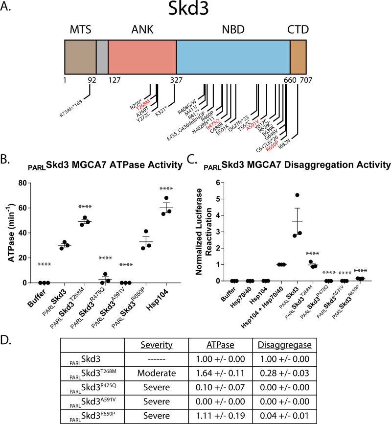
    1. Baumann U, Fernández-Sáiz V, Rudelius M, Lemeer S, Rad R, Knorn AM, Slawska J, Engel K, Jeremias I, Li Z, Tomiatti V, Illert AL, Targosz BS, Braun M, Perner S, Leitges M, Klapper W, Dreyling M, Miething C, Lenz G, Rosenwald A, Peschel C, Keller U, Kuster B, Bassermann F. Disruption of the PRKCD-FBXO25-HAX-1 Axis attenuates the apoptotic response and drives lymphomagenesis. Nature Medicine. 2014;20:1401–1409. doi: 10.1038/nm.3740. - DOI - PubMed

Publication types

Supplementary concepts