An official website of the United States government
The .gov means it’s official.
Federal government websites often end in .gov or .mil. Before
sharing sensitive information, make sure you’re on a federal
government site.
The site is secure.
The https:// ensures that you are connecting to the
official website and that any information you provide is encrypted
and transmitted securely.
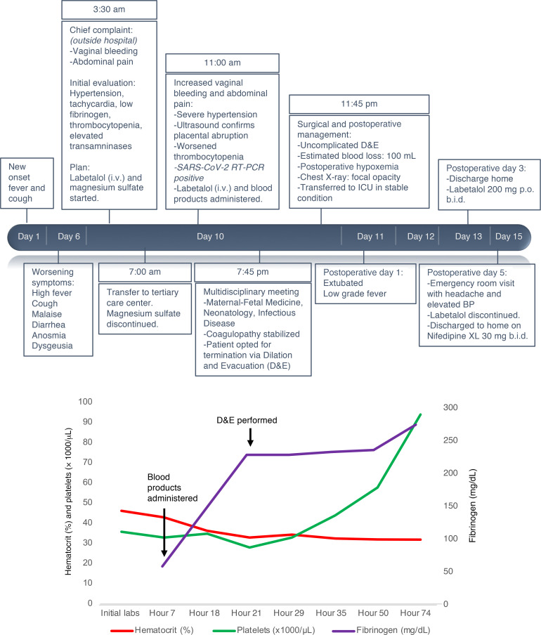
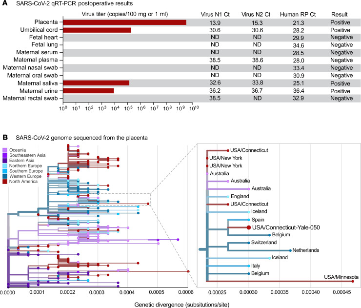
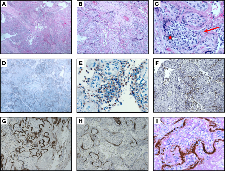
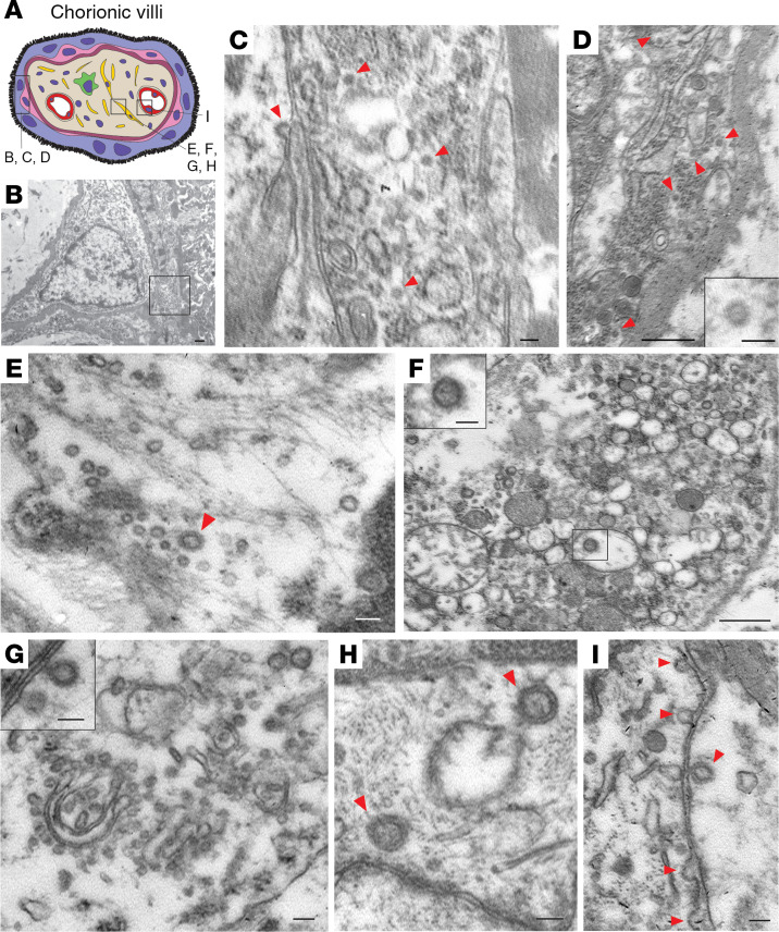
BACKGROUNDThe effects of the novel coronavirus disease 2019 (COVID-19) in pregnancy remain relatively unknown. We present a case of second trimester pregnancy with symptomatic COVID-19 complicated by severe preeclampsia and placental abruption.METHODSWe analyzed the placenta for the presence of severe acute respiratory syndrome coronavirus 2 (SARS-CoV-2) through molecular and immunohistochemical assays and by and electron microscopy and measured the maternal antibody response in the blood to this infection.RESULTSSARS-CoV-2 localized predominantly to syncytiotrophoblast cells at the materno-fetal interface of the placenta. Histological examination of the placenta revealed a dense macrophage infiltrate, but no evidence for the vasculopathy typically associated with preeclampsia.CONCLUSIONThis case demonstrates SARS-CoV-2 invasion of the placenta, highlighting the potential for severe morbidity among pregnant women with COVID-19.FUNDINGBeatrice Kleinberg Neuwirth Fund and Fast Grant Emergent Ventures funding from the Mercatus Center at George Mason University. The funding bodies did not have roles in the design of the study or data collection, analysis, and interpretation and played no role in writing the manuscript.
Conflict of interest: The authors have declared that no conflict of interest exists.
Figures
Figure 1. Case timeline.
( A )…
Figure 1. Case timeline.
( A ) Clinical course of a 35-year-old G3P1011 at 22…
Figure 1. Case timeline.
(A) Clinical course of a 35-year-old G3P1011 at 22 weeks’ gestation with COVID-19 associated with preeclampsia and placental abruption. Timeline from the onset of symptoms to the immediate postpartum period including resuscitation products, hypertension management, and surgical preparation for D&E. BP, blood pressure. (B) Patient’s platelet count (green line), hematocrit (red line), and serum fibrinogen (purple line) throughout hospitalization. Major clinical events are highlighted with arrows.
Figure 2. Examination of SARS–CoV-2 RNA in…
Figure 2. Examination of SARS–CoV-2 RNA in maternal and fetal tissue.
( A ) SARS–CoV-2…
Figure 2. Examination of SARS–CoV-2 RNA in maternal and fetal tissue.
(A) SARS–CoV-2 qRT-PCR results of fetal and maternal samples using the CDC assay which consists of the N1 and N2 primers and probes targeting the coronavirus nucleocapsid and the RP primers and probe targeting human RNase P as an internal control. Ct values from both N1 and N2 must be below 38 for the result to be positive, as internally validated. Virus titers are shown as the average calculation from N1 and N2 Ct values. For the tissues, 80–160 mg was used for extractions, and for the swabs in viral transport media and other liquid samples, 0.25–0.4 mL was used. ND, not detected. (B) The SARS–CoV-2 genome sequenced from the infected placenta was combined with 289 other genomes available from GISAID from around the world. The phylogenetic tree was constructed using IQ-TREE (http://www.iqtree.org/) within the Nextstrain Augur pipeline, and the results were visualized using Auspice (28). Genetic divergence is shown as substitutions per site from the root. An enlarged view of the 18 genomes in the clade that contains the SARS–CoV-2 genome sequenced from the placenta (USA/Connecticut-Yale-050) is shown. The clade is defined by 3 nucleotide substitutions, A28881A, G28882A, and G28883C, providing the equivalent of approximately 95% branch support. The consensus SARS–CoV-2 genome from the placenta (Yale-050) can be found using NCBI’s BioProject PRJNA614976 and the phylogenetic data can be visualized at Nexstrain (https://nextstrain.org/community/grubaughlab/CT-SARS-CoV-2/paper2). The Acknowledgments for the sequences obtained from GISAID can be found at Nexstrain (https://github.com/grubaughlab/CT-SARS-CoV-2/tree/master/paper2).
Figure 3. Histopathology of placenta.
( A …
Figure 3. Histopathology of placenta.
( A – C ). Section of placenta stained with…
Figure 3. Histopathology of placenta.
(A–C). Section of placenta stained with H&E showing histiocytic intervillositis. Original magnification, ×40 (A), ×100 (B), ×400 (C). (C) Star indicates intervillous space infiltrated by immune cells. Arrow indicates perivillous fibrin. (D and E). Immunohistochemical stain for CD68 showing the majority of intervillous inflammatory infiltrate positive (brown stain) for this macrophage marker. Original magnification, ×40 (D), ×400 (E). (F) Staining for CD3, a marker of T lymphocytes. Original magnification, ×100. (G and H). Immunohistochemical staining for SARS–CoV-2 spike protein, demonstrating virus localization predominantly in syncytiotrophoblast cells. Original magnification, ×50 (G), ×400 (H). (I) In situ analysis for the presence of SARS–CoV-2 RNA shows strong positive staining within the placenta . Original magnification, ×400.
Figure 4. Electron microscopic images of coronavirus…
Figure 4. Electron microscopic images of coronavirus particles in different placental cell types.
( A …
Figure 4. Electron microscopic images of coronavirus particles in different placental cell types.
(A) Schematic illustration of chorionic villi. Cytotrophoblasts, syncytiotrophoblasts, fibroblasts, and endothelial cells are indicated in pink, purple, yellow, and red, respectively. (B) Cytotrophoblast (on the left with the nucleus) and syncytiotrophoblast (on the right). Scale bar: 500 nm. (C) Enlarged image of boxed area in B. Cytoplasm of a syncytiotrophoblast cell with virus particles (red arrowheads). Scale bar: 100 nm. (D) Cytoplasm of a cytotrophoblast cell with virus particles indicated by red arrowheads. Scale bar: 500 nm and 100 nm (insert). (E–H) Fibroblast with virus particles indicated by red arrowheads. Scale bars: 100 nm (E), 500 nm (F) (insert, 100 nm), 100 nm (G) (insert, 100 nm), 100 nm (H). (I) Endothelial cell within chorionic villus with budding viral particles indicated by red arrowheads. Scale bar: 100 nm.
Chen H, et al. Clinical characteristics and intrauterine vertical transmission potential of COVID-19 infection in nine pregnant women: a retrospective review of medical records. Lancet. 2020;395(10226):809–815. doi: 10.1016/S0140-6736(20)30360-3.
-
DOI
-
PMC
-
PubMed
Steegers EA, von Dadelszen P, Duvekot JJ, Pijnenborg R. Pre-eclampsia. Lancet. 2010;376(9741):631–644. doi: 10.1016/S0140-6736(10)60279-6.
-
DOI
-
PubMed
doi: 10.5858/arpa.2020-0901-sa. Schwartz DA. An analysis of 38 pregnant women with COVID-19, their newborn infants, and maternal-fetal transmission of SARS-CoV-2: maternal coronavirus infections and pregnancy outcomes [published online March 17, 2020]. Arch Pathol Lab Med .
-
DOI
-
PubMed
Breslin N, et al. COVID-19 infection among asymptomatic and symptomatic pregnant women: Two weeks of confirmed presentations to an affiliated pair of New York City hospitals. Am J Obstet Gynecol MFM. 2020;2(2):100118.
-
PMC
-
PubMed
Zhang Y, et al. Coagulopathy and antiphospholipid antibodies in patients with COVID-19. N Engl J Med. 2020;382(17):e38. doi: 10.1056/NEJMc2007575.
-
DOI
-
PMC
-
PubMed