. 2020 Nov;92(11):2792-2803.
doi: 10.1002/jmv.26212. Epub 2020 Jul 17.

Elucidation of cellular targets and exploitation of the receptor-binding domain of SARS-CoV-2 for vaccine and monoclonal antibody synthesis

Affiliations

Elucidation of cellular targets and exploitation of the receptor-binding domain of SARS-CoV-2 for vaccine and monoclonal antibody synthesis

Abdul Mannan Baig et al. J Med Virol. 2020 Nov.

Abstract

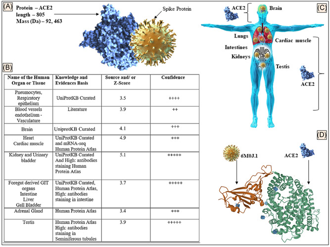
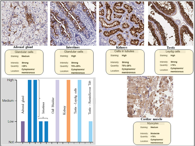
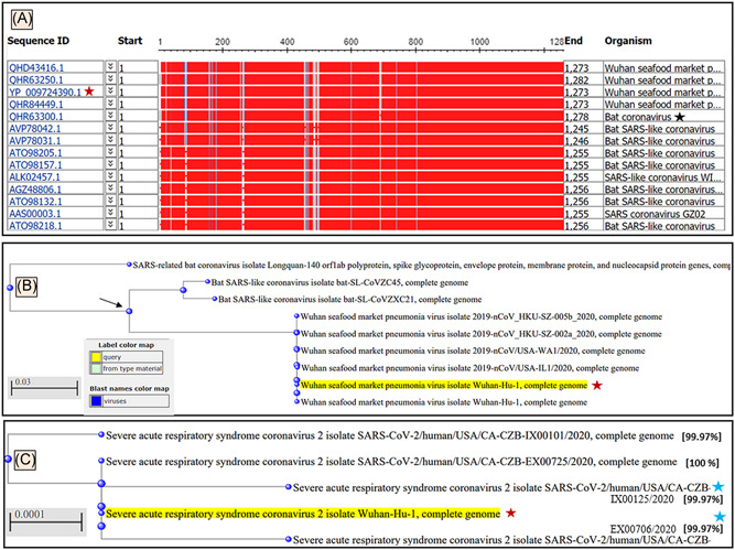
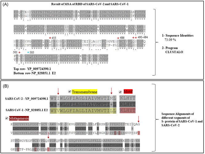
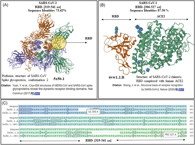
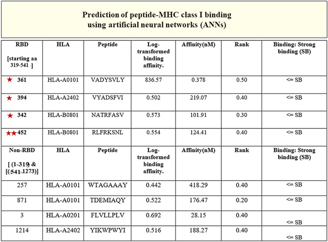
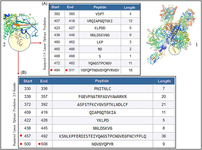
The pandemic caused by novel severe acute respiratory syndrome coronavirus (SARS-CoV-2) has resulted in over 452 822 deaths in the first 20 days of June 2020 due to the coronavirus virus disease 2019 (COVID-19). The SARS-CoV-2 uses the host angiotensin-converting enzyme 2 (ACE2) receptor to gain entry inside the human cells where it replicates by using the cell protein synthesis mechanisms. The knowledge of the tissue distribution of ACE2 in human organs is therefore important to predict the clinical course of the COVID-19. Also important is the understanding of the viral receptor-binding domain (RBD), a region within the spike (S) proteins, that enables the entry of the virus into the host cells to synthesize vaccine and monoclonal antibodies (mAbs). We performed an exhaustive search of human protein databases to establish the tissues that express ACE2 and performed an in-depth analysis like sequence alignments and homology modeling of the spike protein (S) of the SARS-CoV-2 to identify antigenic regions in the RBD that can be exploited to synthesize vaccine and mAbs. Our results show that ACE2 is widely expressed in human organs that may explain the pulmonary, systemic, and neurological deficits seen in COVID-19 patients. We show that though the S protein of the SARS-CoV-2 is a homolog of S protein of SARS-CoV-1, it has regions of dissimilarities in the RBD and transmembrane segments. We show peptide sequences in the RBD of SARS-CoV-2 that can bind to the major histocompatibility complex alleles and serve as effective epitopes for vaccine and mAbs synthesis.

Keywords: 2019-nCoV; BSL-4; COVID-19; MERS virus; SARS virus; SARS-CoV-2; Wuhan coronavirus outbreak; bat virus; biological agents; vaccine and antibody against SARS-CoV-2; viral pandemics; zoonotic infections.


Conflict of interest statement

The authors declare that there are no conflict of interests.

Figures

Figure 1
Figure 1
A, Details of the ACE2 protein showing schematic interaction with S protein. B, The distribution of ACE2 protein in different tissues based on the evidence of microarray, the protein expression data, and published literature. A scoring and confidence level of tissue distribution is also shown. C, Animated image to show the ACE2 distribution in organs of the human body. D, SARS‐CoV‐2 spike protein (6m0j.1) bound to the human ACE2 complex (https://doi.org/10.2210/pdb6M0J/PDB) is shown (data retrieved from References , , and , respectively in accord with the policy of the database resource, which enables the third parties to have access to the data shown). ACE2, angiotensin‐converting enzyme 2; GIT, gastrointestinal tract; SARS‐CoV‐2, severe acute respiratory syndrome coronavirus‐2
Figure 2
Figure 2
Immunocytochemistry data of quantitative expression, location, and staining intensity of ACE2 in human tissues. Protein expression overview of ACE2 showed it to be expressed highly in the intestines, gall bladder, kidney, and testicular tissue. Human Protein Atlas (HPA) did not have neuronal tissue staining in its archive (data retrieved from the HPA database in accord with the policy of the database resource retrieval which enables the third parties to have access to the data shown, http://www.proteinatlas.org/ENSG00000130234-ACE2/tissue)
Figure 3
Figure 3
A, Graphic summary of sequences producing significant alignments of severe acute respiratory syndrome coronavirus‐2 (SARS‐CoV‐2) isolate Wuhan‐Hu‐1, complete genome. B, The phylogenetic tree developed for SARS‐CoV‐2 Wuhan‐Hu‐1 virus in the form of a rectangular cladogram showed its root of origins in the taxon of coronaviruses. B, The bat coronavirus, SARS‐CoV‐1 and SARS‐CoV‐2 Wuhan‐Hu‐1 virus showed a common ancestor. The BLASTn results of SARS‐CoV‐2 Wuhan‐Hu‐1 virus (Wuhan seafood coronavirus, GenBank ID: QHD34416.1) nucleotides showed bat coronavirus (black star) and bat SARS‐like CoV as the top homologs of this virus. C, The phylogenetic tree of the SARS‐CoV‐2 Wuhan‐Hu‐1 virus (yellow highlighted with red stars) compared with the recently deposited genomes of diverse clinical isolate (May 2020) show the occurrence of mutation (99.97% sequence identities) at early (top node) proximal node and recently after the outbreak (blue stars) coronavirus (wild type, black star)
Figure 4
Figure 4
A, Multiple sequence alignment (MSA) results of the receptor‐binding domain (RBD) (319th‐541st aa) (top row) of severe acute respiratory syndrome coronavirus‐2 (SARS‐CoV‐2) (accession# YP_009724390) and the RBD of S protein (306th‐527th aa) of SARS‐CoV‐1 (accession# NP_828851) showed 73.09% of sequence identities. Note five (red stars) of the six amino acid residues mutated in (top row) in RBD of SARS‐CoV‐2. B, Except for the motif, segments of transmembrane regions and points of mutagenesis were noted in between the S protein of and SARS‐CoV‐2 (top row) and of SARS‐CoV‐1 (bottom row)
Figure 5
Figure 5
Homology modeling results of receptor‐binding domain (RBD) of severe acute respiratory syndrome coronavirus‐1 (SARS‐CoV‐1) and SARS‐CoV‐2. A, The results for SARS‐CoV‐2 RBD (319th‐541st aa) showed a template‐based model of perfusion structure of RBD of S protein of SARS‐CoV‐1. B, On homology modeling, the RBD of SARS‐CoV‐1 (306th‐527th aa) developed a template‐based model of SARS‐CoV‐2 RBD (brown ribbons) in complex with ACE2 (green ribbons) with PDB ID (6vw.1). C, The sequence of RBD of SARS‐CoV‐2 aligned with the template (5b5x chain A, B, C) sequences show the near‐identical amino acid similarities between the B and C chains of the template and the Seqres S protein (top row) (template‐based models were developed by SWISS‐MODEL automated and accessible at the database: https://swissmodel.expasy.org/templates/6vw1.1 and PDB ID at https://www.rcsb.org/3d-view/6VW1/1)
Figure 6
Figure 6
The peptides (third column) identified in the RBD (319‐541 aa) region of the S of severe acute respiratory syndrome coronavirus‐2 (SARS‐CoV‐2) (stars) had SB predictions (seventh column). The binding prediction to the MHC class I allele (second column) with log‐transformed binding affinity, nM affinity, and rank (fourth, fifth, and sixth column, respectively) are shown. The non‐RBD resident sequences with SB and nM affinity are shown in the last four columns that were predicted to bind the MHC class I allele. Note the double stars (452‐459 aa) of which 455 is a known residue in the RBD of S protein that engages with the ACE2 receptor. HLA, human leukocyte antigen; MHC, major histocompatibility complex; RBD, receptor‐binding domain; SB, strong binding
Figure 7
Figure 7
A, BepiPred 1.0 prediction software in the automated Immune Epitope Database and Analysis Resource (IEDB) server was used that predicted the location of linear B‐cell epitopes using a combination of a hidden Markov model and a propensity scale method. Results are based on a large benchmark calculation containing close to 85 B‐cell epitopes. Note stars (494‐511 aa), which contain 501 and 505 amino acids that are known to engage with ACE2 receptor. B, BepiPred‐2.0: Sequential B‐Cell Epitope Predictor automated serve was used to predict B‐cell epitopes from receptor‐binding domain (RBD) of the S of protein of SARS‐CoV‐2, using a random forest algorithm trained on epitopes and non‐epitope amino acids that are determined from crystal structures. Note stars (457‐492 aa) which contain 486, 501, and 505 amino acids that are known to engage with ACE2 receptor. The structure of SARS‐CoV‐2 RBD (PDB ID: 6vsb.1) is shown (1) with the peptides predicted (A, B) to be located within the RBD (two zoomed circles) of the template‐based model of SARS‐CoV‐2

References

    1. US Centers for Disease Control and Prevention. Coronavirus disease 2019 (Covid‐19): situation summary. https://www.cdc.gov/coronavirus/2019-nCoV/summary.html. Accessed June 1, 2020.
    1. Watkins J. Preventing a COVID‐19 pandemic. BMJ. 2020;368:m810. 10.1136/bmj.m810 - DOI - PubMed
    1. Gates B. Responding to Covid‐19—a once‐in‐a‐century pandemic? N Engl J Med. 2020;382:1677‐1679. 10.1056/NEJMp2003762 - DOI - PubMed
    1. Day M. Covid‐19: surge in cases in Italy and South Korea makes pandemic look more likely. BMJ. 2020;368:m751. 10.1136/bmj.m751 - DOI - PubMed
    1. Wahba L, Jain N, Fire AZ, et al. A new coronavirus associated with human respiratory disease in China. Nature. 2020;5. 10.1038/s41586-020-2008-3 - DOI - PMC - PubMed

MeSH terms