

Mitochondria and microbiota dysfunction in COVID-19 pathogenesis
- PMID: 32574708
- PMCID: PMC7837003
- DOI: 10.1016/j.mito.2020.06.008
Mitochondria and microbiota dysfunction in COVID-19 pathogenesis
Abstract
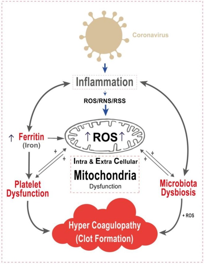
The COVID-19 pandemic caused by the coronavirus (SARS-CoV-2) has taken the world by surprise into a major crisis of overwhelming morbidity and mortality. This highly infectious disease is associated with respiratory failure unusual in other coronavirus infections. Mounting evidence link the accelerated progression of the disease in COVID-19 patients to the hyper-inflammatory state termed as the "cytokine storm" involving major systemic perturbations. These include iron dysregulation manifested as hyperferritinemia associated with disease severity. Iron dysregulation induces reactive oxygen species (ROS) production and promotes oxidative stress. The mitochondria are the hub of cellular oxidative homeostasis. In addition, the mitochondria may circulate "cell-free" in non-nucleated platelets, in extracellular vesicles and mitochondrial DNA is found in the extracellular space. The heightened inflammatory/oxidative state may lead to mitochondrial dysfunction leading to platelet damage and apoptosis. The interaction of dysfunctional platelets with coagulation cascades aggravates clotting events and thrombus formation. Furthermore, mitochondrial oxidative stress may contribute to microbiota dysbiosis, altering coagulation pathways and fueling the inflammatory/oxidative response leading to the vicious cycle of events. Here, we discuss various cellular and systemic incidents caused by SARS-CoV-2 that may critically impact intra and extracellular mitochondrial function, and contribute to the progression and severity of the disease. It is crucial to understand how these key modulators impact COVID-19 pathogenesis in the quest to identify novel therapeutic targets that may reduce fatal outcomes of the disease.
Keywords: Extracellular mitochondria; Hyper-inflammation; Hypercoagulability; Iron; Microbiota; Oxidative stress; Platelet mitochondria.
Copyright © 2020 The Authors. Published by Elsevier B.V. All rights reserved.
Conflict of interest statement
Declaration of Competing Interest The authors declare that they have no known competing financial interests or personal relationships that could have appeared to influence the work reported in this paper.
Figures
References
-
- Al Amir Dache Z., Otandault A., Tanos R., Pastor B., Meddeb R., Sanchez C., Arena G., Lasorsa L., Bennett A., Grange T., El Messaoudi S., Mazard T., Prevostel C., Thierry A.R. Blood contains circulating cell-free respiratory competent mitochondria. FASEB J. 2020;34:3616–3630. - PubMed
-
- Bär F., Bochmann W., Widok A., Von Medem K., Pagel R., Hirose M., Yu X., Kalies K., König P., Böhm R., Herdegen T., Reinicke A.T., Büning J., Lehnert H., Fellermann K., Ibrahim S., Sina C. Mitochondrial gene polymorphisms that protect mice from colitis. Gastroenterology. 2013;145:1055–1063.e3. - PubMed
-
- Bessman N.J., Mathieu J.R.R., Renassia C., Zhou L., Fung T.C., Fernandez K.C., Austin C., Moeller J.B., Zumerle S., Louis S., Vaulont S., Ajami N.J., Sokol H., Putzel G.G., Arvedson T., Sockolow R.E., Lakhal-Littleton S., Cloonan S.M., Arora M., Peyssonnaux C., et al. Dendritic cell–derived hepcidin sequesters iron from the microbiota to promote mucosal healing. Science (80-.) 2020;368:186–189. - PMC - PubMed
Publication types
MeSH terms
Substances
Grants and funding
LinkOut - more resources
Full Text Sources
Other Literature Sources
Medical
Miscellaneous
