Case Reports
. 2020 Jul:57:102833.
doi: 10.1016/j.ebiom.2020.102833. Epub 2020 Jun 20.

Alveolar macrophage dysfunction and cytokine storm in the pathogenesis of two severe COVID-19 patients

Affiliations
Case Reports

Alveolar macrophage dysfunction and cytokine storm in the pathogenesis of two severe COVID-19 patients

Chaofu Wang et al. EBioMedicine. 2020 Jul.

Abstract

Background: The novel coronavirus pneumonia COVID-19 caused by SARS-CoV-2 infection could lead to a series of clinical symptoms and severe illnesses, including acute respiratory distress syndrome (ARDS) and fatal organ failure. We report the fundamental pathological investigation in the lungs and other organs of fatal cases for the mechanistic understanding of severe COVID-19 and the development of specific therapy in these cases.

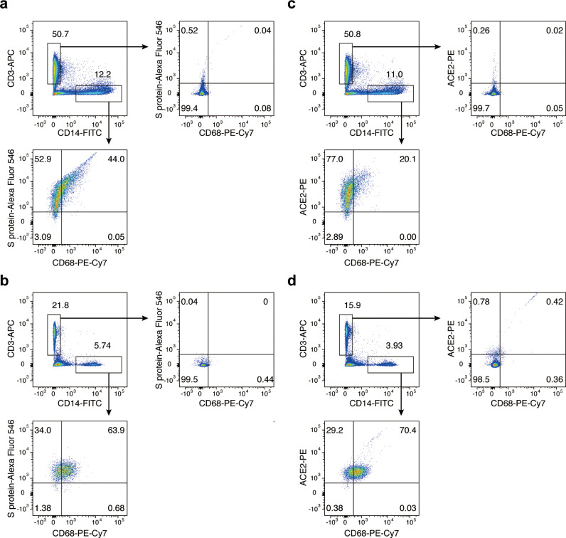
Methods: The autopsy and pathological investigations of specimens were performed on bodies of two deceased cases with COVID-19. Gross anatomy and histological investigation by Hematoxylin and eosin (HE) stained were reviewed on each patient. Alcian blue/periodic acid-Schiff (AB-PAS) staining and Masson staining were performed for the examinations of mucus, fibrin and collagen fiber in lung tissues. Immunohistochemical staining was performed on the slides of lung tissues from two patients. Real-time PCR was performed to detect the infection of SARS-CoV-2. Flow cytometry analyses were performed to detect the direct binding of S protein and the expression of ACE2 on the cell surface of macrophages.

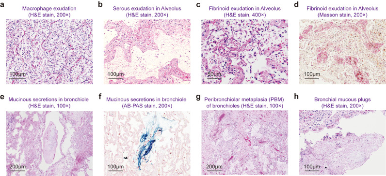
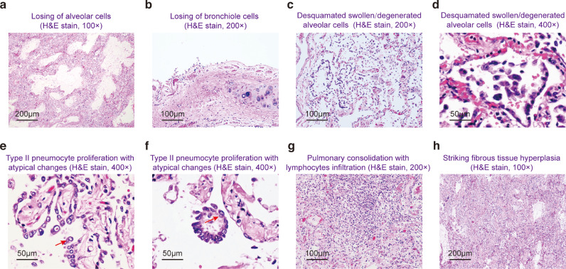
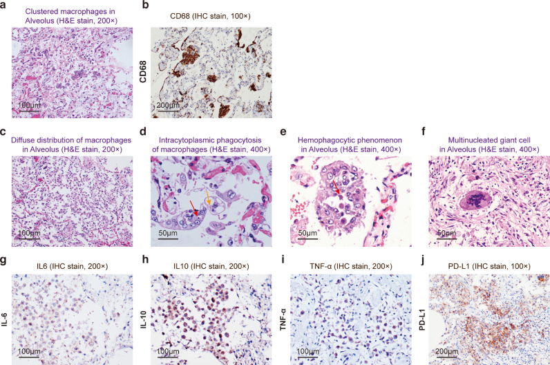
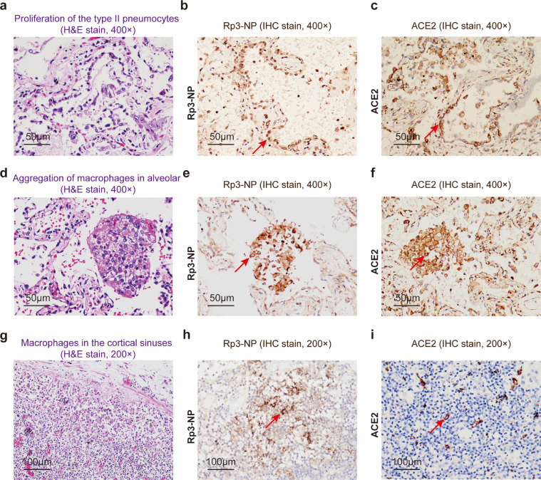
Findings: The main pathological features in lungs included extensive impairment of type I alveolar epithelial cells and atypical hyperplasia of type II alveolar cells, with formation of hyaline membrane, focal hemorrhage, exudation and pulmonary edema, and pulmonary consolidation. The mucous plug with fibrinous exudate in the alveoli and the dysfunction of alveolar macrophages were characteristic abnormalities. The type II alveolar epithelial cells and macrophages in alveoli and pulmonary hilum lymphoid tissue were infected by SARS-CoV-2. S protein of SARS-CoV-2 directly bound to the macrophage via the S-protein-ACE2 interaction.

Interpretation: Infection of alveolar macrophage by SARS-CoV-2 might be drivers of the "cytokine storm", which might result in damages in pulmonary tissues, heart and lung, and lead to the failure of multiple organs .

Funding: Shanghai Guangci Translational Medical Research Development Foundation, Shanghai, China.

Keywords: Alveolar macrophage; COVID-19; Cytokine storm; Pathology; SARS-CoV-2.


Conflict of interest statement

Declaration of Competing Interest The authors declare no competing interests.

Figures

Fig 1
Fig. 1
Extensive exudate and mucinous secretion. (a) A large number of macrophages in alveolar cavities (H&E stain, 200 ×). (b-d) Serous (B, H&E stain, 200 ×) and fibrinoid (c, H&E stain, 400 ×, d, Masson stain, 200 ×) exudation. Mucinous secretions in bronchioles (e, H&E stain, 100 ×) with blue staining by AB-PAS stain (f, 200 ×). Peribronchiolar metaplasia (PBM) of bronchioles and terminal bronchioles (g, H&E stain, 100 ×) as well as bronchial mucous plugs formation (h, H&E stain, 200 ×) are visible.
Fig 2
Fig. 2
Damage of respiratory tracts and reparative changes. (a-b) Intensive sloughing of alveolar (a, H&E stain, 100 ×) and bronchiole (b, H&E stain, 200 ×) epithelial cells. (c-f) Desquamated swollen and degenerated alveolar cells in alveoli (H&E stain, c, 200 × and d, 400 ×); type II pneumocyte proliferation with atypical changes (e and f, H&E stain, 400 ×): enlarged nuclei, clearing of nuclear chromatin and prominent nucleoli. Inclusion bodies are indicated by arrow. (g-h) Pulmonary consolidation with infiltration of lymphocytes (g, H&E stain, 200 ×) and striking fibrous tissue hyperplasia (h, H&E stain, 100 ×).
Fig 3
Fig. 3
Aggregated alveolar macrophages. (a-f) A large number of mononuclear and multinucleate macrophages in alveoli in varied forms: in clusters (a, H&E stain, 200 ×) and immunohistochemistry staining of CD68 (b); Diffuse distribution (c, H&E stain, 200 ×); Intracytoplasmic phagocytosis (d, H&E stain, 400 ×, indicated by red arrow) and Spherical acidophilic hyaline degeneration bodies (indicated by orange arrow); Hemophagocytic phenomenon (e, H&E stain, 400 × indicated by red arrow) and multinucleated giant cell (f, H&E stain, 400 ×). (g-j) Expression of chemokine and inflammatory cytokines: IL-6 (g, 200 ×), IL-10 (h,200 ×), TNFα (i, 200 ×) and extensive expression of PD-L1 (j, 100 ×)
Fig 4
Fig. 4
COVID-19-infected cells in lung and pulmonary hilum lymph nodes. Proliferation of the type II pneumocytes by H&E stain (a, 400 ×) and expression of Rp3-NP by immunohistochemistry in type II pneumocytes (b, 400 ×, arrow); Membrane positive of ACE2 expression in type II pneumocytes and macrophages respectively (c, 400 ×, arrow and f, 400 ×, arrow); Aggregation of macrophages in alveolar by H&E stain (d, 400 ×) and expression of Rp3-NP of SARS-CoV-2 (e, 400 ×, arrow); Macrophages in the cortical sinuses of pulmonary hilum lymph nodes (g, 200 ×), expression of Rp3-NP of SARS-CoV-2 (h, 200 ×, arrow) and ACE2 (i, 200 ×, arrow).
Fig 5
Fig. 5
SARS-CoV-2 Spike protein interaction with CD68 macrophage. (a-b) Flow cytometry analysis showing the distribution of S protein bound immune cells in lung tissues (a) and peripheral blood. (b) CD14 markers were used for labeling monocytes/ macrophages while CD3 for T cells. (c-d) Flow cytometry analysis showing the expression of ACE2 on the surface of monocytes/macrophages, but not of T cells in lung tissues (c) and peripheral blood (d).

References

    1. Huang C, Wang Y, Li X. Clinical features of patients infected with 2019 novel coronavirus in Wuhan, China. Lancet. 2020;395(10223):497–506. - PMC - PubMed
    1. Guan WJ, Ni ZY, Hu Y. Clinical characteristics of coronavirus disease 2019 in China. N Engl J Med. 2020 - PMC - PubMed
    1. Zhou F, Yu T, Du R. Clinical course and risk factors for mortality of adult inpatients with COVID-19 in Wuhan, China: a retrospective cohort study. Lancet. 2020;395(10229):1054–1062. - PMC - PubMed
    1. Chen N, Zhou M, Dong X. Epidemiological and clinical characteristics of 99 cases of 2019 novel coronavirus pneumonia in Wuhan, China: a descriptive study. Lancet. 2020;395(10223):507–513. - PMC - PubMed
    1. Kunisch E, Fuhrmann R, Roth A, Winter R, Lungershausen W, Kinne RW. Macrophage specificity of three anti-CD68 monoclonal antibodies (KP1, EBM11, and PGM1) widely used for immunohistochemistry and flow cytometry. Ann Rheum Dis. 2004;63(7):774–784. - PMC - PubMed

Publication types

MeSH terms