Review
. 2020 Jun 19;12(6):1624.
doi: 10.3390/cancers12061624.

Exploring the Therapeutic Potential of Membrane Transport Proteins: Focus on Cancer and Chemoresistance

Affiliations
Review

Exploring the Therapeutic Potential of Membrane Transport Proteins: Focus on Cancer and Chemoresistance

Shekoufeh Almasi et al. Cancers (Basel). .

Abstract

Improving the therapeutic efficacy of conventional anticancer drugs represents the best hope for cancer treatment. However, the shortage of druggable targets and the increasing development of anticancer drug resistance remain significant problems. Recently, membrane transport proteins have emerged as novel therapeutic targets for cancer treatment. These proteins are essential for a plethora of cell functions ranging from cell homeostasis to clinical drug toxicity. Furthermore, their association with carcinogenesis and chemoresistance has opened new vistas for pharmacology-based cancer research. This review provides a comprehensive update of our current knowledge on the functional expression profile of membrane transport proteins in cancer and chemoresistant tumours that may form the basis for new cancer treatment strategies.

Keywords: cancers; chemoresistance; ion channels; membrane transporters; pumps.


Conflict of interest statement

The authors declare no conflict of interest.

Figures

Figure 1
Figure 1
Different types of ion transport. (A) Active and secondary transport: Primary active transporter uses ATP to move ions across the membrane [A and B], against their electrochemical gradients to create an electrochemical gradient. Secondary active transporter uses the electrochemical gradient generated by primary active transporters to move one molecule down its gradient [B] while transporting another molecule against its electrochemical gradient [C]. (B) Uniporter, antiporter, and symporter: Uniporter carries one molecule or ion in one direction. Antiporter carries two different molecules or ions in opposite directions. Symporter also carries two different molecules or ions in the same direction.
Figure 2
Figure 2
Na+-, K+-ATPase overall structure. (A) Na+-, K+-ATPase consists of a catalytic α subunit and a regulatory β subunit. The α subunit consists of 10 transmembrane helices, harboring 3 different cytoplasmic domains: the actuator responsible for dephosphorylation (shown in red); the nucleotide-binding, responsible for ATP binding (shown in blue); and the phosphorylation domains (shown in cyan). The β subunit consists of one transmembrane helix with a large glycosylated extracellular domain (shown in hexagon orange boxes). ECM = extracellular milieu; CYT = cytoplasm. (B) Overall domain architecture of Na+/K+ transporter in the Na+-bound state (Protein Data Bank [PDB] code 4HQJ). Catalytic α subunit is colored in blue, β subunit is shown in yellow, and Na+ ions are shown in red.
Figure 3
Figure 3
The overall structure of SERCA (Sarco/endoplasmic reticulum Ca2+ ATPase) pump. (A) The topology of SERCA showing 10 transmembrane segments (TMS (transmembrane segment) 11 is found only in SERCA2b), with a large cytoplasmic N-terminal, large cytoplasmic loops, and a luminal C-terminal. (B) Overall 3D-architecture of SERCA transporter in the E2-state complexed with a Thapsigargin derivative Boc-(phi)Tg (Protein Data Bank [PDB] code 3NAN). Cytoplasmic and luminal loops are shown in gray, TMSs are shown in blue, and Tg inhibitor is shown in red. CYT = cytoplasm; ER lumen = endoplasmic reticulum lumen.
Figure 4
Figure 4
Overall structure of the V0 domain of V-ATPase. (A) Topology of V0 domain showing its 9 transmembrane segments with large cytoplasmic N-terminal and C-terminal domains. (B) The overall architecture of the V0 domain of V-ATPase (Protein Data Bank [PDB] code 6C6L), showing all known components of the V0 domain, including subunits a (in red), d (in cyan), e (in blue), f (in pink), and the c-ring (in wheat). CYT = cytoplasm; ER lumen = endoplasmic reticulum lumen. Modified from Roh et al. 2018.
Figure 5
Figure 5
Topology diagram and 3D structure of TRPM2. (A) Topology of TRPM2 showing its 6 transmembrane segments (TMS), a channel pore between TMS 5 and 6, and cytoplasmic N- and C-termini. N-terminal harbors 4 TRPM homology domains (shown in gray). C-terminal has a short coiled-coil region (CCR, shown in yellow) followed by the NUDT9H domain (shown in orange) which is the homolog of soluble mitochondrial ADPRase NUDT9. A functional TRPM2 is composed of four homotetramers TRPM2 (x4) and requires binding and hydrolysis of ADP-ribose (ADPR) by NUDT9-H. ECM = extracellular milieu; CYT = cytoplasm. (B) Overall domain architecture of TRPM2 (Protein Data Bank [PDB] code 6CO7), showing the extracellular view of four units of TRPM2 surrounding the channel pore (shown in different colors: blue, purple, marine, and light blue, “—” in a clockwise direction).
Figure 6
Figure 6
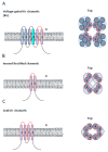
Structural classification of K+ channels. (A) Left: the voltage-sensitive (Kv) and calcium-sensitive K+ (Kca) channels show a similar structure with six transmembrane segments (TMS) and a pore-domain formed by TMS 5 and 6, shown in red. The Kv channels differ in that they contain a voltage sensor in TMS4, shown in cyan. Right: A top view of Kv and Kca channels, showing the six TMS of each of the four subunits and their corresponding pore-forming loops, shown in red. The functional channel is a tetramer protein (x4). (B) Left: a lateral view of monomers of an inward rectifier potassium channel (Kir), showing two TMS connected by a pore-forming loop, shown in red. Right: a top view of Kir channel, showing the convergence of four units of Kir channels to the channel pore. The functional channel is a tetramer protein (x4). (C) Left: topology of a two-pore domain potassium channel (K2P), showing four TMS and two pore domains. Right: a top view of K2P channel, showing the convergence of two units of K2P to form the channel pore. The functional channel is a dimer protein (x2).
Figure 7
Figure 7
NaV channel structure. (A) The topology of the α subunit of NaV channel, showing 24 transmembrane segments (TMS) and four domains (D1–4). Each domain consists of 6 TMS, a pore between the 5/6 TMS, and TMS4 as a voltage sensor (shown in cyan). (B) Overall 3D-architecture of the α-subunit of eukaryotic NaV channel (Protein Data Bank [PDB] code 5XOM). Left: the side view of the ΝaV channel domains D1 (red), D2 (salman), D3 (smudge), and D4 (purple) are shown with N- and C-terminal domains colored in orange and yellow, respectively. Glycosylations located in the extracellular loops of D1 and D3 are represented by green sticks. Right: the extracellular view of the NaV channel showing four domains surrounding the channel pore. ECM = extracellular milieu; CYT = cytoplasm.

References

    1. Kulbacka J., Choromanska A., Rossowska J., Wezgowiec J., Saczko J., Rols M.P. Cell membrane transport mechanisms: Ion channels and electrical properties of cell membranes. Adv. Anat. Embryol. Cell Biol. 2017;227:39–58. doi: 10.1007/978-3-319-56895-9_3. - DOI - PubMed
    1. Prevarskaya N., Skryma R., Shuba Y. Ion channels in cancer: Are cancer hallmarks oncochannelopathies? Physiol. Rev. 2018;98:559–621. doi: 10.1152/physrev.00044.2016. - DOI - PubMed
    1. Yamashita N., Hamada H., Tsuruo T., Ogata E. Enhancement of voltage-gated Na+ channel current associated with multidrug resistance in human leukemia cells. Cancer Res. 1987;47:3736–3741. - PubMed
    1. Lee S.C., Deutsch C., Beck W.T. Comparison of ion channels in multidrug-resistant and -sensitive human leukemic cells. Proc. Natl. Acad. Sci. USA. 1988;85:2019–2023. doi: 10.1073/pnas.85.6.2019. - DOI - PMC - PubMed
    1. Huang Y., Sadee W. Membrane transporters and channels in chemoresistance and -sensitivity of tumor cells. Cancer Lett. 2006;239:168–182. doi: 10.1016/j.canlet.2005.07.032. - DOI - PubMed

LinkOut - more resources