

Effect of SOX2 Repression on Corneal Endothelial Cells
- PMID: 32575737
- PMCID: PMC7352647
- DOI: 10.3390/ijms21124397
Effect of SOX2 Repression on Corneal Endothelial Cells
Abstract
Purpose: Human corneal endothelial cells (hCECs) pump out water from the stroma and maintain the clarity of the cornea. The sex-determining region Y-box 2 (SOX2) participates in differentiation during the development of the anterior segment of the eye and is found in the periphery of wounded corneas. This study was performed to investigate the effect of SOX2 repression on hCECs.
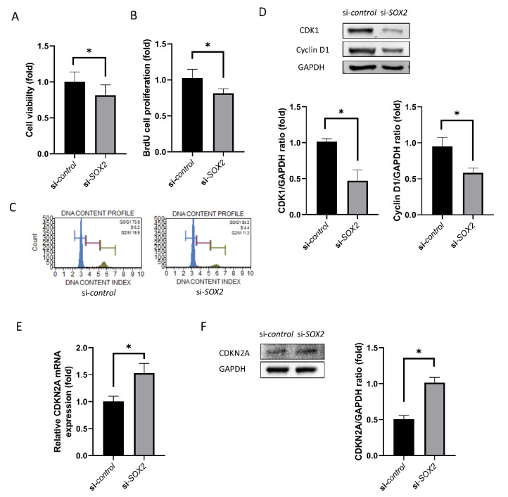
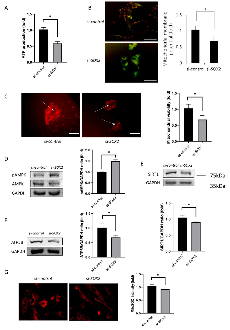
Methods: Cultured hCECs were transfected by siRNA for SOX2. The wound healing rate and cell viability were measured. The cell proliferation-associated protein level was evaluated by Western blotting and RT-PCR. The energy production and mitochondrial function were measured, and cell shape and WNT signaling were assessed.
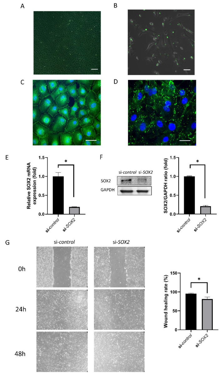
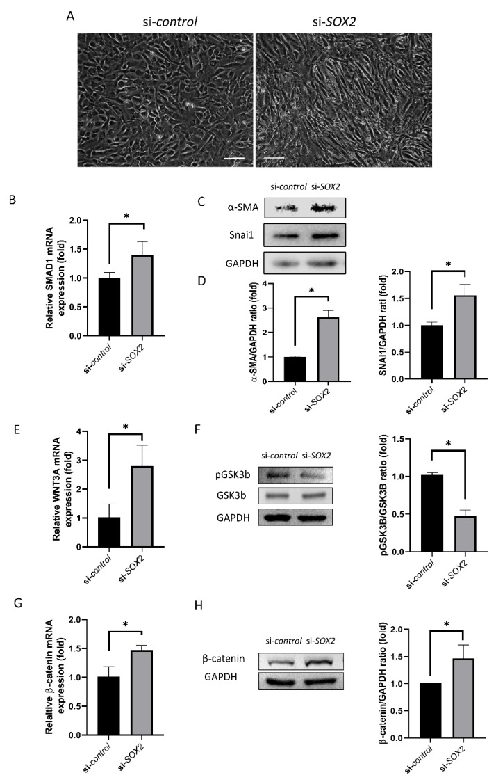
Results: Upon transfecting the cultured cells with siRNA for SOX2, the SOX2 level was reduced by 80%. The wound healing rate and viability were also reduced. Additionally, CDK1, cyclin D1, SIRT1, and ATP5B levels were reduced, and CDKN2A and pAMPK levels were increased. Mitochondrial oxidative stress and mitochondrial viability decreased, and the cell shape became elongated. Furthermore, SMAD1, SNAI1, WNT3A, and β-catenin levels were increased.
Conclusion: SOX2 repression disrupts the normal metabolism of hCECs through modulating WNT signaling and mitochondrial functions.
Keywords: SOX2; WNT signaling; human corneal endothelial cells.
Conflict of interest statement
The authors declare no conflict of interest.
Figures
Similar articles
-
SOX2 Activation Using CRISPR/dCas9 Promotes Wound Healing in Corneal Endothelial Cells.Stem Cells. 2018 Dec;36(12):1851-1862. doi: 10.1002/stem.2915. Epub 2018 Oct 15. Stem Cells. 2018. PMID: 30270540
-
COL8A2 Regulates the Fate of Corneal Endothelial Cells.Invest Ophthalmol Vis Sci. 2020 Sep 1;61(11):26. doi: 10.1167/iovs.61.11.26. Invest Ophthalmol Vis Sci. 2020. PMID: 32931574 Free PMC article.
-
miR-126-3p sensitizes glioblastoma cells to temozolomide by inactivating Wnt/β-catenin signaling via targeting SOX2.Life Sci. 2019 Jun 1;226:98-106. doi: 10.1016/j.lfs.2019.04.023. Epub 2019 Apr 10. Life Sci. 2019. PMID: 30980849
-
[Transplantation of corneal endothelial cells].Nippon Ganka Gakkai Zasshi. 2002 Dec;106(12):805-35; discussion 836. Nippon Ganka Gakkai Zasshi. 2002. PMID: 12610838 Review. Japanese.
-
Mechanism of Proliferation of Cultured Human Corneal Endothelial Cells.Cornea. 2017 Nov;36 Suppl 1:S41-S45. doi: 10.1097/ICO.0000000000001337. Cornea. 2017. PMID: 28902018 Review.
Cited by
-
Long non-coding RNA HOXA11-AS protects the barrier function of corneal endothelial cells by sponging microRNA-155 to alleviate corneal endothelial injury.Am J Transl Res. 2022 Dec 15;14(12):8489-8503. eCollection 2022. Am J Transl Res. 2022. PMID: 36628203 Free PMC article.
-
Long Non-coding RNA Rhabdomyosarcoma 2-Associated Transcript Regulates Angiogenesis in Endothelial Cells.Front Physiol. 2021 Oct 21;12:729157. doi: 10.3389/fphys.2021.729157. eCollection 2021. Front Physiol. 2021. PMID: 34744768 Free PMC article.
-
miR-30c-1 encourages human corneal endothelial cells to regenerate through ameliorating senescence.Aging (Albany NY). 2021 Mar 19;13(7):9348-9372. doi: 10.18632/aging.202719. Epub 2021 Mar 19. Aging (Albany NY). 2021. PMID: 33744867 Free PMC article.
References
-
- Matsubara M., Tanishima T. Wound-healing of the corneal endothelium in the monkey: A morphometric study. Jpn. J. Ophthalmol. 1982;26:264–273. - PubMed
-
- Fujikawa L.S., Wickham M.G., Binder P.S. Wound healing in cultured corneal endothelial cells. Investig. Ophthalmol. Vis. Sci. 1980;19:793–801. - PubMed
MeSH terms
Substances
Grants and funding
LinkOut - more resources
Full Text Sources
Research Materials
Miscellaneous
