. 2020 Jun 20;21(12):4403.
doi: 10.3390/ijms21124403.

Investigating the Role of PPARβ/δ in Retinal Vascular Remodeling Using Pparβ/ δ-Deficient Mice

Affiliations

Investigating the Role of PPARβ/δ in Retinal Vascular Remodeling Using Pparβ/ δ-Deficient Mice

Sze Yuan Ho et al. Int J Mol Sci. .

Abstract

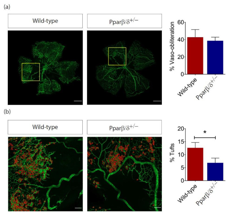
Peroxisome proliferator-activated receptor (PPAR)β/δ is a member of the nuclear receptor superfamily of transcription factors, which plays fundamental roles in cell proliferation and differentiation, inflammation, adipogenesis, and energy homeostasis. Previous studies demonstrated a reduced choroidal neovascularization (CNV) in Pparβ/δ-deficient mice. However, PPARβ/δ's role in physiological blood vessel formation and vessel remodeling in the retina has yet to be established. Our study showed that PPARβ/δ is specifically required for disordered blood vessel formation in the retina. We further demonstrated an increased arteriovenous crossover and wider venous caliber in Pparβ/δ-haplodeficient mice. In summary, these results indicated a critical role of PPARβ/δ in pathological angiogenesis and blood vessel remodeling in the retina.

Keywords: PPARβ/δ; angiogenesis; arteriovenous crossover; blood vessel remodeling; pericytes; vessel caliber.


Conflict of interest statement

S.Y.H., None; Y.P.K., None; B.Y.Q., None; A.T., None; H.M., None; A.V.B., None; N.S.T., None; C.M.C.G., Bayer (C, F), Novartis (C, F), Roche (F), GlaxoSmith Kline (F); T.Y.W., Bayer (C, F), Novartis (C, F), Abbott (C, F), Allergan (C, F), Genentech (C, F), Roche (C, F), Pfizer (C, F); W.W., None; and X.W., None. The funders had no role in the design of the study, in the collection, analyses, or interpretation of data, in the writing of the manuscript, or in the decision to publish the results.

Figures

Figure 1
Figure 1
Peroxisome proliferator-activated receptor (PPAR)β/δ is highly expressed in mouse retina. (a) Representative Western blot of PPARβ/δ, retinal pigment epithelium-specific protein 65 kDa (RPE65) and glyceraldehyde-3-phosphate dehydrogenase (GADPH) in the retina (n = 4) and choroid/RPE (n = 4) compartments of adult C57BL/6 mice. Relative gene expression of Pparβ/δ in (b) the retina (n = 10) and choroid/RPE (n = 3) compartments of C57BL/6 mice; (c) the retina of fasted (n = 4) and re-fed (n = 4) adult C57BL/6 mice; (d) the retina of P2 (n = 4) and P21 (n = 4) C57BL/6 mice, as determined by quantitative real-time polymerase chain reaction (RT-qPCR) analysis. Relative expressions of (e) Pparβ/δ and (f) Carnitine palmitoyltransferase 1A (Cpt1a) in P12 hyperoxic retina of C57BL/6 mice subjected to oxygen-induced retinopathy (OIR) (n = 4) as compared to those in age-matched normoxic retina (n = 4), as determined by RT-qPCR analysis. Relative expressions of (g) Pparβ/δ and (h) Cpt1a in P13 hypoxic retina of C57BL/6 mice subjected to OIR (n = 4) as compared to those in age-matched normoxic retina (n = 4), as determined by RT-qPCR analysis. Gene expressions are quantified relative to the housekeeping gene, Gadph, as detailed in the Materials and Methods. Data are expressed as mean ± standard error of the mean (SEM). Unpaired, two-tailed t-test was used for statistical analysis; *** p < 0.001, * p < 0.05.
Figure 2
Figure 2
Pparβ/δ deletion does not affect normal blood vessel formation. (a) Representative images and quantification of retinal vessel density of P10 Pparβ/δ+/− (n = 4) and wild type (n = 3) retina. Scale bar: 50 µm. (b) Representative images and quantitative analysis of vessel outgrowth from P10 aortic rings isolated from Pparβ/δ+/− (n = 10 explants) and wild-type littermate controls (n = 5 explants). Scale bar: 200 µm. (c) Representative images and quantitative analysis of vessel outgrowth from P3 choroid explants isolated from Pparβ/δ+/− (n = 49 explants) and wild-type littermate controls (n = 34 explants). Scale bar: 200 µm. Data are expressed as mean ± SEM. Unpaired, two-tailed t-test was used for statistical analysis.
Figure 3
Figure 3
PPARβ/δ inhibition by 10h does not affect angiogenesis. (a) Relative gene expression of Angiopoietin-like 4 (ANGPTL4), CPT1A, and Pyruvate Dehydrogenase Kinase 4 (PDK4) in human retinal microvascular endothelial cells (ECs) treated with 100 nM of 10h for 24 h compared to that in dimethyl sulfoxide (DMSO) vehicle control, as determined by RT-qPCR analysis (n = 3). (b) Representative images and quantitative analysis of vessel outgrowth from P3 C57BL/6 aortic ring explants treated with 100 nM of 10 h (n = 9 explants) and DMSO control (n = 11 explants). Scale bar: 200 µm. (c) Representative images and quantitative analysis of vessel outgrowth from P3 C57BL/6 choroidal explants treated with 100nM of 10h (n = 15 explants) and DMSO control (n = 34 explants). Scale bar: 200 µm. Data are expressed as mean ± SEM. Unpaired, two-tailed t-test was used for statistical analysis; *** p < 0.001.
Figure 4
Figure 4
PPARβ/δ mediates retina blood vessel remodeling. (a) Representative retina images of flatmounted P10 Pparβ/δ+/− and wild type retinas, where arteriovenous crossings are highlighted by white arrowheads. Corresponding quantification of arteriovenous crossings in Pparβ/δ+/− (n = 4) and wild type (n = 5) retinas. Scale bar: 500 µm. (b) Representative images of the vein (red box) and artery (yellow box) in P10 Pparβ/δ+/− and wild type retinas. Corresponding quantification of the vessel diameter of the radial veins and arteries of Pparβ/δ+/− (n = 4) and wild type retinas (n = 5). Two points of at least 4 veins/arteries were measured per retina. (c) Representative confocal images for NG2 (red) and CD31 (green) staining of the retinal vasculature in adult Pparβ/δ−/− and wild type retinas. Scale bar: 50 µm. Corresponding quantification of the vessel density and pericyte coverage in wild type (n = 3) and Pparβ/δ−/− (n = 5) retinas. Relative gene expression of (d) Vegf, (e) Tgfβ1, (f) Tie2, and (g) Pdgfrβ mRNA of adult Pparβ/δ−/− and wild type retinas (n ≥ 3), as determined by RT-qPCR analysis. Data are expressed as mean ± SEM. Unpaired, two-tailed t-test was used for statistical analysis; * p < 0.05.
Figure 5
Figure 5
PPARβ/δ is specifically required for pathological retinal angiogenesis. (a) Representative immunofluorescence images of CD1 stained (green) retinal flatmount of P17 Pparβ/δ+/− and wild type retinas subjected to OIR. Scale bar: 500 μm. Corresponding quantification of retinal vaso-obliteration in P17 OIR retinas (n = 6). (b) Pathological neovascular tufts (delineated in higher magnification by red boundary line) and the corresponding quantification of tufts (normalized to retinal area) in Pparβ/δ+/− and wild type P17 OIR retinas (n ≥ 3). Scale bar: 100 μm. Data are expressed as mean ± SEM. Unpaired, two-tailed t-test was used for statistical analysis; * p < 0.05.

References

    1. Lim L.S., Mitchell P., Seddon J.M., Holz F.G., Wong T.Y. Age-related macular degeneration. Lancet. 2012;379:1728–1738. doi: 10.1016/S0140-6736(12)60282-7. - DOI - PubMed
    1. Cheung N., Mitchell P., Wong T.Y. Diabetic retinopathy. Lancet. 2010;376:124–136. doi: 10.1016/S0140-6736(09)62124-3. - DOI - PubMed
    1. Wong T.Y., Scott I.U. Clinical practice. Retinal-vein occlusion. N. Engl. J. Med. 2010;363:2135–2144. doi: 10.1056/NEJMcp1003934. - DOI - PubMed
    1. Kokame G.T., Decarlo T.E., Kaneko K.N., Omizo J.N., Lian R. Anti-Vascular Endothelial Growth Factor Resistance in Exudative Macular Degeneration and Polypoidal Choroidal Vasculopathy. Ophthalmol. Retina. 2019;3:744–752. doi: 10.1016/j.oret.2019.04.018. - DOI - PubMed
    1. Cabral T., Mello L.G.M., Lima L., Polido J., Regatieri C.V., Belfort R., Mahajan V.B. Retinal and choroidal angiogenesis: A review of new targets. Int. J. Retina Vitreous. 2017;3:31. doi: 10.1186/s40942-017-0084-9. - DOI - PMC - PubMed

Substances

LinkOut - more resources