Review
. 2020 Jun 21;9(6):1941.
doi: 10.3390/jcm9061941.

Glia-Derived Extracellular Vesicles in Parkinson's Disease

Affiliations
Review

Glia-Derived Extracellular Vesicles in Parkinson's Disease

Bianca Marchetti et al. J Clin Med. .

Abstract

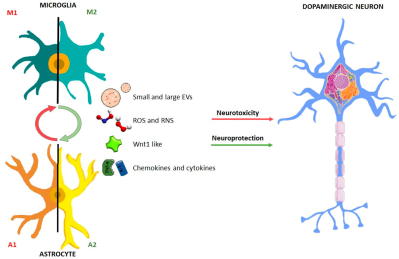
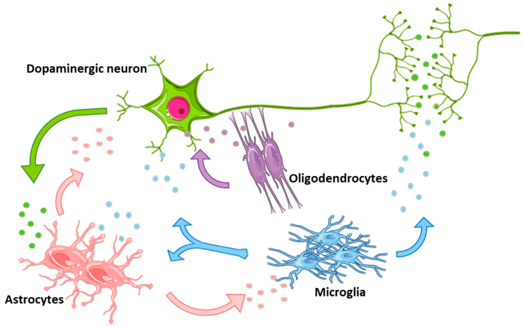
Glial cells are fundamental players in the central nervous system (CNS) development and homeostasis, both in health and disease states. In Parkinson's disease (PD), a dysfunctional glia-neuron crosstalk represents a common final pathway contributing to the chronic and progressive death of dopaminergic (DAergic) neurons of the substantia nigra pars compacta (SNpc). Notably, glial cells communicating with each other by an array of molecules, can acquire a "beneficial" or "destructive" phenotype, thereby enhancing neuronal death/vulnerability and/or exerting critical neuroprotective and neuroreparative functions, with mechanisms that are actively investigated. An important way of delivering messenger molecules within this glia-neuron cross-talk consists in the secretion of extracellular vesicles (EVs). EVs are nano-sized membranous particles able to convey a wide range of molecular cargoes in a controlled way, depending on the specific donor cell and the microenvironmental milieu. Given the dual role of glia in PD, glia-derived EVs may deliver molecules carrying various messages for the vulnerable/dysfunctional DAergic neurons. Here, we summarize the state-of-the-art of glial-neuron interactions and glia-derived EVs in PD. Also, EVs have the ability to cross the blood brain barrier (BBB), thus acting both within the CNS and outside, in the periphery. In these regards, this review discloses the emerging applications of EVs, with a special focus on glia-derived EVs as potential carriers of new biomarkers and nanotherapeutics for PD.

Keywords: Parkinson’s disease; biomarkers; cell-to-cell communication; exosomes; extracellular vesicles; glia; nanotherapeutics.


Conflict of interest statement

The authors declare no conflict of interest.

Figures

Figure 1
Figure 1
Dual role of glial cells in the regulation of the balance between neurotoxicity and neuroprotection in Parkinson’s disease. Astrocyte and microglial schematic drawing illustrating the dual facet of glial cells. Upon basal ganglia injury, glial cells acquire an activated “harmful” so-called astrocyte (A1) and microglial (M1) phenotype, characterized by up-regulated release of a number of pro-inflammatory mediators, including cytokines and chemokines, associated by generation of elevated levels of reactive oxygen (ROS) and nitrogen species (RNS), in turn generating a vicious cycle of inflammation and dopaminergic degeneration. Glial cells are also endowed with neuroprotective factors, including Wnt1, that incite neuroprotection and repair of the dysfunctional neurons. The potential glia-glia and glia-neuron crosstalk via small and large vesicles is illustrated (for detail, see the text).
Figure 2
Figure 2
Schematic representation of glial cells and neurons communicating with each other by secretion of extracellular vesicles (EVs). Depending on the microenvironmental conditions these vesicles may mediate either harmful or neuroprotective effects. The colors of EVs reflect the donor cell type: green for neurons, light blue for microglia, pink for astrocytes and violet for oligodendrocytes.
Figure 3
Figure 3
Contribution of extracellular vesicles secreted by glial cells in glia-neuron crosstalk; (A) Schematic representation of a glial cell releasing EVs, both exosomes (via fusion of MVBs with the plasma membrane) and shedding vesicles (directly released outside the cell). (B) In neurodegenerative diseases, EVs have been seen transporting unfolded/aggregated proteins or miRNAs contributing to spread the pathology. These “pathological” EVs may be used as specific biomarkers of disease. (C) On the contrary, physiological or modified EVs may transport protective biomolecules and may be used as therapeutic tools in neurodegenerative conditions.

References

    1. Barres B.A. The Mystery and Magic of Glia: A Perspective on Their Roles in Health and Disease. Neuron. 2008;60:430–440. doi: 10.1016/j.neuron.2008.10.013. - DOI - PubMed
    1. Bélanger M., Magistretti P.J. The role of astroglia in neuroprotection. Dialogues Clin. Neurosci. 2009;11:281–295. - PMC - PubMed
    1. Sofroniew M.V., Vinters H.V. Astrocytes: Biology and pathology. Acta Neuropathol. 2010;119:7–35. doi: 10.1007/s00401-009-0619-8. - DOI - PMC - PubMed
    1. Gallo F., Morale M.C., Avola R., Marchetti B. Cross-talk between luteinizing hormone-releasing hormone (LHRH) neurons and astroglial cells: Developing glia release factors that accelerate neuronal differentiation and stimulate LHRH release from GT(1-1) neuronal cell line and LHRH neurons induce astroglia proliferation. Endocrine. 1995;3:863–874. doi: 10.1007/BF02738891. - DOI - PubMed
    1. Marchetti B. Cross-talk signals in the CNS Role of neurotrophic and hormonal factors adhesion molecules and intercellular signaling agents in luteinizing hormone-releasing hormone LHRH -astroglial interactive network. Front. Biosci. 1997;2:A177. doi: 10.2741/A177. - DOI - PubMed

LinkOut - more resources