. 2020 Jul 23;63(14):7740-7765.
doi: 10.1021/acs.jmedchem.0c00570. Epub 2020 Jul 14.

Novel Thienopyrimidine Inhibitors of Leishmania N-Myristoyltransferase with On-Target Activity in Intracellular Amastigotes

Affiliations

Novel Thienopyrimidine Inhibitors of Leishmania N-Myristoyltransferase with On-Target Activity in Intracellular Amastigotes

Andrew S Bell et al. J Med Chem. .

Abstract

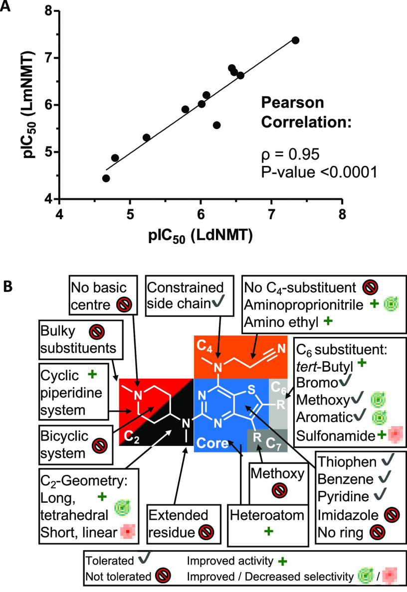
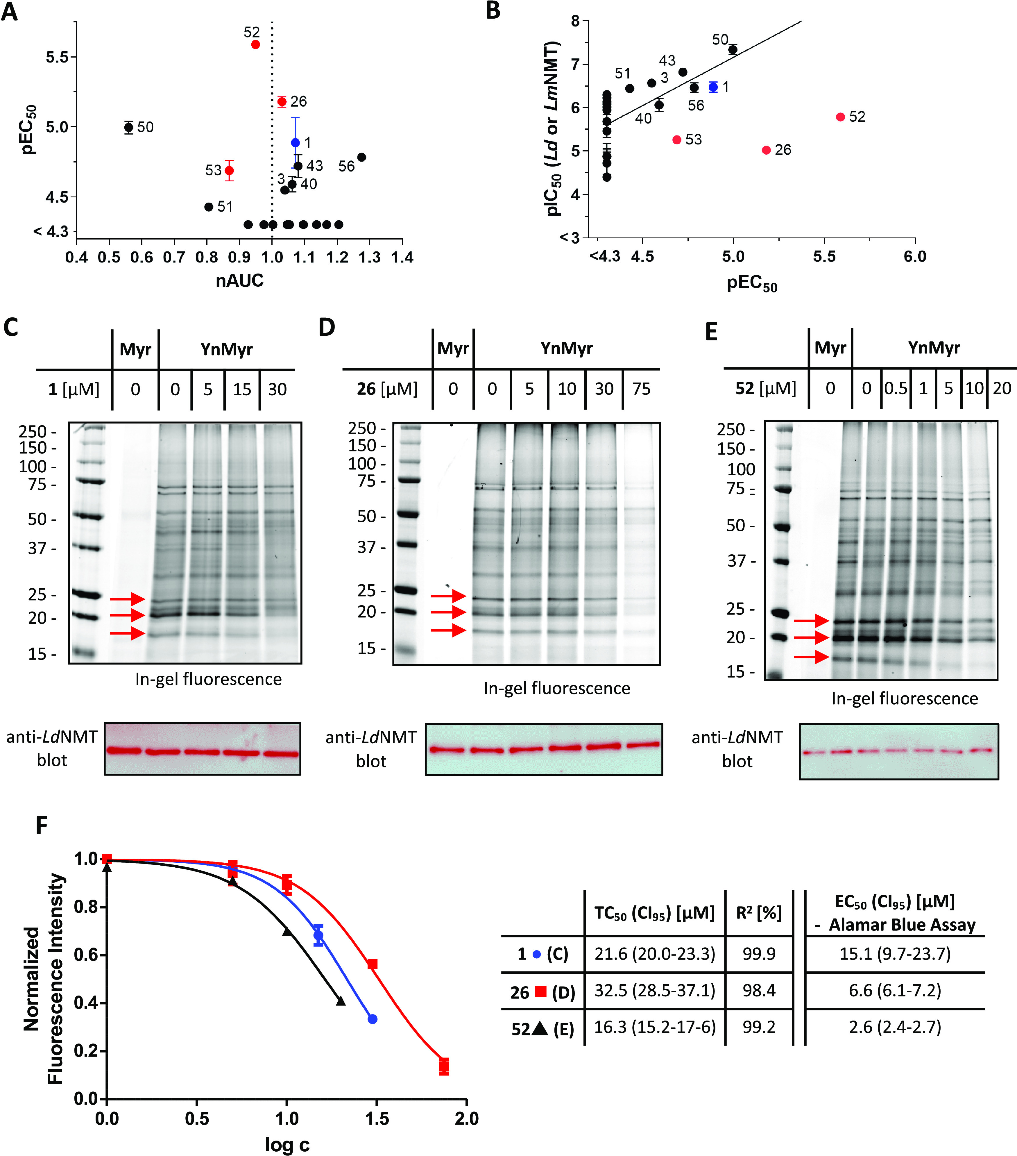
The leishmaniases, caused by Leishmania species of protozoan parasites, are neglected tropical diseases with millions of cases worldwide. Current therapeutic approaches are limited by toxicity, resistance, and cost. N-Myristoyltransferase (NMT), an enzyme ubiquitous and essential in all eukaryotes, has been validated via genetic and pharmacological methods as a promising anti-leishmanial target. Here we describe a comprehensive structure-activity relationship (SAR) study of a thienopyrimidine series previously identified in a high-throughput screen against Leishmania NMT, across 68 compounds in enzyme- and cell-based assay formats. Using a chemical tagging target engagement biomarker assay, we identify the first inhibitor in this series with on-target NMT activity in leishmania parasites. Furthermore, crystal structure analyses of 12 derivatives in complex with Leishmania major NMT revealed key factors important for future structure-guided optimization delivering IMP-105 (43), a compound with modest activity against Leishmania donovani intracellular amastigotes and excellent selectivity (>660-fold) for Leishmania NMT over human NMTs.


Conflict of interest statement

The authors declare no competing financial interest.

Figures

Figure 1
Figure 1
Binding pocket of compound 1 in LmNMT. (A) Ribbon rendering of LmNMT color ramped from amino (blue) to carboxy (red) terminus. The atoms of the myristoyl-CoA and 1 ligands are shown in space-filling representations and colored by atom types C (gray), O (red), N (blue), P (magenta), and S (yellow). (B) Structure of 1 (also known as IMP-083). (C) Cylinder rendering of the ligands emphasizing the proximal and distal binding sites for a pair of molecules of 1, which pack against one another. (D) Binding pocket for 1 showing the Cα and side chains of 1-interacting residues together with the C-terminal residue Leu421 whose α-carboxylate makes an ion-pairing interaction with the tertiary amino group of the proximal ligand. Water molecules in the neighborhood of 1 are shown as red spheres, and polar protein–ligand interactions are denoted by dashed lines. PDB ID: 4cgo.
Figure 2
Figure 2
(A) Correlation of LmNMT-based and LdNMT-based pIC50 values obtained with the CPM assay. The Pearson coefficient (ρ) and P-value are shown. (B) Schematic representation of the SAR results. The four structural segments of the inhibitor are highlighted. All symbols are annotated in the lowest box.
Scheme 1
Scheme 1. General Scheme for the Synthesis of 1 and Related Analogues
Reagents and conditions: (i) amine (1.0 equiv), N,N-diisopropylethylamine (DIPEA, 1.2 equiv), EtOAc, room temperature (rt), 18 h; (ii) amine (4–10 equiv), EtOAc, microwave, 120–200 °C, 30–90 min; (iii) 1-chloroethyl chloroformate (1.0 equiv), DCE, reflux, 1 h; (iv) MeOH, reflux, 1 h; and (v) aldehyde (1.0 equiv), NaBH(OAc)3, DCE, rt 3–18 h.
Figure 3
Figure 3
Comparison of the modes of binding of selected inhibitors with varied cores or C2 substitution. MyrCoA is not shown in the binding site, for clarity. Stereo renderings can be found in Figure S3. (A) 1 and 3 emphasizing the highly similar binding modes of the proximal ligand pair, which differ only in the core substitution of the thienopyrimidine for a quinazoline. Protein carbons: light green or green; ligand carbons: gray, dark gray; and waters: crimson or red. PDB ID 4cgo, 6qda. (B) 1, 10, and 12 exploring N-substitutions on the piperidine ring. As before, the proximal ligands are closely superimposable. The same is true of the distal ligands, with the exception of the piperidine ring, which is significantly displaced in the ethyl-substituted 12. Protein carbons: light green, green, lawn green; ligand carbons: gray, dark gray, light gray; and waters: crimson, red, violet red. PDB ID 4cgo, 6qdb, 6qdc. (C) 1 and 2 probing the significance of linear versus cyclic amine extensions from C2 of the core. The proximal ligand’s binding mode is altered in 2, with tilting of the thienopyrimidine core, dual conformations of the C2 amine extension, and repositioning of the C4 aminopropionitrile. No distal ligand was observed in the LmNMT complex with 2. Protein carbons: light green or green; ligand carbons: gray, dark gray; and waters: crimson or red. PDB ID 4cgo, 6qd9. (D) Chemical structures.
Figure 4
Figure 4
Comparison of the modes of binding of selected inhibitors with varied C4 or C6 substitution. For clarity, MyrCoA is not shown in the binding site. Stereo renderings can be found in Figure S4. (A) 1 and 43 demonstrate similar binding interactions despite the rigidification of the aminopropionitrile. The distal ligand does not appear, and 43 demonstrates dual conformations of the C2 substituent. Protein carbons: light green, green; ligand carbons: gray, dark gray; and waters: crimson, red, violet red. PDB ID 4cgo, 6qdd. (B) Chemical structures of relevant ligands. (C, D) tert-Butyl C6 substitution is accommodated in the proximal ligands for 51 and 52. However, the distal ligand conformation is inverted in 51 and dual C2 conformations are observed in 52, which is present at partial occupancy PDB ID 4cgo, 6qde, 6qdf. (E) 2 and 56 have analogous binding modes with an additional hydrogen bond in 56 mediated by the bromine substituent. PDB ID 4cgo, 6qdg.
Figure 5
Figure 5
(A) Plot of pIC50 against clog P. Diagonal lines represent areas of corresponding lipophilic efficiency (LipE) values. (B) Plot of LipE against selectivity (selectivity = IC50 (HsNMT)/IC50 (LdNMT)). (C) Schematic representation of fragment merging strategy between 3 and DDD85646(11) to generate 64 and related analogues. (D) Crystal structure of 1 and 64 in complex with LmNMT. 64 principally occupies the site of the proximal ligand in 1 extending through the sulfonamide group, which forms hydrogen bonds with the LmNMT backbone amide. PDB ID 4cgo, 6qdh.
Figure 6
Figure 6
(A) Plot of anti-L. donovani amastigote activity (pEC50) against macrophage toxicity (normalized area under the curve, nAUC). Thienopyrimidine 1 is highlighted in blue. (B) Plot of Leishmania NMT pIC50 (either Ld or Lm) against cell-based pEC50. A significant correlation (ρ = 0.75, P = 0.0006) is evident if 26, 52, and 53 (highlighted in red) are excluded. Compound 1 is highlighted in blue. (C–E) In-gel fluorescence and anti-LdNMT Western blot results for the cell-based tagging assay. (F) Fluorescence intensities of several individual bands between 25 and 15 kDa were quantified in (C–E) and normalized to the corresponding intensities of the control samples (without inhibitor) and the anti-LdNMT blot results. Tagging IC50 values (TC50) were extracted by nonlinear regression with a sigmoidal dose–response model (constraints: bottom = 0; top = 1.0) to the mean normalized intensity.
Figure 7
Figure 7
Comparison between ligands 1 and 43.

References

    1. Torres-Guerrero E.; Quintanilla-Cedillo M. R.; Ruiz-Esmenjaud J.; Arenas R. Leishmaniasis: A Review. F1000Research 2017, 6, 750.10.12688/f1000research.11120.1. - DOI - PMC - PubMed
    1. WHO, Leishmaniasis. https://www.who.int/news-room/fact-sheets/detail/leishmaniasis (accessed 14 June, 2020).
    1. Hefnawy A.; Berg M.; Dujardin J. C.; De Muylder G. Exploiting Knowledge on Leishmania Drug Resistance to Support the Quest for New Drugs. Trends Parasitol. 2017, 33, 162–174. 10.1016/j.pt.2016.11.003. - DOI - PubMed
    1. Yamazaki K.; Kaneko Y.; Suwa K.; Ebara S.; Nakazawa K.; Yasuno K. Synthesis of Potent and Selective Inhibitors of Candida Albicans N-Myristoyltransferase Based on the Benzothiazole Structure. Bioorg. Med. Chem. 2005, 13, 2509–2522. 10.1016/j.bmc.2005.01.033. - DOI - PubMed
    1. Fang W.; Robinson D. A.; Raimi O. G.; Blair D. E.; Harrison J. R.; Lockhart D. E. A.; Torrie L. S.; Ruda G. F.; Wyatt P. G.; Gilbert I. H.; Van Aalten D. M. F. N -Myristoyltransferase Is a Cell Wall Target in Aspergillus Fumigatus. ACS Chem. Biol. 2015, 10, 1425–1434. 10.1021/cb5008647. - DOI - PMC - PubMed

Publication types

MeSH terms