

The "slide technique"-a novel free-hand method of subaxial cervical pedicle screw placement
- PMID: 32576178
- PMCID: PMC7313203
- DOI: 10.1186/s12891-020-03420-0
The "slide technique"-a novel free-hand method of subaxial cervical pedicle screw placement
Abstract
Background: Cervical Pedicle Screw (CPS) placement is a challenging work due to the high risk of neurovascular complications. Although there have been several different free-hand or navigation assisted techniques for CPS placement, perforations may occur during screw insertion, especially lateral perforation. The objective of this manuscript is to describe a novel free-hand technique for subaxial CPS placement (C3-C7) and to evaluate if it decreases the chances of perforation.
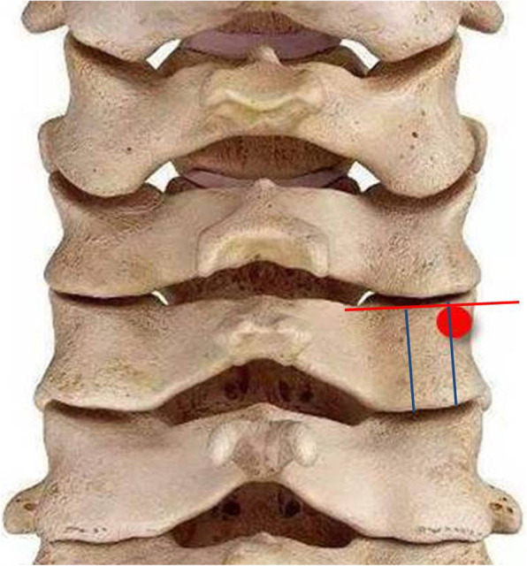
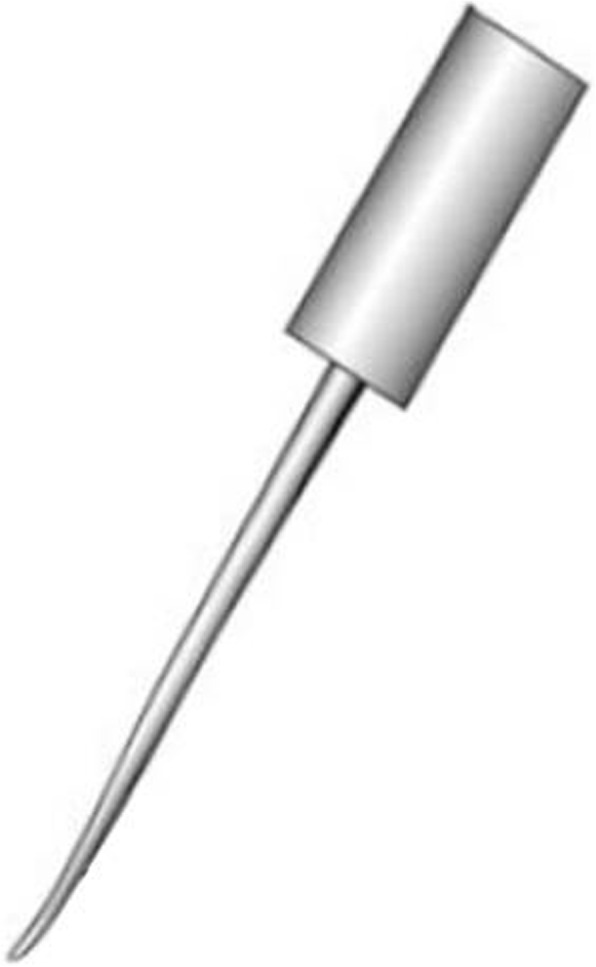
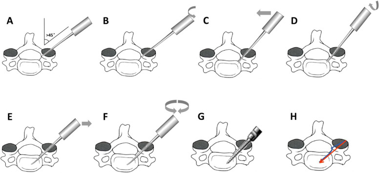
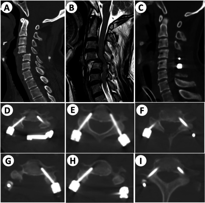
Methods: Thirty-two patients undergoing surgery with CPS instrumentation (C3-C7) at our institute between June 2017 and December 2018 were included in this study. All the patients had cervical trauma, and pedicle screw insertion was performed according to the free-hand "slide technique". The lamina, lateral mass and facet joint of the target area were exposed and the optimal entry point was found on the lateral mass posterior surface. A pedicular probe was then inserted and gently advanced. During the pedicle probe insertion, the cortex of the medial margin of the pedicle acted as a slide to permit the safe insertion of the screw. If the pedicle screw pathway was intact, the screw of the appropriate size was carefully placed. Three-dimensional (3D) CT imaging reconstruction was performed in all the patients after surgery, and screw perforations were graded with the Gertzbein-Robbins classification.
Results: Thirty-two patients who met the inclusion criteria were included in this study. A total of 257 CPSs (C3-7) were inserted, of which 41 CPSs were in C3, 61 CPSs were in C4, 55 CPSs were in C5, 53 CPSs were in C6, and 47 CPSs were in C7. The diameter and length of CPSs were 3.5 mm and 22-26 mm respectively. According to the Gertzbein-Robbins classification, grade 0, 231 screws; grade 1, 19 screws; and grade 2, 7 screws. No neurovascular complications occurred stemming from malpositioning of pedicle screws. Among perforated screws (26 screws), there were 16 lateral perforations, 5 medical perforations, and 4 inferior perforations.
Conclusions: The initial usage result shows the "slide technique" is a safe, effective and cost-effective technique for pedicle screw placement in the cervical spine. This is the first report of such a technique, and further studies are needed.
Keywords: Cervical pedicle screw (CPS); Cervical spine trauma; Free-hand; Slide technique.
Conflict of interest statement
The authors declare that they have no competing interests.
Figures
References
-
- Sjöström L, Karlström G, Pech P, Rauschning W. Indirect Spinal Canal Decompression in Burst Fractures Treated With Pedicle Screw Instrumentation. Spine. 1996;21(1):113–23. - PubMed
-
- Carl AL, Tromanhauser SG, Roger DJ. Pedicle Screw Instrumentation for Thoracolumbar Burst Fractures and Fracture-Dislocations. Spine. 1992;17(8):317–24. - PubMed
-
- Kwan MK, Chiu CK, Gani SMA, CCY W. Accuracy and Safety of Pedicle Screw Placement in Adolescent Idiopathic Scoliosis Patients: A Review of 2020 Screws using computed tomography assessment. Spine. 2017;42(5):326–35. - PubMed
-
- Oda I, Abumi K, Ito M, Kotani Y, Oya T, Hasegawa K, et al. Palliative Spinal Reconstruction Using Cervical Pedicle Screws for Metastatic Lesions of the Spine. Spine. 2006;31(13):1439–44. - PubMed
MeSH terms
Grants and funding
LinkOut - more resources
Full Text Sources
Medical
Miscellaneous
