. 2020 Jul 7;117(27):15884-15894.
doi: 10.1073/pnas.2003473117. Epub 2020 Jun 23.

HGT in the human and skin commensal Malassezia: A bacterially derived flavohemoglobin is required for NO resistance and host interaction

Affiliations

HGT in the human and skin commensal Malassezia: A bacterially derived flavohemoglobin is required for NO resistance and host interaction

Giuseppe Ianiri et al. Proc Natl Acad Sci U S A. .

Abstract

The skin of humans and animals is colonized by commensal and pathogenic fungi and bacteria that share this ecological niche and have established microbial interactions. Malassezia are the most abundant fungal skin inhabitant of warm-blooded animals and have been implicated in skin diseases and systemic disorders, including Crohn's disease and pancreatic cancer. Flavohemoglobin is a key enzyme involved in microbial nitrosative stress resistance and nitric oxide degradation. Comparative genomics and phylogenetic analyses within the Malassezia genus revealed that flavohemoglobin-encoding genes were acquired through independent horizontal gene transfer events from different donor bacteria that are part of the mammalian microbiome. Through targeted gene deletion and functional complementation in Malassezia sympodialis, we demonstrated that bacterially derived flavohemoglobins are cytoplasmic proteins required for nitric oxide detoxification and nitrosative stress resistance under aerobic conditions. RNA-sequencing analysis revealed that endogenous accumulation of nitric oxide resulted in up-regulation of genes involved in stress response and down-regulation of the MalaS7 allergen-encoding genes. Solution of the high-resolution X-ray crystal structure of Malassezia flavohemoglobin revealed features conserved with both bacterial and fungal flavohemoglobins. In vivo pathogenesis is independent of Malassezia flavohemoglobin. Lastly, we identified an additional 30 genus- and species-specific horizontal gene transfer candidates that might have contributed to the evolution of this genus as the most common inhabitants of animal skin.

Keywords: Malassezia; flavohemoglobin; horizontal gene transfer.


Conflict of interest statement

The authors declare no competing interest.

Figures

Fig. 1.
Fig. 1.
Evidence for independent HGT events of the flavohemoglobin-encoding genes in Malassezia from the Actinobacteria. (A) Maximum likelihood phylogeny consisting of 2,155 flavohemoglobin protein sequences. Two groups (clades 1 and 2) of horizontally transferred flavohemoglobin genes (YHB1 and YHB101) in Malassezia are colored in orange. Other tree branches are colored according to the key on the Top Left, representing other major groups of organisms. The phylogeny was visualized using iTOL v3.6.1 (61) and rooted at the midpoint. (B and C) Zoomed views of the ML phylogeny showing in more detail the position of Malassezia flavohemoglobins from clades 1 and 2, and their putative bacterial donor lineages. (D) Results of topological constraint tests that significantly rejected the monophyletic origin for both Malassezia flavohemoglobins clades, providing additional support for independent HGT events.
Fig. 2.
Fig. 2.
Evolutionary trajectory of flavohemoglobin-encoding genes in Malassezia after their acquisition via HGT from different donor bacteria lineages. (A) Phylogenetic relationship of Malassezia species with available genome sequence inferred from the concatenation of 246 single-copy proteins. Color codes assigned to the different phylogenetic clades (named A to D) are kept consistent in all figures. The tree was rooted at the midpoint and white circles in the tree nodes indicate full UFboot and SH-aLRT branch support. The proposed evolutionary events that led to the final arrangement of the flavohemoglobin-encoding genes reported in B are shown in the phylogenetic tree, as given in the key; double arrows indicate relocation of the YHB101 gene in subtelomeric position. (B) Chromosomal regions encompassing the YHB1 gene in Malassezia. Genes are shown as arrows denoting the direction of transcription, and orthologs are represented in the same color. Nonsyntenic genes are shown in white, and small arrows in black represent tRNAs. The YHB1 gene is shown as red arrows outlined in bold in the center. The end of a scaffold is represented by a forward slash. For M. yamatoensis and M. slooffiae, yellow bars indicate the absence of the YHB1 gene in otherwise syntenic regions, and those in green indicate instances where another flavohemoglobin-encoding gene, named YHB101 and represented as orange arrows outlined in bold, was acquired by an independent HGT event. A defective YHB1 gene in Malassezia nana CBS9557 is denoted by the Greek symbol ψ. Gene codes in red or blue are as they appear in M. globosa (prefix “MGL_”) or M. sympodialis (prefix “MSYG_”) genome annotations, respectively; those in black were named based on top BLASTp hits in S. cerevisiae; and “hyp” represent hypothetical proteins. Black circle represents the end of a chromosome. Scaffold/chromosomal locations and accession numbers are given for each region in SI Appendix, Table S1.
Fig. 3.
Fig. 3.
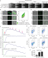
M. sympodialis flavohemoglobins are involved in nitrosative stress resistance and NO degradation. (A) Stress sensitivity assay of M. sympodialis WT, yhb1Δ mutant, and complementing strains yhb1Δ + YHB1 and yhb1Δ + YHB101 on mDixon agar supplemented with the NO donor agent DETA NONOate and NaNO2, and with hydrogen peroxide. (B) GFP expression in the M. sympodialis yhb1Δ mutant, and complementing strains yhb1Δ + YHB1 and yhb1Δ + YHB101, and respective GFP signal analyzed through FACS. (C) NO consumption assay by M. sympodialis WT, yhb1Δ mutant, and complementing strains yhb1Δ + YHB1 and yhb1Δ + YHB101; the blue trace indicates the NO level over a period of 15 min. NO and Malassezia (Y = yeast) injections are indicated by purple arrows. In this experiment, 10 µL (Y), 20 µL (×2) and 30 µL (×3), and 40 µL (×4) and 50 µL (×5) of Malassezia cellular suspensions were injected. (D) Representative fluorescent staining of intracellular NO with DAF-FM DA in M. sympodialis WT and two independent yhb1Δ mutants, and (E and F) quantification of the NO signal by flow cytometry; spontaneous fluorescence of M. sympodialis was used as background to detect specifically DAF-FM DA signal. Asterisks indicates statistically significant differences (* = P < 0.05, ** = P < 0.01) according to the unpaired Student’s t test with Welch’s correction. “ns” is not significant.
Fig. 4.
Fig. 4.
Transcriptomic profile of M. sympodialis strains under NO-accumulating conditions. (A) MA plot displaying the transcriptomic changes of the M. sympodialis yhb1Δ compared to the M. sympodialis WT strain. Red dots indicate differentially expressed genes for FDR < 0.05. The most up-regulated and down-regulated genes are indicated, along with the YHB1 gene, which represents an internal control as its down-regulation is expected because the gene is deleted. (B) MA plot displaying the transcriptomic changes of M. sympodialis WT grown in the presence of NaNO2 compared to the untreated control. Red dots indicate differentially expressed genes for FDR < 0.05; the most up-regulated and down-regulated genes are indicated. (C) Gene Ontology classification relative to the RNA-seq condition reported in B. Up-regulated genes are indicated in red, and down-regulated genes are indicated in green. (D) Venn diagrams showing comparison of the up-regulated and down-regulated genes relative to RNA-seq conditions reported in A and B; the panel on the Right shows a heatmap of the log2 FC of the shared up-regulated (red) and down-regulated (green) genes. Predicted allergens are indicated with one asterisk, and two asterisks indicate a predicted secreted lipase.
Fig. 5.
Fig. 5.
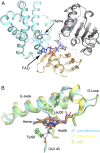
Three-dimensional X-ray crystal structure of the M. yamatoensis flavohemoglobin Yhb101. (A) The globin domain (cyan) binds a heme molecule. The reductase domain consists of a FAD-binding domain (gray) and a NAD-binding domain (tan) that bind a FAD molecule. (B) An overlay of flavohemoglobin globin domains from fungus, bacteria, and yeast: globin domains of M. yamatoensis (PDB ID 6O0A; blue), S. cerevisiae (PDB ID 4G1V; yellow), and E. coli (PDB ID 1GVH; green) show structural similarity.
Fig. 6.
Fig. 6.
Malassezia genes acquired through HGT from bacteria. HGT candidates identified in the genomes of the 15 Malassezia species (represented on the Top according to their phylogenetic classification) are shown as different lines in the presence–absence matrix, with the closest ortholog in S. cerevisiae indicated in parenthesis, where available. For each HGT candidate, the presence of the gene in a genome is indicated by orange square, and the intensity of the color is correlated with the gene copy number (numbers in white). HGT candidates occurring in multiple Malassezia species are shown in the Top half of the matrix, whereas those that are species-specific HGT candidates are shown in the Bottom half of the matrix, and color coded as shown in the key. Asterisks indicate HGT candidate genes identified in the previous study (3). The bacterially derived gene encoding an aliphatic amidase identified in M. nana CBS9557 seems to be another instance of a pseudogene in this strain (indicated as ψ).

References

    1. Findley K., et al. ., Topographic diversity of fungal and bacterial communities in human skin. Nature 498, 367–370 (2013). - PMC - PubMed
    1. Theelen B., et al. ., Malassezia ecology, pathophysiology, and treatment. Med. Mycol. 56 (suppl. 1), S10–S25 (2018). - PubMed
    1. Wu G., et al. ., Genus-wide comparative genomics of Malassezia delineates its phylogeny, physiology, and niche adaptation on human skin. PLoS Genet. 11, e1005614 (2015). - PMC - PubMed
    1. Gaitanis G., Magiatis P., Hantschke M., Bassukas I. D., Velegraki A., The Malassezia genus in skin and systemic diseases. Clin. Microbiol. Rev. 25, 106–141 (2012). - PMC - PubMed
    1. Sparber F., et al. ., The skin commensal yeast Malassezia triggers a type 17 response that coordinates anti-fungal immunity and exacerbates skin inflammation. Cell Host Microbe 25, 389–403.e6 (2019). - PubMed

Publication types

MeSH terms

LinkOut - more resources