Loosening of the mesothelial barrier as an early therapeutic target to preserve peritoneal function in peritoneal dialysis
- PMID: 32576713
- PMCID: PMC7321674
- DOI: 10.23876/j.krcp.20.052
Loosening of the mesothelial barrier as an early therapeutic target to preserve peritoneal function in peritoneal dialysis
Abstract
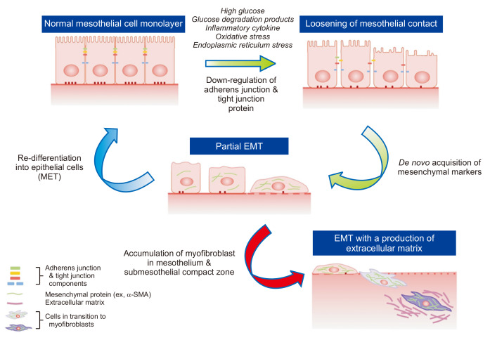
Phenotype transition of peritoneal mesothelial cells (MCs) including the epithelial-to-mesenchymal transition (EMT) is regarded as an early mechanism of peritoneal dysfunction and fibrosis in peritoneal dialysis (PD), producing proinflammatory and pro-fibrotic milieu in the intra-peritoneal cavity. Loosening of intercellular tight adhesion between adjacent MCs as an initial process of EMT creates the environment where mesothelium and submesothelial tissue are more vulnerable to the composition of bio-incompatible dialysates, reactive oxygen species, and inflammatory cytokines. In addition, down-regulation of epithelial cell markers such as E-cadherin facilitates de novo acquisition of mesenchymal phenotypes in MCs and production of extracellular matrices. Major mechanisms underlying the EMT of MCs include induction of oxidative stress, pro-inflammatory cytokines, endoplasmic reticulum stress and activation of the local renin-angiotensin system. Another mechanism of peritoneal EMT is mitigation of intrinsic defense mechanisms such as the peritoneal antioxidant system and anti-fibrotic peptide production in the peritoneal cavity. In addition to use of less bio-incompatible dialysates and optimum treatment of peritonitis in PD, therapies to prevent or alleviate peritoneal EMT have demonstrated a favorable effect on peritoneal function and structure, suggesting that EMT can be an early interventional target to preserve peritoneal integrity.
Keywords: Adhesion molecule; Epithelial-to-mesenchymal transition; Peritoneal fibrosis; Peritoneal mesothelial cells.
Conflict of interest statement
The author has no conflicts of interest to declare.
Figures
Similar articles
-
Olive leaf extract counteracts epithelial to mesenchymal transition process induced by peritoneal dialysis, through the inhibition of TGFβ1 signaling.Cell Biol Toxicol. 2019 Apr;35(2):95-109. doi: 10.1007/s10565-018-9438-9. Epub 2018 Jul 6. Cell Biol Toxicol. 2019. PMID: 29978330
-
Transition of mesothelial cell to fibroblast in peritoneal dialysis: EMT, stem cell or bystander?Perit Dial Int. 2015 Jan-Feb;35(1):14-25. doi: 10.3747/pdi.2014.00188. Perit Dial Int. 2015. PMID: 25700459 Free PMC article. Review.
-
Paricalcitol attenuates TGF-β1-induced phenotype transition of human peritoneal mesothelial cells (HPMCs) via modulation of oxidative stress and NLRP3 inflammasome.FASEB J. 2019 Feb;33(2):3035-3050. doi: 10.1096/fj.201800292RR. Epub 2018 Oct 24. FASEB J. 2019. PMID: 30354670
-
Shorter daily dwelling time in peritoneal dialysis attenuates the epithelial-to-mesenchymal transition of mesothelial cells.BMC Nephrol. 2014 Feb 20;15:35. doi: 10.1186/1471-2369-15-35. BMC Nephrol. 2014. PMID: 24555732 Free PMC article. Clinical Trial.
-
Epithelial-to-mesenchymal transition of the mesothelial cell--its role in the response of the peritoneum to dialysis.Nephrol Dial Transplant. 2006 Jul;21 Suppl 2:ii2-7. doi: 10.1093/ndt/gfl183. Nephrol Dial Transplant. 2006. PMID: 16825254 Review.
Cited by
-
TGF-β1 Receptor Inhibitor SB525334 Attenuates the Epithelial to Mesenchymal Transition of Peritoneal Mesothelial Cells via the TGF-β1 Signaling Pathway.Biomedicines. 2021 Jul 19;9(7):839. doi: 10.3390/biomedicines9070839. Biomedicines. 2021. PMID: 34356903 Free PMC article.
-
PPARγ alleviates peritoneal fibrosis progression along with promoting GLUT1 expression and suppressing peritoneal mesothelial cell proliferation.Mol Cell Biochem. 2022 Jul;477(7):1959-1971. doi: 10.1007/s11010-022-04419-y. Epub 2022 Apr 5. Mol Cell Biochem. 2022. PMID: 35380292 Free PMC article.
-
Mitochondrial Dysfunction Plays a Relevant Role in Pathophysiology of Peritoneal Membrane Damage Induced by Peritoneal Dialysis.Antioxidants (Basel). 2021 Mar 13;10(3):447. doi: 10.3390/antiox10030447. Antioxidants (Basel). 2021. PMID: 33805753 Free PMC article.
-
Epithelial-mesenchymal transition in organ fibrosis development: current understanding and treatment strategies.Burns Trauma. 2022 Apr 8;10:tkac011. doi: 10.1093/burnst/tkac011. eCollection 2022. Burns Trauma. 2022. PMID: 35402628 Free PMC article. Review.
-
Inhibition of calpain9 attenuates peritoneal dialysis-related peritoneal fibrosis.Front Pharmacol. 2022 Dec 1;13:962770. doi: 10.3389/fphar.2022.962770. eCollection 2022. Front Pharmacol. 2022. PMID: 36532773 Free PMC article.
References
Publication types
LinkOut - more resources
Full Text Sources