. 2020 Jun 23;10(1):10142.
doi: 10.1038/s41598-020-67039-w.

Mesenchymal Stromal Cell Bioreactor for Ex Vivo Reprogramming of Human Immune Cells

Affiliations

Mesenchymal Stromal Cell Bioreactor for Ex Vivo Reprogramming of Human Immune Cells

Ashley Allen et al. Sci Rep. .

Erratum in

Abstract

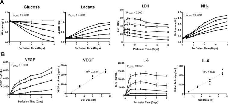
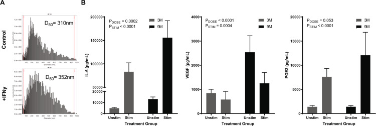
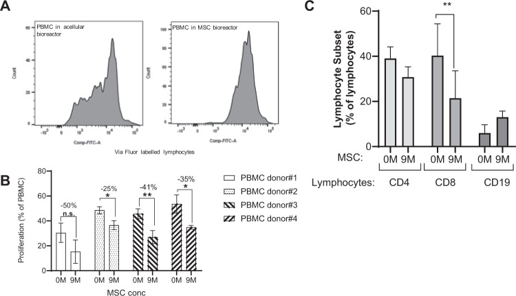
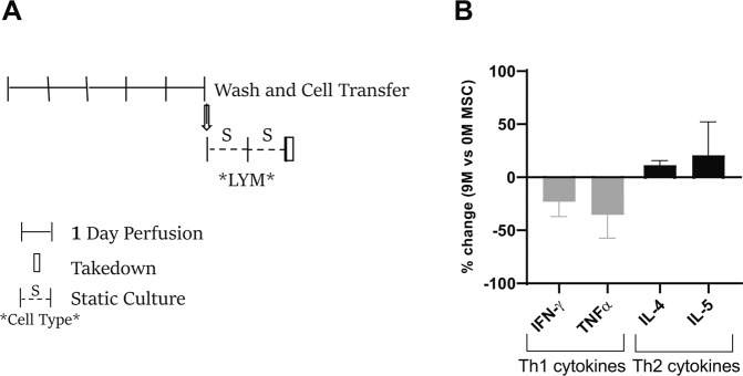
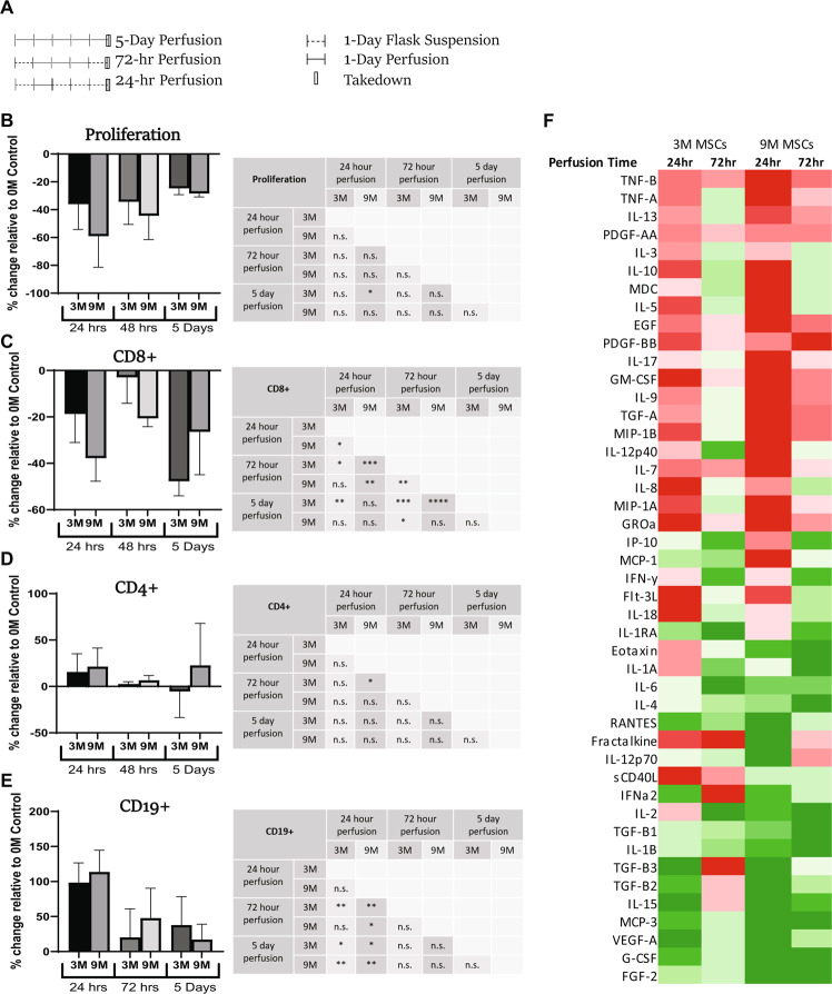
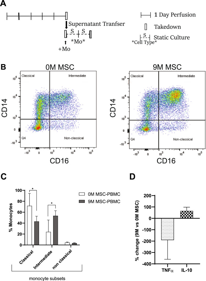
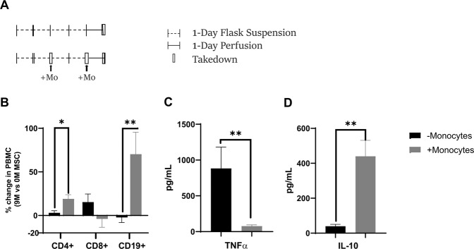
Bone marrow mesenchymal stromal cells (MSCs) have been studied for decades as potent immunomodulators. Clinically, they have shown some promise but with limited success. Here, we report the ability of a scalable hollow fiber bioreactor to effectively maintain ideal MSC function as a single population while also being able to impart an immunoregulatory effect when cultured in tandem with an inflamed lymphocyte population. MSCs were seeded on the extraluminal side of hollow fibers within a bioreactor where they indirectly interact with immune cells flowing within the lumen of the fibers. MSCs showed a stable and predictable metabolite and secreted factor profile during several days of perfusion culture. Exposure of bioreactor-seeded MSCs to inflammatory stimuli reproducibly switched MSC secreted factor profiles and altered microvesicle composition. Furthermore, circulating, activated human peripheral blood mononuclear cells (PBMCs) were suppressed by MSC bioreactor culture confirmed by a durable change in their immunophenotype and function. This platform was useful to study a model of immobilized MSCs and circulating immune cells and showed that monocytes play an important role in MSC driven immunomodulation. This coculture technology can have broad implications for use in studying MSC-immune interactions under flow conditions as well as in the generation of ex vivo derived immune cellular therapeutics.


Conflict of interest statement

The authors declare no competing interests.

Figures

Figure 1
Figure 1
Dose-dependent MSC Metabolism and Measurement of Secreted Factors During 8-Day Microreactor Continuous Perfusion. Microreactors seeded with 0 (◆), 1 ×10⁶ (∙), 3 ×10⁶ (▪), 6 ×10⁶ (▴), and 9 ×10⁶ (▼) viable MSCs were incubated for 2 hours at 37 °C followed by a 24 hour hold at room temperature. Microreactors were then perfused with media containing 10% FBS, 1% antibiotic-antimycotic, 0.2% gentamicin and sampled over 8 days. (A) Glucose (g/L), Lactate (g/L), LDH (U/L), and NH3 (mmol/L) were quantified via CedEx Bio measurement on thawed samples (A). Measurement of secreted factors in these samples was performed via ELISA for IL-6 (pg/mL) and VEGF (pg/mL) (B). Graphs show average values for each cell dose group + standard deviations for each. Two-way ANOVA was performed for all timepoints with n = 3 (Days 1-4) showing a significant impact of dose on device metabolism and secreted factor performance. Correlation of cell dose to device IL-6 and VEGF performance at 24 hours of perfusion is shown for individual devices.
Figure 2
Figure 2
Dynamic Modulation of MSC Bioreactor Output with Introduction of Inflammatory Stimulus. Microreactors were seeded and perfused in a 50-mL circuit containing inflammatory stimuli. Particle Size Analysis of MSC extracellular vesicles (MSC-EVs) produced by bioreactors seeded with 3 ×10⁶ cells after 12 hours of perfusion with or without IFNγ stimulation (5 ng/mL) (A). Secreted factors (IL-6, VEGF, and PGE2) were measured via ELISA after seeding microreactors with 3 ×10⁶ (gray) or 9 ×10⁶ (black) viable MSCs, incubating for two hours at 37 °C, hold for 24 hours at RT, and perfusion with or without an inflammatory cocktail (10 ng/mL IFNγ, 1 ng/mL IL-1β, and 5 ng/mL TNFα) for 48 hours (B). Graphs show average values for each cell dose group + standard deviations for each (n = 2 donors, n = 3/donor). Two Way ANOVA was performed on each set of samples to determine effects of dose (PDOSE) and stimulation (PSTIMULATION) on MSC output.
Figure 3
Figure 3
Secreted Factors following Perfusion of Activated PBMCs through MSC-Bioreactor Circuits. PBMCs activated with PHA (5 ug/mL) and supplemented with IL-2 (100 ng/mL) were perfused through bioreactor circuits containing 0 M or 9 M (n = 3) and medium was recovered. Both (A) factor concentration and (B) extracellular vesicle characteristics were modulated with MSC treatment.
Figure 4
Figure 4
Prevention of lymphocyte activation in MSC bioreactors. PBMCs were labeled with ViaFluor, stimulated (PHA/IL-2) and perfused for 5-days via circuits containing microreactors seeded with 0, or 9 ×10⁶ MSCs per device. MSC treatment was shown to inhibit T-cell proliferation as demonstrated by ViaFluor labelling (A). This was observed for four PBMC donors (n = 4 donors, n = 3/donor) (B). Percent decrease in PBMC proliferation with MSC (9 M) is indicated. Perfusion via MSC-seeded microreactors (9 M) exhibited changes in CD4, CD8, and CD19 + lymphocytes when compared to acellular controls (0 M) (n = 2 donors, n = 3/donor) (C). Graphs show average values for each cell dose group + standard deviations for two donors. A student’s t-test was performed on each set. **p ≤ 0.01 *p ≤ 0.05.
Figure 5
Figure 5
MSC bioreactor reprogramming of T cell cytokine responses. Stimulated (PHA/IL-2) PBMCs perfused in a bioreactor (+/− MSC) for 5-days were plated on a 6-well tissue culture dish for two additional days (A). Lymphocyte secreted factors (in the supernatant) were assayed using multiplex cytokine panel. Th1/Th2 specific cytokines are shown as a percentage change with MSC (9 M) treatment relative to acellular (0 M MSC) (n = 1 donor, 3 runs/donor) (B).
Figure 6
Figure 6
Dose and duration effects of ex vivo MSC perfusion on human lymphocytes (A) Stimulated (PHA/IL-2) PBMCs were perfused for either 24 hours, 72 hours or for 5 days through circuits containing microreactors seeded with either 0, 3, or 9 ×10⁶ MSCs per device (0 M, 3 M, 9 M) (n = 2 donors, n ≥ 3/donor) (5 day historical only has one donor). The 24- and 72-hour perfusion groups were first placed into static culture for 24 hours prior to perfusion initiation. Each group was perfused for the designated time and then placed into static culture until collection on Day 5. Relative to 0 M control MSC treatment was shown to inhibit lymphocyte proliferation in all conditions (B), with a trend correlating with MSC dose response. CD8 + T cell proliferation was also inhibited by perfusion (C) while B-cell proliferation increased (E) in a dose and duration dependent manner for each subpopulation. A student’s t-test was performed on each set. ****p ≤ 0.0001 ***p ≤ 0.001 **p ≤ 0.01 *p ≤ 0.05. n.s. = not significant. Graphs show average values for each cell dose + standard deviation. (F) Culture media samples were collected at Day 5 and analyzed via multiplex. Measurement of percent change was calculated by determining the output of any condition relative to the 0 M control. The absolute value of the percent change was then charted into columns according to MSC dose and perfusion duration. Comparative analysis of intensities were calculated within each row with darker colors representing larger values. Red blocks indicate decreases in percent change while green blocks indicate increases. Of all conditions, the 9 M MSC 24-hour perfusion group showed the largest changes in analyte values (n = 1 donor).
Figure 7
Figure 7
Transfer of MSC bioreactor/PBMC perfusate alter primary human monocyte differentiation. MSC reprogrammed PBMCs (PHA/IL-2 activated) perfusate/supernatant from a 5-day bioreactor (with −/+ MSC) was added on monocytes cultured on a cell-repellent tissue culture dish for two days (A). After two days, monocytes were stained using CD14 and CD16 antibodies and dot blots are shown (B) and percentage change in monocyte subsets with and without MSC-reprogrammed PBMC is shown (n = 2 donors, n = 3/donor) (C). The levels of TNFα and IL-10 in the supernatant of monocytes after two-days of addition of PBMCs perfusate from circuits with or without MSCs is plotted as percentage change with MSC addition (n = 2 donors, n = 3/donor) (D).
Figure 8
Figure 8
Role of monocytes in MSC bioreactor immune reprogramming. PHA/IL-2 stimulated PBMCs were cultured in static tissue culture flasks for 4-days. On day 2 and day 4, monocytes (~2.8 M/ bioreactor) were added into PBMC flask (in the monocyte addition group in gray). At day 4, after addition of monocytes PBMCs were perfused in bioreactor for 24 hours via circuits seeded with 0, or 9 ×10⁶ MSCs per device (A). Following 24-hours of perfusion, the cells were assayed via flow cytometry (B) and supernatant was assayed for TNFα and IL-10 levels both without and with monocyte addition (C). Overall, the addition of monocytes to the PBMCs enhanced the MSC immunomodulatory effects as shown by changes in lymphocyte population, TNFα and IL-10 levels (D). A student’s t-test was performed on each set (**p ≤ 0.01 *p ≤ 0.05). Graphs show average values + standard deviation (n = 1 donors, n = 3/donor).

References

    1. Parekkadan B, et al. Bone marrow stromal cell transplants prevent experimental enterocolitis and require host CD11b+ splenocytes. Gastroenterology. 2011;140:966–75. doi: 10.1053/j.gastro.2010.10.013. - DOI - PMC - PubMed
    1. Bouchlaka MN, et al. Human Mesenchymal Stem Cell-Educated Macrophages Are a Distinct High IL-6-Producing Subset that Confer Protection in Graft-versus-Host-Disease and Radiation Injury Models. Biol. Blood Marrow Transplant. 2017;23:897–905. doi: 10.1016/j.bbmt.2017.02.018. - DOI - PMC - PubMed
    1. Francois M, Romieu-Mourez R, Li M, Galipeau J. Human MSC suppression correlates with cytokine induction of indoleamine 2,3-dioxygenase and bystander M2 macrophage differentiation. Mol. Ther. 2012;20:187–95. doi: 10.1038/mt.2011.189. - DOI - PubMed
    1. Chiesa S, et al. Mesenchymal stem cells impair in vivo T-cell priming by dendritic cells. Proc. Natl. Acad. Sci. USA. 2011;108:17384–9. doi: 10.1073/pnas.1103650108. - DOI - PMC - PubMed
    1. Abbas AK, Murphy KM, Sher A. Functional diversity of helper T lymphocytes. Nature. 1996;383:787–93. doi: 10.1038/383787a0. - DOI - PubMed

Publication types