Case Reports
. 2020 Apr 14;8(6):962-966.
doi: 10.1002/ccr3.2795. eCollection 2020 Jun.

Unusual iodine-131 postablation whole-body scintigraphy patterns in patients after robot-assisted/endoscopic thyroidectomy: Case series

Affiliations
Case Reports

Unusual iodine-131 postablation whole-body scintigraphy patterns in patients after robot-assisted/endoscopic thyroidectomy: Case series

Mi Ra Kim et al. Clin Case Rep. .

Abstract

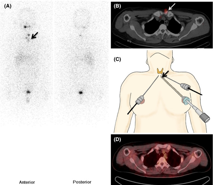
Robot-assisted/endoscopic thyroidectomy causes irritation due to instruments or implantation of thyroid tissue on the anterior chest wall and lower neck. We present three patients who exhibited unexpected focal lesions on postablation Iodine-131 single-photon emission computed tomography/computed tomography without biochemical and structural diseases. Meticulous surgical techniques are important to prevent complications.

Keywords: Iodine‐131 whole‐body scintigraphy (WBS); endoscopic thyroidectomy; positron emission tomography/computed tomography (PET/CT); robot‐assisted thyroidectomy; single‐photon emission computed tomography/computed tomography (SPECT/CT); thyroid carcinoma; unusual patterns.


Conflict of interest statement

The authors declare that there are no conflicts of interest with respect to the research, authorship, and/or publication of this article.

Figures

Figure 1
Figure 1
A 54‐y‐old woman with Hürthle cell carcinoma underwent endoscopic total thyroidectomy with left central lymph node dissection through a bilateral breast and modified left axillary approach. A, Remnant thyroid tissue with another area of focal uptake around the left lower neck (black arrow) noted on whole‐body scintigraphy (WBS) after 481‐MBq radioiodine therapy. Several physiologic foci are seen in the nasal cavity, oral cavity, liver, bladder along with urine contamination, and remnant thyroid tissue. B, Single‐photon emission computed tomography/computed tomography (SPECT‐CT) images demonstrate this uptake around the left sternoclavicular junction (white arrow). C, Skin incisions were performed around the areola and medial to the left anterior axillary fold in this patient. The lesion in question (black arrow) was potentially caused by irritation due to surgical instruments that were all directed to the midline of the sternal notch as well shown on this illustration. D, No abnormal uptake seen in the corresponding area on positron emission tomography/computed tomography (PET‐CT) images
Figure 2
Figure 2
A 36‐y‐old woman with papillary thyroid carcinoma underwent robot‐assisted total thyroidectomy with left central neck lymph node dissection. She was treated with 370‐MBq radioiodine therapy. A, Postradioiodine therapy whole‐body scintigraphy (WBS) shows unusual uptake near the left lower neck (black arrow) with remnant thyroid tissue. Several physiologic foci are seen in the oral cavity, liver, bowel, bladder, and remnant thyroid tissue. B, Single‐photon emission computed tomography/computed tomography (SPECT‐CT) images show uptake around the left sternoclavicular junction (white arrow). C, The lesion indicated by the arrow in this illustration is the affected area caused by the bilateral axillo‐breast surgical approach, which was compatible with the uptake observed in Figure 2A and B. D, No area of abnormal uptake in the corresponding region as seen on positron emission tomography/computed tomography (PET‐CT) images
Figure 3
Figure 3
A 42‐y‐old woman with multiple papillary thyroid carcinomas underwent robot‐assisted total thyroidectomy. A and B, Postradioiodine therapy whole‐body scintigraphy (WBS) and single‐photon emission computed tomography/computed tomography (SPECT‐CT) performed after the patient received 592‐MBq radioiodine therapy. Focal areas of subcutaneous uptake are seen in the upper center area of the right breast including the residual thyroid tissue (black and white arrows). Several physiologic foci are seen in the nasal cavity, oral cavity, salivary gland, liver, bowel with feces, bladder, and remnant thyroid tissue on postradioiodine therapy WBS. C, The lesion in this illustration is potentially related to the physical stress caused by subcutaneous tunneling from the circumareolar area to the suprasternal notch where blunt dissection was performed to create a working space for thyroidectomy (black arrow). D, No abnormal uptake in the corresponding area on positron emission tomography/computed tomography (PET‐CT) images

References

    1. Tae K, Ji YB, Song CM, Ryu J. Robotic and endoscopic thyroid surgery: evolution and advances. Clin Exp Otorhinolaryngol. 2019;12(1):1‐11. - PMC - PubMed
    1. Lee KE, Kim HY, Park WS, et al. Postauricular and axillary approach endoscopic neck surgery: a new technique. World J Surg. 2009;33(4):767‐772. - PubMed
    1. Ikeda Y, Takami H, Tajima G, et al. Total endoscopic thyroidectomy: axillary or anterior chest approach. Biomed Pharmacother. 2002;56(Suppl 1):72s‐78s. - PubMed
    1. Jung CH, Kim BY, Kim JW, et al. False‐positive F‐18 FDG PET/CT from foreign body reaction on anterior chest wall after endoscopic total thyroidectomy via axillo‐breast approach for thyroid cancer: two case reports. Clin Nucl Med. 2011;36(11):1036‐1038. - PubMed
    1. Kim HR, Hong HS, Lee EH, et al. Ultrasonographic findings of foreign‐body granulomas after endoscopic thyroidectomy via an axillo‐breast approach. J Korean Soc Radiol. 2012;66(6):507‐517.

Publication types

LinkOut - more resources