The Old Supracondylar Fracture of Femur Treated by Gradual Deformity Correction Using the Ilizarov Technique Followed by the Second-Stage Internal Fixation in an Elderly Patient With Osteoporosis
- PMID: 32577319
- PMCID: PMC7289063
- DOI: 10.1177/2151459320931673
The Old Supracondylar Fracture of Femur Treated by Gradual Deformity Correction Using the Ilizarov Technique Followed by the Second-Stage Internal Fixation in an Elderly Patient With Osteoporosis
Abstract
Background: The supracondylar nonunion of femur in elderly individuals is rare and challenging to manage. Nothing in English literatures or guidelines is available regarding this particular fracture characterized by osteoporosis, soft-tissue contracture, shortening, and joint stiffness. We report a case of an elderly patient with a supracondylar nonunion of the femur, which was successfully treated using staged Ilizarov techniques and dual plating.
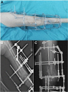
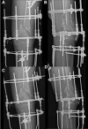
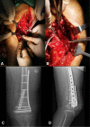
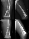
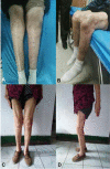
Case presentation: An 84-year-old female patient was admitted to our orthopedic department for her pain and soft-tissue swelling around the right knee with claudication and shortening deformity of the affected extremity. She denied any specific history of trauma and had sought traditional Chinese medical attention for 6 months before she presented to our hospital. Diagnosis of the right femoral supracondylar nonunion was made based on the X-ray and computed tomography. Ilizarov external fixator was carried out for successive and slow distraction and gradual correction of the shortening deformity, in consideration of the nonunion was still present. Subsequently, internal fixation with dual plating of the distal femur was performed. Excellent function and patient satisfaction were observed at 6 months of follow-up.
Conclusion: The protocol of Ilizarov technique with subsequent internal fixation of dual plating seems to be an efficient solution to the supracondylar nonunion of femur in elderly patients with osteoporosis. The advantage of the protocol is that it allows knee joint motion, avoids neurovascular complications, and gentle correction of soft-tissue contractures.
Keywords: Ilizarov technique; dual plating; elderly patients; internal fixation; nonunion; old fracture; osteoporosis; supracondylar fracture of femur.
© The Author(s) 2020.
Conflict of interest statement
Declaration of Conflicting Interests: The author(s) declared no potential conflicts of interest with respect to the research, authorship, and/or publication of this article.
Figures

Similar articles
-
Use of Ilizarov external fixation for a periprosthetic supracondylar femur fracture.J Arthroplasty. 1999 Jan;14(1):118-21. doi: 10.1016/s0883-5403(99)90214-0. J Arthroplasty. 1999. PMID: 9926965
-
Distal femoral derotational osteotomy with external fixation for correction of excessive femoral anteversion in patients with cerebral palsy.J Pediatr Orthop B. 2015 Sep;24(5):425-32. doi: 10.1097/BPB.0000000000000168. J Pediatr Orthop B. 2015. PMID: 25794115
-
Retrograde Intramedullary Nailing Hardware Failure of a Supracondylar Distal Femur Fracture With Intercondylar Extension.Cureus. 2022 Jun 24;14(6):e26276. doi: 10.7759/cureus.26276. eCollection 2022 Jun. Cureus. 2022. PMID: 35911273 Free PMC article.
-
Ilizarov Gradual Distraction Correction for Distal Tibial Severe Varus Deformity Resulting from Epiphyseal Fracture: Case Report and Literature Review.J Foot Ankle Surg. 2021 Jan-Feb;60(1):204-208. doi: 10.1053/j.jfas.2020.09.004. Epub 2020 Sep 19. J Foot Ankle Surg. 2021. PMID: 33187902 Review.
-
Similar outcomes of locking compression plating and retrograde intramedullary nailing for periprosthetic supracondylar femoral fractures following total knee arthroplasty: a meta-analysis.Knee Surg Sports Traumatol Arthrosc. 2017 Sep;25(9):2921-2928. doi: 10.1007/s00167-016-4050-0. Epub 2016 Feb 20. Knee Surg Sports Traumatol Arthrosc. 2017. PMID: 26897137 Review.
Cited by
-
Role of microsurgical techniques combined with Ilizarov techniques in limb salvage and functional reconstruction of thermal‑crush injuries of the hand: A case report.Exp Ther Med. 2024 May 21;28(1):291. doi: 10.3892/etm.2024.12580. eCollection 2024 Jul. Exp Ther Med. 2024. PMID: 38827471 Free PMC article.
References
-
- Kempf I, Grosse A, Abalo C. Locked intramedullary nailing. Its application to femoral and tibial axial, rotational, lengthening, and shortening osteotomies. Clin Orthop Relat Res. 1986;212:165–173. - PubMed
-
- Yadav SS. Double oblique diaphyseal osteotomy. A new technique for lengthening deformed and short lower limbs. J Bone Joint Surg Br. 1993;75(6):962–966. - PubMed
-
- Bhave A, Shabtai L, Ong PH, Standard SC, Paley D, Herzenberg JE. Custom knee device for knee contractures after internal femoral lengthening. Orthopedics. 2015, 38(7): e567–e572. - PubMed
Publication types
LinkOut - more resources
Full Text Sources

