. 2020 Apr 29;7(2):e132.
doi: 10.14440/jbm.2020.320. eCollection 2020.

A simple method for long-term vital-staining of ciliated epidermal cells in aquatic larvae

Affiliations

A simple method for long-term vital-staining of ciliated epidermal cells in aquatic larvae

Jörn von Döhren et al. J Biol Methods. .

Abstract

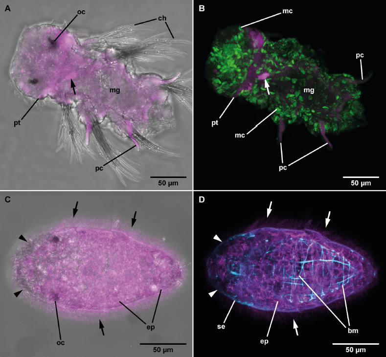
Observing the process of growth and differentiation of tissues and organs is of crucial importance for the understanding of the evolution of organs in animals. Unfortunately, it is notoriously difficult to continuously monitor developmental processes due to the extended time they take. Long-term labeling of the tissues of interest represents a promising alternative to raise these pivotal data. In the case of the prototroch, a band of ciliated cells typical of marine, planktotrophic trochophora larvae, we were able to apply a long-term fluorescent vital-staining to the prototroch cells that remains detectable throughout further larval life. We were able to stain ciliated cells of planktonic larvae from different spiralian clades by using long-chain dialkylcarbocyanine dyes that are detectable in different fluorescent emission spectra in combination with a non-ionic surfactant. The larvae survived and developed normally, their ciliated cells retaining the originally applied fluorescent labels. Combined with additional fluorescent staining of the larvae after fixation, we provide an easy, versatile, and broadly applicable method to investigate the processes of the differentiation of epidermal organs in various aquatic larvae.

Keywords: DiI; Spiralia; cLSM; fluorescent staining; prototroch.


Conflict of interest statement

Competing interests: The authors have declared that no competing interests exist.

Figures

Figure 1.
Figure 1.
Young larvae after staining with DiI (A) or DiI/Pluronic F-127 (B and C) or DiD/Pluronic F-127 (D), cLSM (magenta: DiI, yellow: anti α-tubulin, red: DiD). A. Platynereis dumerilii, 24-hours old larva, maximum projection (apical view): the ciliated prototroch cells (pt) are stained. Note the large yolk vesicles in the four gastrulated macromeres (asterisks). B. Mytilus edulis, 21-hours old larva, maximum projection (anterior to upper left): only epidermal cell (ep) with cilia (ci) show DiI signal, unciliated regions of the epidermis are unstained (asterisk). C. Cephalothrix oestrymnica, 2-day old larva, maximum projection of 5 middle sections (4.4 μm; anterior to upper left): only the epidermal cell (ep) layer including the apical tuft (arrows) is stained; cells of the midgut epithelium (mg) are unstained. Cells of the apical plate (ap) extend deeply into the larva (arrowhead). D. Thalassema thalassema, 44-hours old larva, maximum projection (anterior is up): fluorescent DiD signals detectable in the ciliated prototroch (pt) and in some ciliated cells of the apical plate (ap). Note the small, dot-shaped signal visible in an unciliated part of the larva (arrow).
Figure 2.
Figure 2.
Advanced larvae, some days after staining with DiI (A and B) or DiI/Pluronic F-127 (C and D), cLSM (magenta: DiI, green: EdU, cyan: phalloidin). A. Platynereis dumerilii, 4-day old larva, maximum projection (anterior to upper left corner): DiI signal is most prominent in the prototroch (pt), although some weak fluorescence is seen in the midgut (mg); the parapodial cirri (pc) next to the chaetae (ch) show autofluorescence due to glandular tissue. Note the strong, cell-shaped, internalized DiI signal (arrow). B. Same as in (A): Mitotically active cells (mc) are seen throughout the larval body, indicating ongoing development after DiI incubation. Note the strong, cell-shaped, internalized DiI signal (arrow). C. Cephalothrix oestrymnica, 8-day old larva (anterior to left side): The posterior part of the epidermis, up to the level of the larval ocelli (oc) show DiI signal, whereas the anterior-most part is unstained (arrowheads). Note the DiI signal visible in the cilia (arrows) of the primary epidermal cells (ep). D. Same as in (C): Phalloidin staining of F-actin reveals the body wall muscles (bm) and the outlines of the primary (ep) and secondary epidermis (se) cells. The primary epidermis shows DiI labelling, whereas the secondary epidermis is unstained (arrowheads). Note the DiI signal visible in the cilia (arrows) of the primary epidermis (ep).
Figure 3.
Figure 3.
Advanced larvae some days after staining with DiI/Pluronic (A-C, anterior to top right corner) or DiD/Pluronic (D), cLSM (magenta: DiI, yellow: anti α-tubulin, green: nuclei, red: DiD, cyan: phalloidin). A. Mytilus edulis, 6-day old larva: The most prominent DiI signals are seen in epidermis (ep) of the velum (ve) and along the ventral side, no signal is detectable on the dorsal side (arrow). The midgut (mg) is filled with unicellular planktonic algae. B. Same as in (A): DiI signal is prominent in the ciliated epidermis (ep) of the velum (ve) and along the ventral side encompassing the mouth (mo) and the anal opening (ao). No DiI signal is seen in the unciliated epidermis (arrow). The midgut (mg) is filled with algae that show intense chlorophyll autofluorescence (arrowhead). C. Same as in (A): The primary epidermis cells (ep) of the velum (ve) and on the ventral side show DiI signal. No DiI signal is detected in the midgut (mg) and the dorsal pallial epithelium (pe, arrow). D. Thalassema thalassema, 4-day old larva (anterior is up): The ciliated prototroch (pt) shows prominent DiD signal. Labeling of F-actin with phalloidin shows the prominent ring-muscle underneath the prototroch (pm), the buccal muscles (bu) and some additional circular (cm) and longitudinal muscle (lm) strands. Ingested unicellular planktonic algae in the midgut (mg) show an intense chlorophyll autofluorescence (arrowheads).

References

    1. Smart TI, Von Dassow G. (2009) Unusual development of the mitraria larva in the polychaete Owenia collaris. Biol Bull 217: 253-268. doi: 10.1086/BBLv217n3p253. PMID: - DOI - PubMed
    1. Martín-Durán JM, Passamaneck YJ, Martindale MQ, Hejnol A. (2016) The developmental basis for the recurrent evolution of deuterostomy and protostomy. Nat Ecol Evol 1: 5. doi: 10.1038/s41559-016-0005. PMID: - DOI - PubMed
    1. Hejnol A, Schnabel R. (2006) What a couple of dimensions can do for you: Comparative developmental studies using 4D microscopy--examples from tardigrade development. Integr Comp Biol 46: 151-161. doi: 10.1093/icb/icj012. PMID: - DOI - PubMed
    1. Lyons DC, Perry KJ, Henry JQ. (2017) Morphogenesis along the animal-vegetal axis: fates of primary quartet micromere daughters in the gastropod Crepidula fornicata. BMC Evol Biol 17: 217. doi: 10.1186/s12862-017-1057-1. PMID: - DOI - PMC - PubMed
    1. Stach T, Winter J, Bouquet J, Chourrout D, Schnabel R. (2008) Embryology of a planktonic tunicate reveals traces of sessility. Proc Natl Acad Sci USA 105: 7229-7234. doi: 10.1073/pnas.0710196105. PMID: - DOI - PMC - PubMed

LinkOut - more resources