. 2020 Jun 10;6(24):eaba8399.
doi: 10.1126/sciadv.aba8399. eCollection 2020 Jun.

A single dose of ChAdOx1 MERS provides protective immunity in rhesus macaques

Affiliations

A single dose of ChAdOx1 MERS provides protective immunity in rhesus macaques

Neeltje van Doremalen et al. Sci Adv. .

Abstract

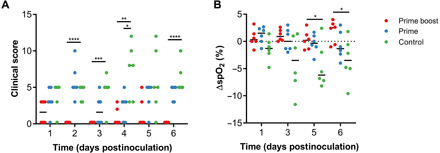
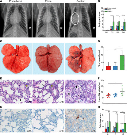
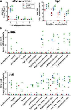
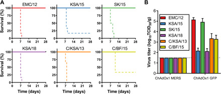
Developing a vaccine to protect against the lethal effects of the many strains of coronavirus is critical given the current global pandemic. For Middle East respiratory syndrome coronavirus (MERS-CoV), we show that rhesus macaques seroconverted rapidly after a single intramuscular vaccination with ChAdOx1 MERS. The vaccine protected against respiratory injury and pneumonia and reduced viral load in lung tissue by several orders of magnitude. MERS-CoV replication in type I and II pneumocytes of ChAdOx1 MERS-vaccinated animals was absent. A prime-boost regimen of ChAdOx1 MERS boosted antibody titers, and viral replication was completely absent from the respiratory tract tissue of these rhesus macaques. We also found that antibodies elicited by ChAdOx1 MERS in rhesus macaques neutralized six different MERS-CoV strains. Transgenic human dipeptidyl peptidase 4 mice vaccinated with ChAdOx1 MERS were completely protected against disease and lethality for all different MERS-CoV strains. The data support further clinical development of ChAdOx1 MERS.


Figures

Fig. 1
Fig. 1. Vaccination of rhesus macaques with ChAdOx1 MERS elicits a humoral immune response.
Serum samples were collected from NHPs at times of vaccination (−56 and −28 DPI), 14 days later, and at challenge. (A) Overview of experimental timeline. V-M, vaccination with ChAdOx1 MERS; V-G, vaccination with ChAdOx1 GFP; E, exam; C, challenge and exam; N, exam and necropsy. (B) Twofold serial diluted serum samples were tested for MERS-CoV S–specific antibodies using enzyme-linked immunosorbent assay (ELISA). (C) Twofold serial diluted serum samples were tested for neutralizing antibodies against MERS-CoV in VeroE6 cells. Line, geometric mean; dotted line, limit of detection (LoD). Statistical significance between −28 and −14 DPI in the prime-boost group was determined via one-tailed paired Student’s t test. Statistical significance between prime-boost and prime-only groups on 0 DPI was determined via two-tailed unpaired Student’s t test. **P < 0.01; ***P < 0.001.
Fig. 2
Fig. 2. Clinical scoring and spO2 values are improved in ChAdOx1 MERS–vaccinated animals compared to ChAdOx1 GFP–vaccinated animals.
(A) Animals were evaluated daily, and clinical score was assessed using an established scoring sheet. (B) Changes in oxygen saturation from pre-inoculation values [Δ% peripheral capillary oxygen saturation (spO2)] were determined on exam days. Statistical significance between groups was determined via two-tailed unpaired Student’s t test. Line, median; dotted line, baseline value; *P < 0.025; **P < 0.01; ***P < 0.001; ****P < 0.0001.
Fig. 3
Fig. 3. Single-dose vaccination with ChAdOx1 MERS protects rhesus macaques against bronchointerstitial pneumonia caused by MERS-CoV challenge.
Rhesus macaques were vaccinated with a prime-boost or prime-only regimen of ChAdOx1 MERS, or with ChAdOx1 GFP and challenged with MERS-CoV. (A) Ventrodorsal thoracic radiographs collected on 6 DPI. A marker (R) indicates the right side of animal. No pathologic changes were observed in animals vaccinated with ChAdOx1 MERS via a prime-boost or prime-only regimen. Animal vaccinated with ChAdOx1 GFP shows focally extensive area of increased pulmonary opacity and deviation of the cardiac silhouette, highlighted in the circle located in the middle and caudal lung lobes. (B) Thoracic radiographs of each animal were scored per lung lobe, resulting in a maximum score of 18. Values were averaged per group per day (D), and mean with SD is shown (see table S2 for more details). (C) Gross pathology of lungs shows no pathologic changes in ChAdOx1 MERS–vaccinated animals and focally extensive areas of consolidation in left cranial, middle, and caudal lung lobes in control animals (asterisks). (D) Gross lung lesions were scored for each lung lobe, ventral and dorsal. Values were averaged per group, and mean with SD is shown. (E) Lung tissue sections were stained with hematoxylin and eosin. Moderate numbers of lymphocyte accumulation around pulmonary arterioles (asterisks) and mild thickening of alveolar septae by lymphocytes and macrophages (arrows) in lung tissue of animals vaccinated with ChAdOx1 MERS. Marked bronchointerstitial pneumonia with abundant pulmonary edema and fibrin (asterisks), type II pneumocyte hyperplasia (arrows), and increased numbers of alveolar macrophages (arrowheads) in lung tissue of control animals. Magnification, ×200. (F) Lung–to–body weight (BW) ratio was determined for all animals at necropsy. Mean with SD is shown. (G) Lung tissue sections were stained with antibody against MERS-CoV antigen, which is visible as a red-brown staining. No immunoreactivity was found in ChAdOx1 MERS–vaccinated animals, whereas multifocal immunoreactivity of type I and II pneumocytes could be found in lung tissue of ChAdOx1 GFP–vaccinated animals. (H) Lung tissue sections were scored on severity of lesions (0, no lesions; 1, 1 to 10%; 2, 11 to 25%; 3, 26 to 50%; 4, 51 to 75%; and 5, 76 to 100%) and averaged per group. Mean with SD is shown. A, bronchointerstitial pneumonia; B, type II pneumocyte hyperplasia; C, hemorrhages, edema, and fibrin deposits. Statistical significance between groups was determined via two-tailed unpaired Student’s t test. *P < 0.025; **P < 0.01; ***P < 0.001; ****P < 0.0001. Photo credit: Neeltje van Doremalen, NIAID/NIH.
Fig. 4
Fig. 4. Vaccination with ChAdOx1 MERS results in reduced virus replication in the respiratory tract.
(A) Infectious virus titers and viral load were determined in BAL fluid. Individual values are depicted. (B) UpE and mRNA copies were determined in respiratory tract tissues collected at 6 DPI. Individual values are depicted. Line, geometric mean; dotted line, limit of detection.
Fig. 5
Fig. 5. ChAdOx1 MERS provides cross-protection against different MERS-CoV strains in the mouse model.
(A) Survival curves of ChAdOx1 MERS–vaccinated (solid line) and ChAdOx1 GFP–vaccinated (dashed line) hDPP4 mice challenged with MERS-CoV. (B) Infectious virus titers in lung tissue collected at 3 DPI from hDPP4 mice challenged with MERS-CoV. Mean titer with SD is shown.

References

    1. de Wit E., van Doremalen N., Falzarano D., Munster V. J., SARS and MERS: Recent insights into emerging coronaviruses. Nat. Rev. Microbiol. 14, 523–534 (2016). - PMC - PubMed
    1. WHO, Middle East Respiratory Syndrome (2019); www.emro.who.int/health-topics/mers-cov/mers-outbreaks.html.
    1. WHO, (2020); https://www.who.int/docs/default-source/coronaviruse/situation-reports/2....
    1. Bin S. Y., Heo J. Y., Song M.-S., Lee J., Kim E.-H., Park S.-J., Kwon H.-i., Kim S. M., Kim Y.-i., Si Y.-J., Lee I.-W., Baek Y. H., Choi W.-S., Min J., Jeong H. W., Choi Y. K., Environmental contamination and viral shedding in MERS patients during MERS-CoV outbreak in south korea. Clin. Infect. Dis. 62, 755–760 (2016). - PMC - PubMed
    1. Kim K. H., Tandi T. E., Choi J. W., Moon J. M., Kim M. S., Middle East respiratory syndrome coronavirus (MERS-CoV) outbreak in South Korea, 2015: Epidemiology, characteristics and public health implications. J. Hosp. Infect. 95, 207–213 (2017). - PMC - PubMed

Publication types

MeSH terms