. 2019 Aug 23;2(8):4773-4781.
doi: 10.1021/acsanm.9b00606. Epub 2019 Jun 19.

Entropic Trapping of DNA with a Nanofiltered Nanopore

Affiliations

Entropic Trapping of DNA with a Nanofiltered Nanopore

Michelle H Lam et al. ACS Appl Nano Mater. .

Abstract

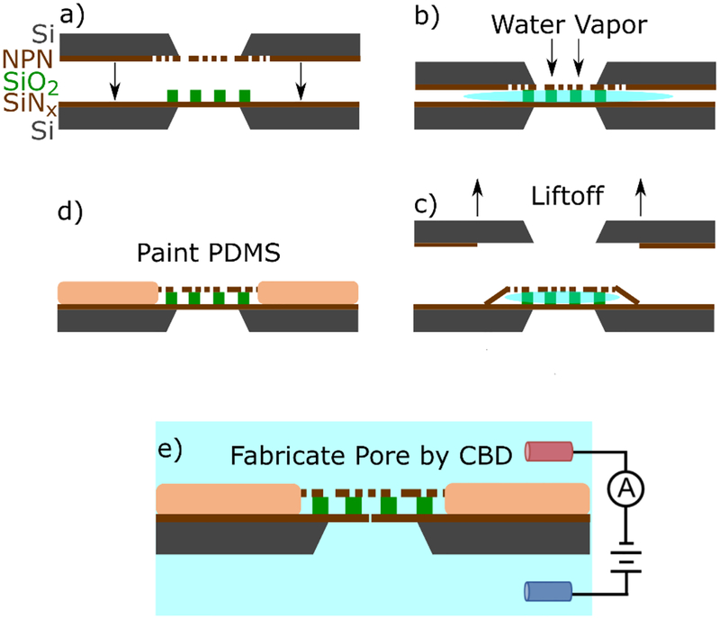
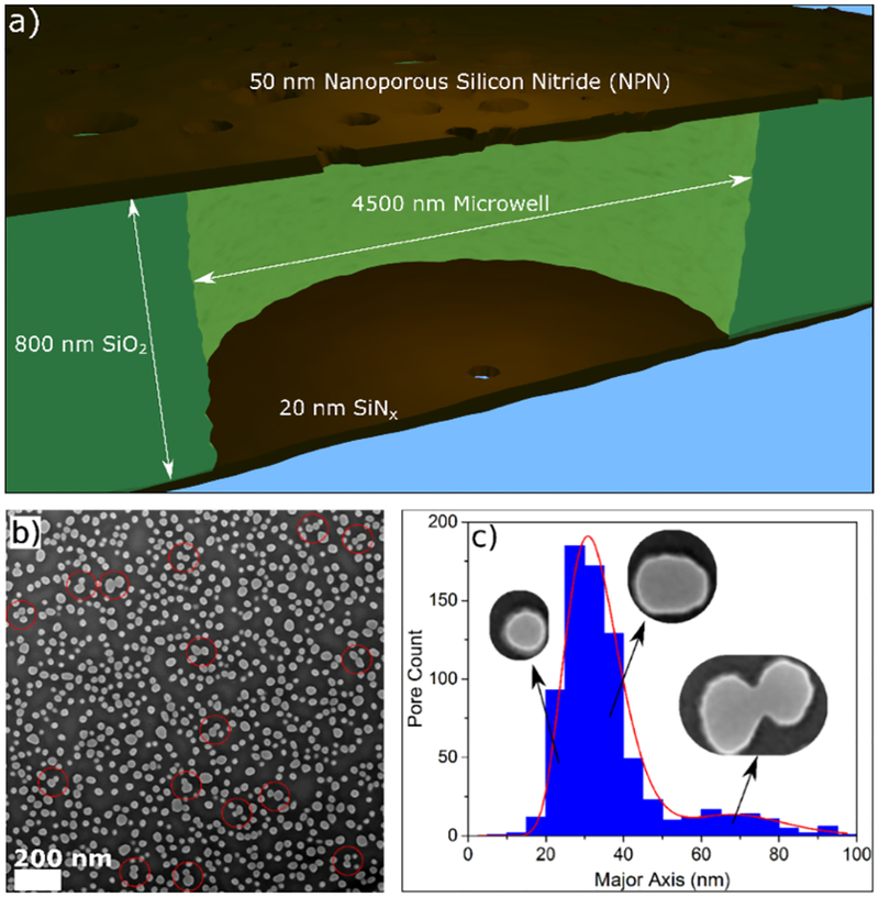
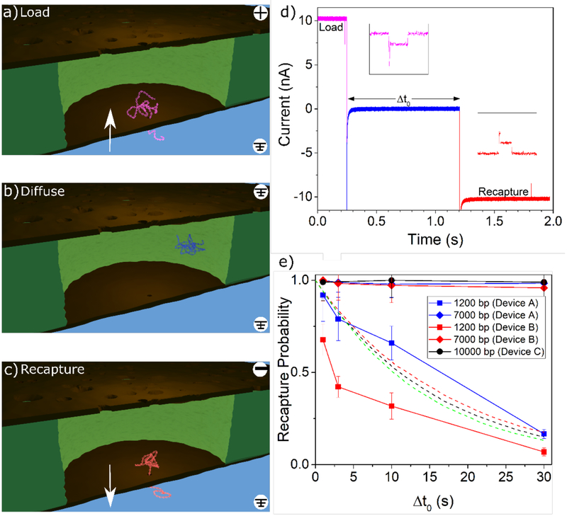
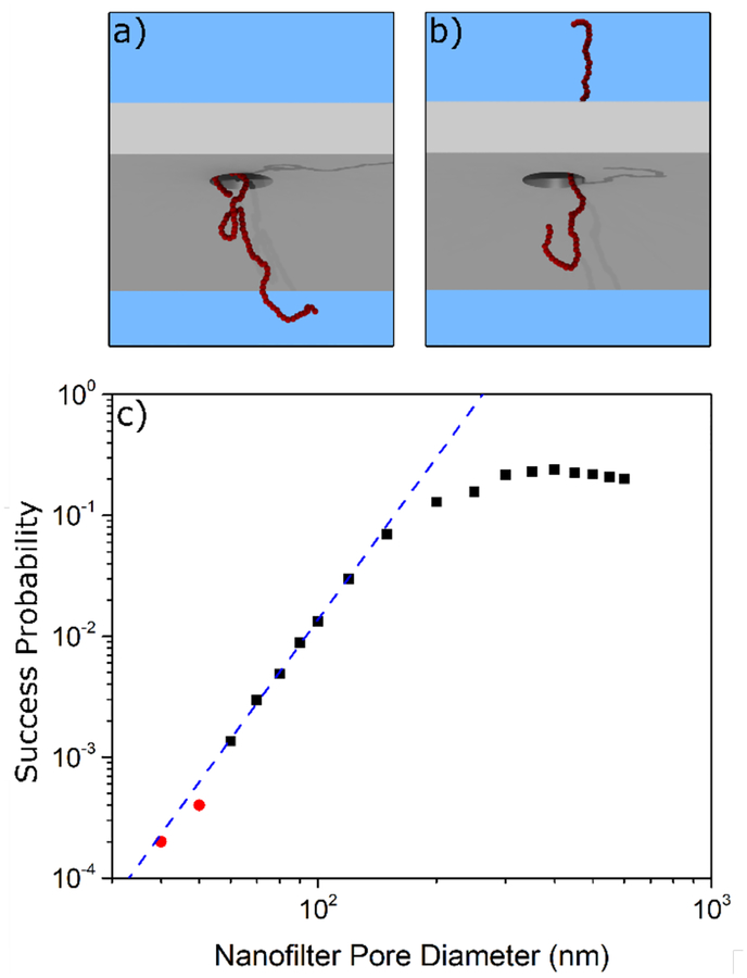
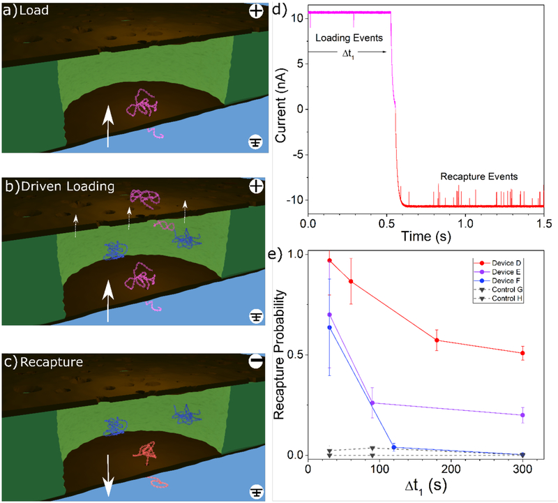
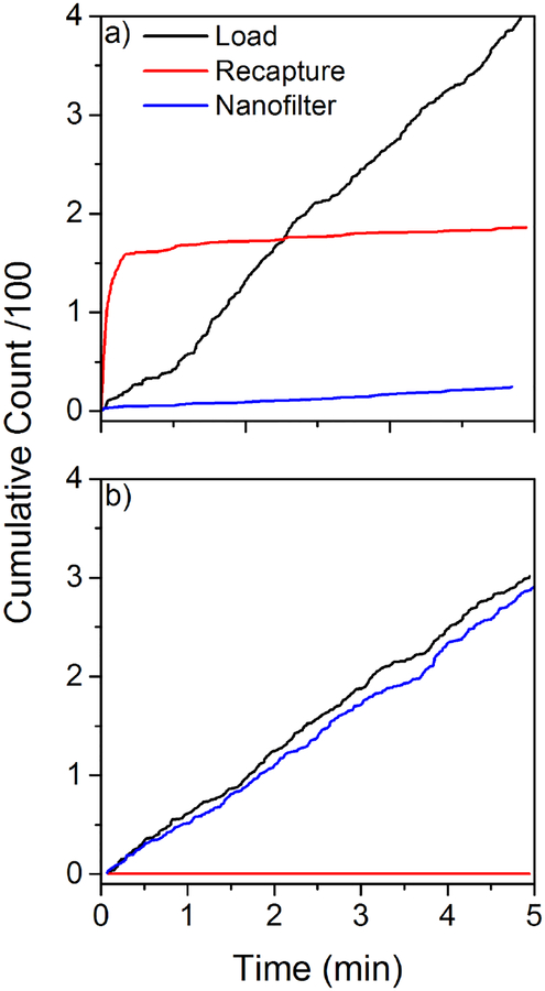
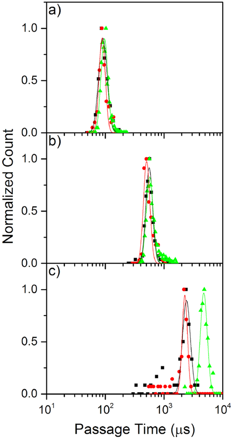
Elucidating the kinetics of DNA passage through a solid-state nanopore is a fertile field of research, and mechanisms for controlling capture, passage, and trapping of biopolymers are likely to find numerous technological applications. Here we present a nanofiltered nanopore device, which forms an entropic cage for DNA following first passage through the nanopore, trapping the translocated DNA and permitting recapture for subsequent reanalysis and investigation of kinetics of passage under confinement. We characterize the trapping properties of this nanodevice by driving individual DNA polymers into the nanoscale gap separating the nanofilter and the pore, forming an entropic cage similar to a "two pores in series" device, leaving polymers to diffuse in the cage for various time lengths, and attempting to recapture the same molecule. We show that the cage results in effectively permanent trapping when the radius of gyration of the target polymer is significantly larger than the radii of the pores in the nanofilter. We also compare translocation dynamics as a function of translocation direction in order to study the effects of confinement on DNA just prior to translocation, providing further insight into the nanopore translocation process. This nanofiltered nanopore device realizes simple fabrication of a femtoliter nanoreactor in which to study fundamental biophysics and biomolecular reactions on the single-molecule level. The device provides an electrically-permeable single-molecule trap with a higher entropic barrier to escape than previous attempts to fabricate similar structures.

Keywords: DNA; entropy; nanoconfinement; nanofabrication; nanopore; nanoporous membrane; nanotechnology.


Conflict of interest statement

Conflicts of Interest KB, GRM, JLM, and VTC declare competing financial interest in the form of a patent on the nanofiltered nanopore device. JLM is a cofounder of SiMPore Inc. All other authors declare no competing financial interest.

Figures

Figure 1:
Figure 1:
A schematic illustrating of the assembly of the entropic trap. a) two chips containing an intact 20 nm thick SiNx sensing membrane decorated with an 800 nm SiO2 spacer containing a hexagonal grid of 4.5 μm microwells is brought into close proximity with a NPN membrane. b) water vapour floods the cavity and provides a weak adhesion force between the two membranes. c) the NPN support chip is lifted off, leaving behind the NPN layer capping the trapping cavities. d) Polydimethylsiloxane (PDMS) is painted around dual membrane stack, and over some of the interface to reduce device capacitance and permanently bond the two membranes. e) A nanopore is formed in one of the cavities at random using controlled breakdown.
Figure 2:
Figure 2:
a) Schematic of a cross-section of a single microwell in the device geometry, not to scale. The 50 nm thick NPN nanofilter, containing pores with an average diameter of 31 ± 9 nm, is separated from a 20 nm thick SiNx membrane by an 800 nm SiO2 spacer containing an array of 4500 nm diameter wells, one of which contains the sensing pore. b) SEM image of the nanofilter membrane, showing a random distribution of pores. The scale bar is 200 nm. Note that in several places, indicated with red circles, neighbouring pores overlap, resulting in a single large oblong merged pore. c) Distribution of pore sizes as measured by their major axis (not average diameter, which ins plotted in Supporting Information Section S2). The data is well fit by two log-normal distributions. The corresponding representative pores in the insets are taken from (b).
Figure 3:
Figure 3:
The diffusive trapping mode. a) DNA is captured by the sensing pore and pulled upwards into the cavity in between the two membranes. b) In the diffusive trapping mode, immediately following capture, the field is turned off, and the captured DNA is allowed to freely diffuse around the cavity under no applied voltage. c) The voltage is reversed and the DNA, if still trapped in the cavity, is recaptured by the sensing pore. Renders are for conceptual illustration only; geometry and DNA are not to scale. d) A representative current trace showing a single capture-recapture event in the diffusive trapping mode, with a 1s delay. Additional events are shown in Supporting Information Section S3. e) Experimental recapture probability trends for varying delay times in the diffusive trapping mode (solid lines). Three different devices (indicated by marker colour) were used for these diffusive experiments. Squares correspond to 1.2 kbp, diamonds to 7 kbp, and circles to 10 kbp. The sensing pores had diameters of 9.5 nm (Device A: 577 loading events for 1.2 kbp, 727 loading events for 7 kbp), 7.5 nm (Device B: 430 loading events for 1.2 kbp, 426 loading events for 7 kbp), and 11.5 nm (Device C: 305 loading events for 10 kbp). Dashed lines show simulation results for 1.2 kbp DNA in devices whose filters contained 40, 75, and 160 nanofilter pores with diameters of 90 nm (green), 80 nm (red), and 70 nm (black), respectively. Error bars are estimated using simple Poisson statistics.
Figure 4:
Figure 4:
a) A snapshot of a 1.2 kbp (with Rg ~ 55 nm) equivalent DNA polymer during a simulated escape attempt by diffusion through the nanofilter. b) The same polymer at a later moment in time as it crosses the membrane. c) The simulated probability that the polymer will successfully cross the membrane before diffusing away from the filter.
Figure 5:
Figure 5:
a) DNA is captured by the sensing pore and pulled into the cavity in between the two membranes. b) In the “driven trapping” mode, other DNA molecules can continue to be captured into the cavity though the sensing pore and can also escape through the nanofilter under the influence an applied voltage. c) The voltage is reversed and any DNA trapped in the cavity is recaptured by the sensing pore. Renders are for conceptual illustration only; geometry and DNA are not to scale. d) A representative current trace showing the transitional region of a driven trapping experiment for an applied voltage of ±200 mV for device D. e) Recapture probability for increasing loading durations in the driven trapping configuration. 7 kbp dsDNA was used for all driven trapping experiments. Three nanofiltered nanopores (circles) and two standard control nanopores (inverted triangles) were used. In alphabetical order, sensing pores had diameters of 10.0 nm (736 loading events), 7.8 nm (90 loading events), 8.1 nm (410 loading events), 7.9 nm (790 loading events), and 8.6 nm (577 loading events). Error bars are estimated using simple Poisson statistics.
Figure 6:
Figure 6:
Cumulative event counts in the three translocation modes (load/back: capture by the sensing pore into the cavity; recapture/red: capture by the sensing pore from the cavity; nanofilter/blue: capture by the sensing pore through the nanofilter) for two devices during 5 minutes loading experiments in the driven trapping mode at 200 mV, showing the extremes of the possible trapping behaviors with 7 kbp dsDNA. a) Cumulative event counts for device D, which is a very efficient trap. b) Cumulative event counts for device F, which is not an efficient trap.
Figure 7:
Figure 7:
Passage times for loading (black squares), recapture (red circles), and nanofilter capture (green triangles) experiments. a) Histograms of passage times of the three capture modes for 1.2 kbp dsDNA capture by a 6.9 nm diameter sensing pore (240 loading events, 89 recapture events, and 4549 nanofilter events). b) Corresponding histograms of passage times for 7 kbp dsDNA using the same pore as (a) (139 loading events, 139 recapture events, and 1276 nanofilter events). c) Corresponding histograms of passage times for 10kbp dsDNA using a 7.2 nm diameter sensing pore (90 loading events, 88 recapture events, and 78 nanofilter events). Only passage times for unfolded, single-level translocation events are included. All experiments are performed in 3.6 M LiCl pH 8 at 200 mV.

References

    1. Fologea D; Gershow M; Ledden B; McNabb DS; Golovchenko JA; Li J Detecting Single Stranded DNA with a Solid State Nanopore. Nano Lett 2005, 5 (10), 1905–1909. 10.1021/nl051199m. - DOI - PMC - PubMed
    1. Derrington IM; Butler TZ; Collins MD; Manrao E; Pavlenok M; Niederweis M; Gundlach JH Nanopore DNA Sequencing with MspA. Proc. Natl. Acad. Sci. U. S. A 2010, 107 (37), 16060–16065. 10.1073/pnas.1001831107. - DOI - PMC - PubMed
    1. Clarke J; Wu H-C; Jayasinghe L; Patel A; Reid S; Bayley H Continuous Base Identification for Single-Molecule Nanopore DNA Sequencing. Nat. Nanotechnol 2009, 4 (4), 265–270. 10.1038/nnano.2009.12. - DOI - PubMed
    1. Mathé J; Visram H; Viasnoff V; Rabin Y; Meller A Nanopore Unzipping of Individual DNA Hairpin Molecules. Biophys. J 2004, 87 (5), 3205–3212. - PMC - PubMed
    1. Bell NAW; Chen K; Ghosal S; Ricci M; Keyser UF Asymmetric Dynamics of DNA Entering and Exiting a Strongly Confining Nanopore. Nat. Commun 2017, 8 (1), 380 10.1038/s41467-017-00423-9. - DOI - PMC - PubMed

LinkOut - more resources