[Preprint]. 2020 Jun 9:2020.06.08.141267.
doi: 10.1101/2020.06.08.141267.

Structural basis of a public antibody response to SARS-CoV-2

Affiliations

Structural basis of a public antibody response to SARS-CoV-2

Meng Yuan et al. bioRxiv. .

Update in

  • Structural basis of a shared antibody response to SARS-CoV-2.
    Yuan M, Liu H, Wu NC, Lee CD, Zhu X, Zhao F, Huang D, Yu W, Hua Y, Tien H, Rogers TF, Landais E, Sok D, Jardine JG, Burton DR, Wilson IA. Yuan M, et al. Science. 2020 Aug 28;369(6507):1119-1123. doi: 10.1126/science.abd2321. Epub 2020 Jul 13. Science. 2020. PMID: 32661058 Free PMC article.

Abstract

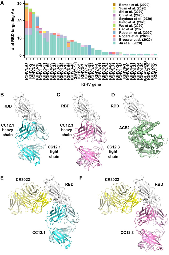
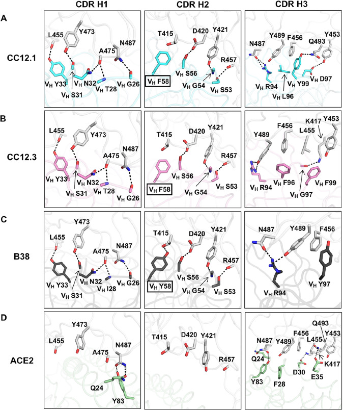
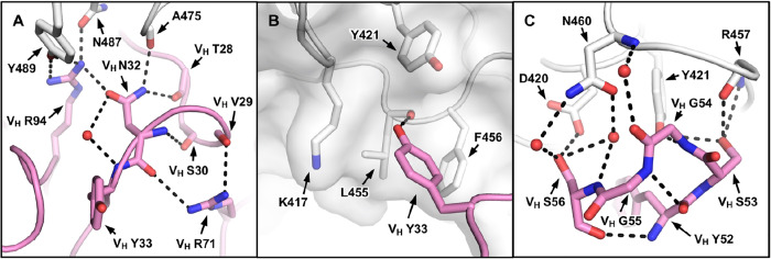
Molecular-level understanding of human neutralizing antibody responses to SARS-CoV-2 could accelerate vaccine design and facilitate drug discovery. We analyzed 294 SARS-CoV-2 antibodies and found that IGHV3-53 is the most frequently used IGHV gene for targeting the receptor binding domain (RBD) of the spike (S) protein. We determined crystal structures of two IGHV3-53 neutralizing antibodies +/- Fab CR3022 ranging from 2.33 to 3.11 Å resolution. The germline-encoded residues of IGHV3-53 dominate binding to the ACE2 binding site epitope with no overlap with the CR3022 epitope. Moreover, IGHV3-53 is used in combination with a very short CDR H3 and different light chains. Overall, IGHV3-53 represents a versatile public VH in neutralizing SARS-CoV-2 antibodies, where their specific germline features and minimal affinity maturation provide important insights for vaccine design and assessing outcomes.


Figures

Figure 1.
Figure 1.. Structures of two IGHV3–53 antibodies.
(A) The distribution of IGHV gene usage is shown for a total of 294 RBD-targeting antibodies (–28). (B-F) Crystal structures of (B) CC12.1 in complex with SARS-CoV-2 RBD, (C) CC12.3 with SARS-CoV-2 RBD, (D) human ACE2 with SARS-CoV-2 RBD (PDB 6M0J) (12), (E) SARS-CoV-2 RBD with CC12.1 and CR3022, and (F) SARS-CoV-2 RBD with CC12.3 and CR3022.
Figure 2.
Figure 2.. Epitopes of IGHV35–3 antibodies.
(A-C) Epitopes of (A) CC12.1, (B) CC12.3, and (C) B38 (PDB 7BZ5) (23). Epitope residues contacting the heavy chain are in orange and the light chain are in yellow. On the left panels, CDR loops are labeled. On the right panels, epitope residues are labeled. For clarity, only representative epitope residues are labeled. Epitope residues that are also involved in ACE2 binding are in red. (D) ACE2-binding residues are shown in blue. On the left panel, ACE2 is shown in green with in semi-transparent representation. On the right panel, ACE2-binding residues are labeled. A total of 16 residues are used for ACE2 binding (12), but only 13 are labeled here since the other three are at the back of the structure in this view and do not interact with the antibodies of interest. (E) Epitope residues for CC12.1, CC12.3, and B38 were identified by PISA (39) and annotated on the SARS-CoV-2 RBD sequence, which is aligned to the SARS-CoV RBD sequence with non-conserved residues highlighted. The 16 ACE2-binding residues were as described previously (12).
Figure 3.
Figure 3.. Interactions between the RBD and the heavy chain CDR loops.
(A-C) Highly similar interaction modes between SARS-CoV-2 RBD and the antibody CDR H1 and H2 loops, but not the H3 loop are observed for in (A) CC12.1, (B) CC12.3, and (C) B38 (PDB 7BZ5) (23). The RBD is in white and antibody residues are in cyan, pink, and dark gray, respectively. Oxygen atoms are in red, and nitrogen atoms in blue. Hydrogen bonds are represented by dashed lines. (D) The interaction between ACE2 (green) and residues of the RBD (PDB 6M0J) (12) that are shown in (A-C).
Figure 4.
Figure 4.. Two IGHV3–53 germline-encoded motifs.
(A) The extensive hydrogen bond network that involves VH N32 of the NY motif in CDR H1 is illustrated. (B) The hydrophobic cage interaction between the RBD and VH Y33 of the NY motif in CDR H1 is shown. (C) The hydrogen bond network that involves the SGGS motif in CDR H2 is highlighted. CC12.3 is shown because its structure is at higher resolution than CC12.1.
Figure 5.
Figure 5.. Constraints on CDR H3 length.
(A) The heavy and light chains of CC12.1 (cyan), as well as the RBD (white) are shown in surface representation, with CDR H3 (red) highlighted in cartoon representation. (B) Same as panel A, except that CC12.3 (pink) is shown. (C) The lengths of CDR H3 in RBD-targeting antibodies that were previously isolated (–28) are analyzed. The distribution of CDR H3 lengths in RBD-targeting IGHV3–53 antibodies and those in non-IGHV3–53-encoded antibodies are compared. A Mann-Whitney U test was performed to compute the p-value.

References

    1. Kissler S. M., Tedijanto C., Goldstein E., Grad Y. H., Lipsitch M., Projecting the transmission dynamics of SARS-CoV-2 through the postpandemic period. Science 10.1126/science.abb5793 (2020). - DOI - PMC - PubMed
    1. Lurie N., Saville M., Hatchett R., Halton J., Developing COVID-19 vaccines at pandemic speed. N Engl J Med 382, 1969–1973 (2020). - PubMed
    1. Andrews S. F., McDermott A. B., Shaping a universally broad antibody response to influenza amidst a variable immunoglobulin landscape. Curr Opin Immunol 53, 96–101 (2018). - PubMed
    1. Parameswaran P. et al., Convergent antibody signatures in human dengue. Cell Host Microbe 13, 691–700 (2013). - PMC - PubMed
    1. Pieper K. et al., Public antibodies to malaria antigens generated by two LAIR1 insertion modalities. Nature 548, 597–601 (2017). - PMC - PubMed

Publication types