[Preprint]. 2020 May 29:2020.05.28.121533.
doi: 10.1101/2020.05.28.121533.

Structures of human antibodies bound to SARS-CoV-2 spike reveal common epitopes and recurrent features of antibodies

Affiliations

Structures of human antibodies bound to SARS-CoV-2 spike reveal common epitopes and recurrent features of antibodies

Christopher O Barnes et al. bioRxiv. .

Update in

Abstract

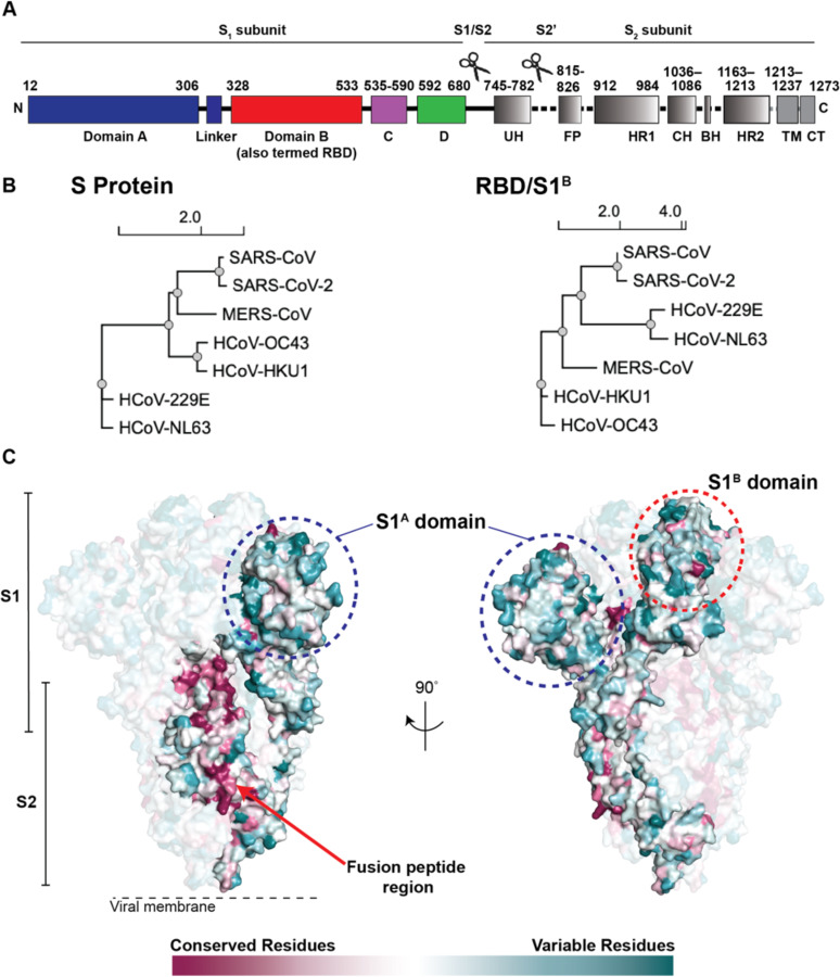
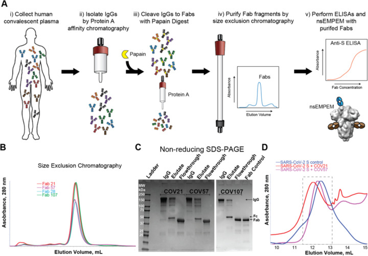
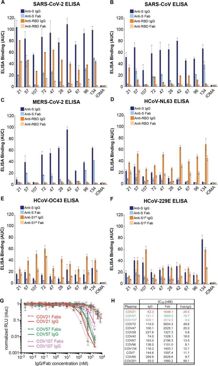
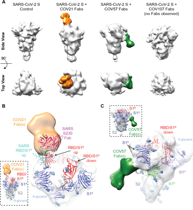
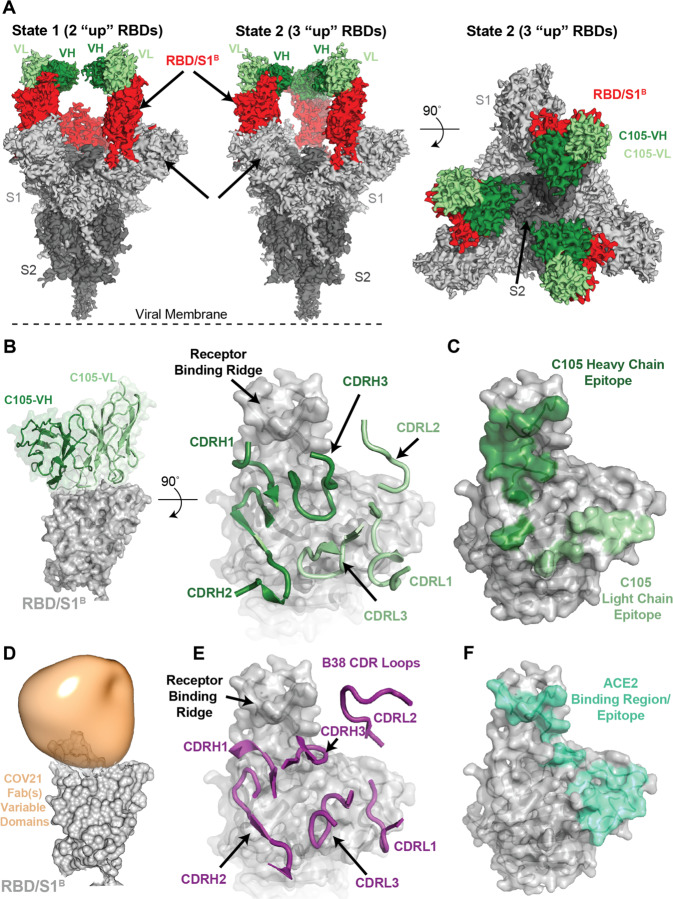
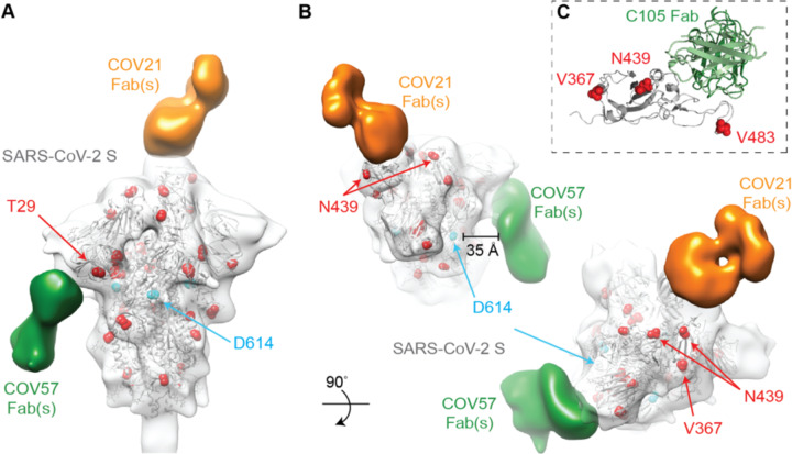
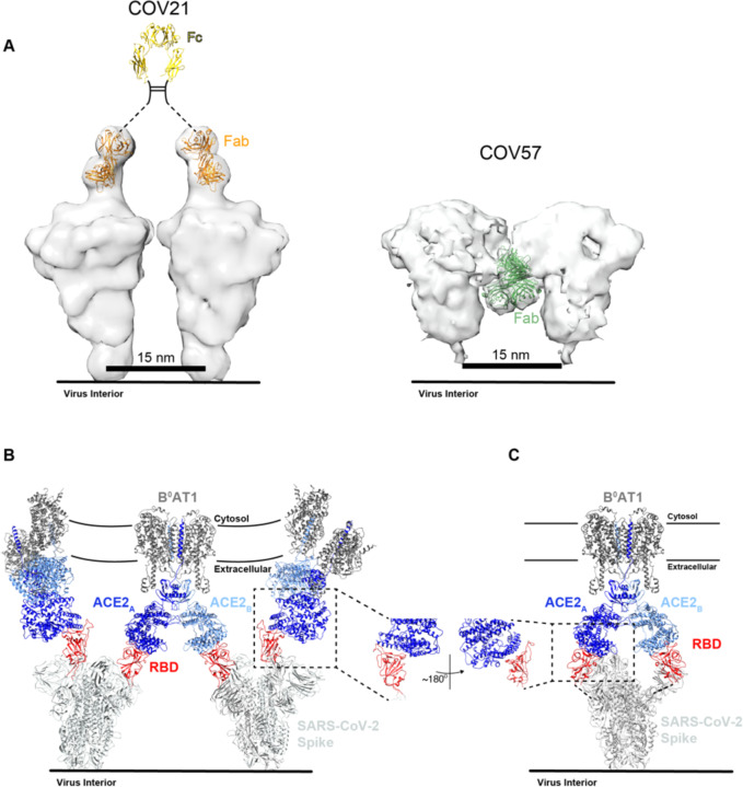
Neutralizing antibody responses to coronaviruses focus on the trimeric spike, with most against the receptor-binding domain (RBD). Here we characterized polyclonal IgGs and Fabs from COVID-19 convalescent individuals for recognition of coronavirus spikes. Plasma IgGs differed in their degree of focus on RBD epitopes, recognition of SARS-CoV, MERS-CoV, and mild coronaviruses, and how avidity effects contributed to increased binding/neutralization of IgGs over Fabs. Electron microscopy reconstructions of polyclonal plasma Fab-spike complexes showed recognition of both S1A and RBD epitopes. A 3.4Å cryo-EM structure of a neutralizing monoclonal Fab-S complex revealed an epitope that blocks ACE2 receptor-binding on "up" RBDs. Modeling suggested that IgGs targeting these sites have different potentials for inter-spike crosslinking on viruses and would not be greatly affected by identified SARS-CoV-2 spike mutations. These studies structurally define a recurrent anti-SARS-CoV-2 antibody class derived from VH3-53/VH3-66 and similarity to a SARS-CoV VH3-30 antibody, providing criteria for evaluating vaccine-elicited antibodies.

Keywords: COVID-19; Coronavirus; ELISA; Fab; IgG; MERS-CoV; SARS-CoV; SARS-CoV-2; convalescent plasma; electron microscopy.


Conflict of interest statement

Declaration of Interests The authors declare no competing interests.

Figures

Figure 1.
Figure 1.. Coronavirus S proteins show localized regions of conservation and variability.
(A) Schematic of SARS-CoV-2 S protein domain architecture. The S1 and S2 subunits are indicated, with scissors representing the locations of proteolytic cleavage sites required for S priming prior to fusion. UH = upstream helix, FP = fusion peptide, HR1 = heptad repeat 1, CH = central helix, BH = β-hairpin, HR2 = heptad repeat 2, TM = transmembrane region, CT = cytoplasmic tail. (B) Phylogenetic trees of selected coronaviruses based on protein sequences of S proteins and RBD/S1B domains. (C) Sequence conservation of 7 human coronaviruses plotted as a surface. The sequence alignment was generated using SARS-CoV-2 (GenBank MN985325.1), SARS-CoV (AAP13441.1), MERS-CoV (JX869059.2), HCoV-OC43 (AAT84362.1), HCoV-229E (AAK32191.1), HCoV-NL63 (AAS58177.1), and HCoV-HKU1 (Q0ZME7.1). Conservation was calculated by ConSurf Database (Landau et al., 2005) and displayed using a surface representation of the structure of the SARS-CoV-2 S protein (PDB code 6VXX).
Figure 2.
Figure 2.. Plasma Fabs bind to SARS-CoV-2 S protein.
(A) Schematic of polyclonal IgG and Fab purification from human plasma for nsEMPEM protocol. (B) SEC profile of Fabs (left) and SDS-PAGE of purified IgGs and Fabs (right) from COV21, COV57, and COV107 plasma samples. (C) SEC demonstration that plasma-derived Fabs from COV21 and COV57 shift the SARS-CoV-2 S protein trimer to a higher apparent molecular weight. No shift was observed when Fabs from COV107 were analyzed by SEC with S protein (data not shown). Fractions pooled and concentrated for nsEMPEM are boxed. See also Figure S1.
Figure 3.
Figure 3.. Convalescent plasma IgG and Fab binding properties demonstrate recognition of diverse coronaviruses and effects of avidity.
Results from ELISAs assessing binding of IgGs and Fabs purified from plasmas from 10 COVID-19 individuals (X-axis) presented as area under the curve (AUC; shown as mean ± S.D. of values derived from experiments conducted in triplicate). Binding was assessed against S and RBD proteins for SARS-CoV-2 (A), SARS-CoV (B), MERS-CoV (C), HCoV-NL63 (D), HCoV-OC43 (E), and HCoV-229E (F). Polyclonal IgGs or Fabs were evaluated at a top concentration of 50 μg/mL and 7 additional 4-fold serial dilutions. Binding of the IgG and Fab from IOMA, an antibody against HIV-1 (Gristick et al., 2016), was used as a control in each assay. (G) In vitro neutralization assays comparing the potencies of purified plasma IgGs and purified plasma Fabs. COV21, COV57, and COV107 plasma Fabs and IgGs are highlighted in the indicated colors; curves for 10 other plasmas (listed in panel H) are gray. (H) Molar IC50 values for purified plasma IgGs and Fabs for the indicated plasmas are listed with the molar ratio for IC50 (Fab) to IC50 (IgG) shown in the right column. See also Figure S2, S3.
Figure 4.
Figure 4.. EM reveals distinct predominant epitopes targeted by convalescent plasma antibodies.
(A) Side and top views for representative 3D reconstructions of four nsEMPEM datasets (S protein alone, S + COV21 Fabs, S + COV57 Fabs, S + COV107 Fabs). Bound Fabs observed in reconstructions from COV21 and COV57 plasmas are highlighted with false coloring as orange and green, respectively. No Fabs were observed in the reconstruction of COV107 Fabs plus S protein. Refined 3D models for SARS-CoV-2 S trimer-polyclonal Fab complexes from COV21 (panel B), and COV57 (panel C) were rigid-body fit with reference structures in Chimera (Goddard et al., 2007; Pettersen et al., 2004), displayed as cartoons (S1A: blue, S1B: red, S2: gray). (B) For COV21, the volume was best-fitted with PDB 6VYB (SARS-CoV-2, one “up” S1B conformation, inset). Overlay of PDB 6NB6 showed similarities in S1B epitope targeting of COV21 Fab (orange) and the human SARS-CoV neutralizing antibody, S230 (magenta, cartoon). (C) COV57 was fitted with PDB 6VXX (closed, prefusion conformation, inset). Fab density (green) was focused on the S1A domain. See also Figure S4.
Figure 5.
Figure 5.. A cryo-EM structure of a monoclonal Fab-S protein complex resembles the COV21 Fab(s)-S reconstruction.
(A) Reconstructed volumes for mAb C105 bound to SARS-CoV-2 S trimers in state 1 (two “up” RBDs, two bound Fabs) and state 2 (three “up” RBDs, three bound Fabs). (B) Left: Cartoon representation of VH-VL domains of C105 bound to an RBD. Right: CDR loops of C105 overlaid on surface representation of the RBD (shown as a gray surface). (C) RBD surface showing contacts by C105 VH-VL (contacts defined as an RBD residue within 7 Å of a VH or VL residue Cα atom). (D) RBD surface fitted with volume representing the variable domains of the COV21 Fab(s) nsEMPEM reconstruction. (E) CDR loops of B38 mAb overlaid on surface representation of the RBD (from PDB code 7BZ5). (F) RBD surface showing contacts by ACE2 (contacts defined as an RBD residue within 7 Å of an ACE2 residue Cα atom) (from PDB code 6VW1). See also Figure S5, S6.
Figure 6.
Figure 6.. Identified S mutations are unlikely to affect epitopes revealed by nsEMPEM and single-particle cryo-EM.
(A,B) Refined 3D model of SARS-CoV-2 S trimer alone was fitted with a reference structure (PDB 6VYB; gray cartoon) with locations of mutations observed in circulating SARS-CoV-2 isolates (Table S3) highlighted (red spheres). Residues affected by mutations that are disordered in the SARS-CoV-2 S structure (V483A) or in regions that are not included in the S ectodomain (signal sequence or cytoplasmic tail) are not shown. Densities corresponding to Fabs were separated, colored, and displayed on the same 3D volume. (C) C105-RBD interaction from the cryo-EM structure of the C105-S complex (Figure 5) showing locations of RBD mutations. V483 is ordered in this structure. See also Tables S3 and S4.
Figure 7.
Figure 7.. S protein epitopes offer different possibilities for avidity effects during IgG and receptor binding.
(A) Left: Model of two adjacent S trimers separated by ~15 nm, as seen on coronaviruses by cryo-electron tomography (Neuman et al., 2011), demonstrating that the orientation of COV21 Fab(s) on S could accommodate inter-spike crosslinking by a single IgG. The Fc portion of the IgG (PDB code 1IGT) was modeled assuming flexibility between the Fabs and the Fc (Sandin et al., 2004) and with the hinge region indicated by a dotted line since it is disordered in crystal structures of intact IgGs (Harris et al., 1992; Harris et al., 1998; Saphire et al., 2001). Right: Example of a model of two adjacent S trimers with bound Fab(s) in the orientation observed in the COV57 Fab(s)-S reconstruction demonstrating that inter-spike crosslinking is unlikely due to the “downward” orientation of the Fab(s), which does not permit linking by an Fc region, and predicted steric clashes between adjacent Fabs. Inter-spike crosslinking is also not possible for other orientations of two adjacent COV57 Fab(s)-S complexes (not shown). (B) Model of S trimers with two RBDs in an “up” position based on a cryo-EM structure of SARS-CoV S trimer (Kirchdoerfer et al., 2018) (PDB code 6CRX) interacting with full-length ACE2 receptors from the cryo-EM structure of soluble SARS-CoV-2 RBDs bound to the dimeric membrane form of ACE2 (Yan et al., 2020) (PDB code 6M17). Inter-spike crosslinking is possible if ACE2 dimers cluster in the membrane. (C) Model of intra-spike crosslinking between dimeric ACE2 and an S protein trimer with two RBDs in an “up” position. The RBDs were rotated by ~180° about their long axes to allow binding of the ACE2 ectodomains. Rotation of the RBD is a possibility since its position is flexible with respect to the remaining part of the S trimer (Walls et al., 2019). In this model, RBDs from a single S trimer could bind the ACE2 dimer in the same configuration as seen in the B°AT1-ACE2-SARS-CoV-2 RBD structure (PDB code 6M17).

References

    1. Adams P.D., Afonine P.V., Bunkoczi G., Chen V.B., Davis I.W., Echols N., Headd J.J., Hung L.W., Kapral G.J., Grosse-Kunstleve R.W., et al. (2010). PHENIX: a comprehensive Python-based system for macromolecular structure solution. Acta Crystallogr D Biol Crystallogr 66, 213–221. - PMC - PubMed
    1. Bell J.M., Chen M., Baldwin P.R., and Ludtke S.J. (2016). High resolution single particle refinement in EMAN2.1. Methods 100, 25–34. - PMC - PubMed
    1. Bianchi M., Turner H.L., Nogal B., Cottrell C.A., Oyen D., Pauthner M., Bastidas R., Nedellec R., McCoy L.E., Wilson I.A., et al. (2018). Electron-Microscopy-Based Epitope Mapping Defines Specificities of Polyclonal Antibodies Elicited during HIV-1 BG505 Envelope Trimer Immunization. Immunity 49, 288–300 e288. - PMC - PubMed
    1. Briney B., Inderbitzin A., Joyce C., and Burton D.R. (2019). Commonality despite exceptional diversity in the baseline human antibody repertoire. Nature 566, 393–397. - PMC - PubMed
    1. Brouwer P.J.M., Caniels T.G., van der Straten K., Snitselaar J.L., Aldon Y., Bangaru S., Torres J.L., Okba N.M.A., Claireaux M., Kerster G., et al. (2020). Potent neutralizing antibodies from COVID-19 patients define multiple targets of vulnerability. bioRxiv 10.1101/2020.05.12.088716 - DOI - PMC - PubMed

Publication types