This is a preprint.
Landscape and Selection of Vaccine Epitopes in SARS-CoV-2
- PMID: 32577654
- PMCID: PMC7302209
- DOI: 10.1101/2020.06.04.135004
Landscape and Selection of Vaccine Epitopes in SARS-CoV-2
Update in
-
Landscape and selection of vaccine epitopes in SARS-CoV-2.Genome Med. 2021 Jun 14;13(1):101. doi: 10.1186/s13073-021-00910-1. Genome Med. 2021. PMID: 34127050 Free PMC article.
Abstract
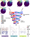
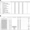
There is an urgent need for a vaccine with efficacy against SARS-CoV-2. We hypothesize that peptide vaccines containing epitope regions optimized for concurrent B cell, CD4+ T cell, and CD8+ T cell stimulation would drive both humoral and cellular immunity with high specificity, potentially avoiding undesired effects such as antibody-dependent enhancement (ADE). Additionally, such vaccines can be rapidly manufactured in a distributed manner. In this study, we combine computational prediction of T cell epitopes, recently published B cell epitope mapping studies, and epitope accessibility to select candidate peptide vaccines for SARS-CoV-2. We begin with an exploration of the space of possible T cell epitopes in SARS-CoV-2 with interrogation of predicted HLA-I and HLA-II ligands, overlap between predicted ligands, protein source, as well as concurrent human/murine coverage. Beyond MHC affinity, T cell vaccine candidates were further refined by predicted immunogenicity, viral source protein abundance, sequence conservation, coverage of high frequency HLA alleles and co-localization of CD4+ and CD8+ T cell epitopes. B cell epitope regions were chosen from linear epitope mapping studies of convalescent patient serum, followed by filtering to select regions with surface accessibility, high sequence conservation, spatial localization near functional domains of the spike glycoprotein, and avoidance of glycosylation sites. From 58 initial candidates, three B cell epitope regions were identified. By combining these B cell and T cell analyses, as well as a manufacturability heuristic, we propose a set of SARS-CoV-2 vaccine peptides for use in subsequent murine studies and clinical trials.
Conflict of interest statement
Conflict of Interest Statement: None
Figures


Similar articles
-
Landscape and selection of vaccine epitopes in SARS-CoV-2.Genome Med. 2021 Jun 14;13(1):101. doi: 10.1186/s13073-021-00910-1. Genome Med. 2021. PMID: 34127050 Free PMC article.
-
Immunoinformatics prediction of overlapping CD8+ T-cell, IFN-γ and IL-4 inducer CD4+ T-cell and linear B-cell epitopes based vaccines against COVID-19 (SARS-CoV-2).Vaccine. 2021 Feb 12;39(7):1111-1121. doi: 10.1016/j.vaccine.2021.01.003. Epub 2021 Jan 18. Vaccine. 2021. PMID: 33478794 Free PMC article.
-
COVID-19 coronavirus vaccine T cell epitope prediction analysis based on distributions of HLA class I loci (HLA-A, -B, -C) across global populations.Hum Vaccin Immunother. 2021 Apr 3;17(4):1097-1108. doi: 10.1080/21645515.2020.1823777. Epub 2020 Nov 11. Hum Vaccin Immunother. 2021. PMID: 33175614 Free PMC article.
-
Development of multi-epitope peptide-based vaccines against SARS-CoV-2.Biomed J. 2021 Mar;44(1):18-30. doi: 10.1016/j.bj.2020.09.005. Epub 2020 Oct 1. Biomed J. 2021. PMID: 33727051 Free PMC article. Review.
-
Identification of Novel Candidate Epitopes on SARS-CoV-2 Proteins for South America: A Review of HLA Frequencies by Country.Front Immunol. 2020 Sep 3;11:2008. doi: 10.3389/fimmu.2020.02008. eCollection 2020. Front Immunol. 2020. PMID: 33013857 Free PMC article. Review.
References
-
- With record-setting speed, vaccine makers take their first shots at the new coronavirus. Science | AAAS https://www.sciencemag.org/news/2020/03/record-setting-speed-vaccine-mak... (2020).
Publication types
Grants and funding
LinkOut - more resources
Full Text Sources
Other Literature Sources
Research Materials
Miscellaneous
