

The role of anterior prefrontal cortex (area 10) in face-to-face deception measured with fNIRS
- PMID: 32577765
- PMCID: PMC7812627
- DOI: 10.1093/scan/nsaa086
The role of anterior prefrontal cortex (area 10) in face-to-face deception measured with fNIRS
Abstract
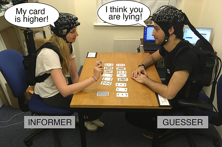
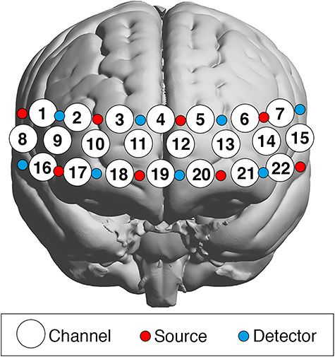
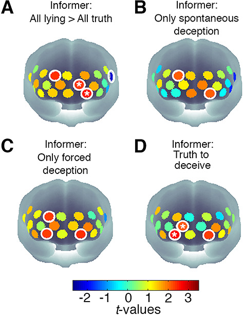
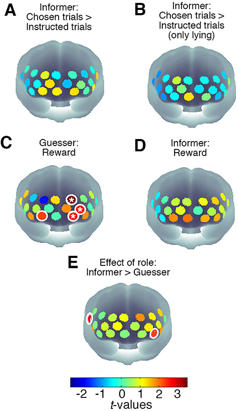
Anterior prefrontal cortex (PFC, Brodmann area 10) activations are often, but not always, found in neuroimaging studies investigating deception, and the precise role of this area remains unclear. To explore the role of the PFC in face-to-face deception, we invited pairs of participants to play a card game involving lying and lie detection while we used functional near infrared spectroscopy (fNIRS) to record brain activity in the PFC. Participants could win points for successfully lying about the value of their cards or for detecting lies. We contrasted patterns of brain activation when the participants either told the truth or lied, when they were either forced into this or did so voluntarily and when they either succeeded or failed to detect a lie. Activation in the anterior PFC was found in both lie production and detection, unrelated to reward. Analysis of cross-brain activation patterns between participants identified areas of the PFC where the lead player's brain activity synchronized their partner's later brain activity. These results suggest that during situations that involve close interpersonal interaction, the anterior PFC supports processing widely involved in deception, possibly relating to the demands of monitoring one's own and other people's behaviour.
Keywords: anterior prefrontal cortex; deception; fNIRS; face-to-face social interactions; hyperscanning.
© The Author(s) 2020. Published by Oxford University Press.
Figures
Similar articles
-
Gender difference in spontaneous deception: A hyperscanning study using functional near-infrared spectroscopy.Sci Rep. 2017 Aug 8;7(1):7508. doi: 10.1038/s41598-017-06764-1. Sci Rep. 2017. PMID: 28790399 Free PMC article.
-
Functional near-infrared spectroscopy to investigate hemodynamic responses to deception in the prefrontal cortex.Brain Res. 2009 Dec 15;1303:120-30. doi: 10.1016/j.brainres.2009.09.085. Epub 2009 Sep 25. Brain Res. 2009. PMID: 19782657
-
Neural correlates of spontaneous deception: A functional near-infrared spectroscopy (fNIRS)study.Neuropsychologia. 2013 Mar;51(4):704-12. doi: 10.1016/j.neuropsychologia.2012.12.018. Epub 2013 Jan 20. Neuropsychologia. 2013. PMID: 23340482 Free PMC article.
-
Cooperative Behavior Evokes Interbrain Synchrony in the Prefrontal and Temporoparietal Cortex: A Systematic Review and Meta-Analysis of fNIRS Hyperscanning Studies.eNeuro. 2022 Apr 13;9(2):ENEURO.0268-21.2022. doi: 10.1523/ENEURO.0268-21.2022. Print 2022 Mar-Apr. eNeuro. 2022. PMID: 35365502 Free PMC article.
-
What Guides Us to Neurally and Behaviorally Align With Anyone Specific? A Neurobiological Model Based on fNIRS Hyperscanning Studies.Neuroscientist. 2020 Apr;26(2):108-116. doi: 10.1177/1073858419861912. Epub 2019 Jul 11. Neuroscientist. 2020. PMID: 31296135 Review.
Cited by
-
Being 'in sync'-is interactional synchrony the key to understanding the social brain?Soc Cogn Affect Neurosci. 2021 Jan 18;16(1-2):1-4. doi: 10.1093/scan/nsaa148. Soc Cogn Affect Neurosci. 2021. PMID: 33104804 Free PMC article.
-
Novel Approaches and Cognitive Neuroscience Perspectives on False Memory and Deception.Front Psychol. 2022 Mar 21;13:721961. doi: 10.3389/fpsyg.2022.721961. eCollection 2022. Front Psychol. 2022. PMID: 35386904 Free PMC article.
-
Increased Interpersonal Brain Synchronization in Romantic Couples Is Associated with Higher Honesty: An fNIRS Hyperscanning Study.Brain Sci. 2023 May 21;13(5):833. doi: 10.3390/brainsci13050833. Brain Sci. 2023. PMID: 37239304 Free PMC article.
-
Prefrontal cortical activation associated with prospective memory while walking around a real-world street environment.Neuroimage. 2022 Sep;258:119392. doi: 10.1016/j.neuroimage.2022.119392. Epub 2022 Jun 15. Neuroimage. 2022. PMID: 35714887 Free PMC article.
-
How choice and motor mimicry affect affiliation: An fNIRS study.Imaging Neurosci (Camb). 2025 Jun 16;3:IMAG.a.40. doi: 10.1162/IMAG.a.40. eCollection 2025. Imaging Neurosci (Camb). 2025. PMID: 40800929 Free PMC article.
References
-
- Abe N., Suzuki M., Tsukiura T., et al. (2006). Dissociable roles of prefrontal and anterior cingulate cortices in deception. Cerebral Cortex, 16(2), 192–9. - PubMed
-
- Abe N., Suzuki M., Mori E., Itoh M., Fujii T. (2007). Deceiving others: distinct neural responses of the prefrontal cortex and amygdala in simple fabrication and deception with social interactions. Journal of Cognitive Neuroscience, 19(2), 287–95. - PubMed
-
- Abe N., Fujii T., Ito A., et al. (2014). The neural basis of dishonest decisions that serve to harm or help the target. Brain and Cognition, 90, 41–9. - PubMed
-
- Amodio D.M., Frith C.D. (2006). Meeting of minds: the medial frontal cortex and social cognition Nature reviews neuroscience, 7(4), 268–77. - PubMed
Publication types
MeSH terms
Grants and funding
LinkOut - more resources
Full Text Sources
Medical
Miscellaneous
