

Use of strain ultrasound elastography versus fine-needle aspiration cytology for the differential diagnosis of thyroid nodules: a retrospective analysis
- PMID: 32578823
- PMCID: PMC7297517
- DOI: 10.6061/clinics/2020/e1594
Use of strain ultrasound elastography versus fine-needle aspiration cytology for the differential diagnosis of thyroid nodules: a retrospective analysis
Abstract
Objective: Fine-needle aspiration cytology is the risk stratification tool for thyroid nodules, and ultrasound elastography is not routinely used for the differential diagnosis of thyroid cancer. The current study aimed to compare the diagnostic parameters of ultrasound elastography and fine-needle aspiration cytology, using surgical pathology as the reference standard.
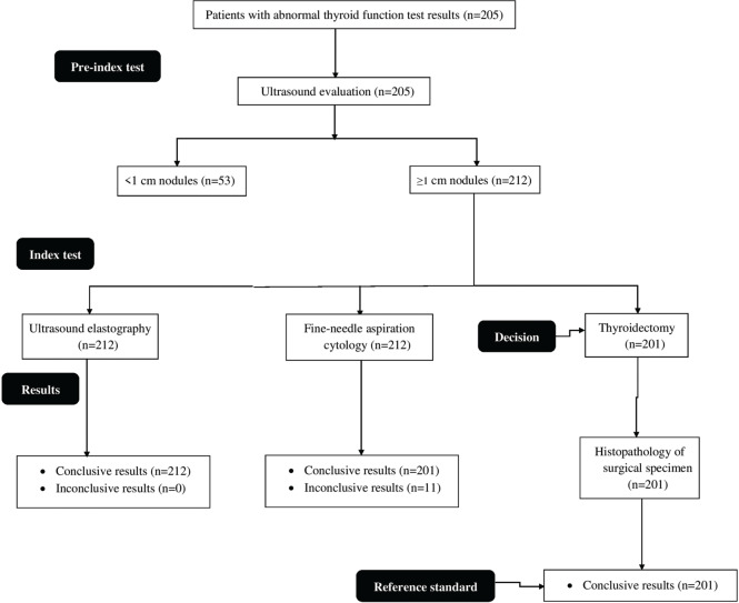
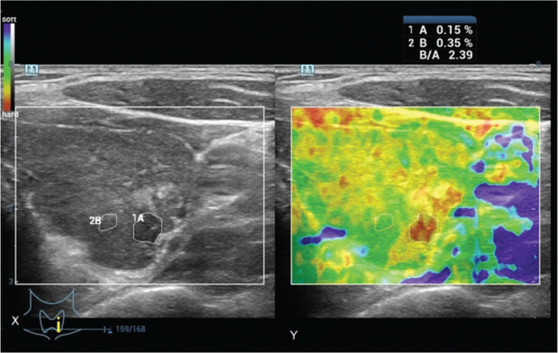
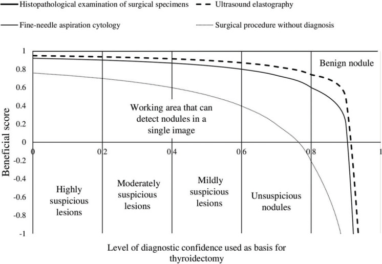
Methods: In total, 205 patients with abnormal thyroid function test results underwent ultrasound-guided fine-needle aspiration cytology on the basis of the American College of Radiology Thyroid Imaging-Reporting and Data System classification and strain ultrasound elastography according to the ASTERIA criteria. Histopathological examination of the surgical specimens was performed according to the 2017 World Health Organization classification system. Moreover, a beneficial score analysis for each modality was conducted.
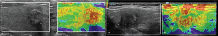
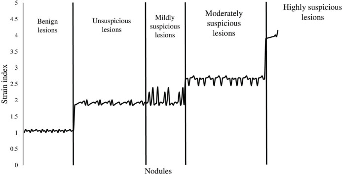
Results: Of 265 nodules, 212 measured ≥1 cm. The strain index value increased from benign to malignant nodules, and the presence of autoimmune thyroid diseases did not affect the results (p>0.05 for all categories). The sensitivities of histopathological examination, ultrasound elastography, and fine-needle aspiration cytology for detection of nodules measuring ≥1 cm were 1, 1, and 0.97, respectively. The working area for detecting nodule(s) in a single image was similar between strain ultrasound elastography and fine-needle aspiration cytology for highly and moderately suspicious nodules. However, for mildly suspicious, unsuspicious, and benign nodules, the working area for detecting nodule(s) in a single image was higher in strain ultrasound elastography than in fine-needle aspiration cytology.
Conclusion: Strain ultrasound elastography for highly and moderately suspicious nodules facilitated the detection of mildly suspicious, unsuspicious, and benign nodules.
Conflict of interest statement
No potential conflict of interest was reported.
Figures
Similar articles
-
Real-time ultrasound elastography in the diagnosis of newly identified thyroid nodules in adults: the ElaTION RCT.Health Technol Assess. 2024 Aug;28(46):1-51. doi: 10.3310/PLEQ4874. Health Technol Assess. 2024. PMID: 39252469 Free PMC article. Clinical Trial.
-
Comparison the accuracy of thyroid sono-elastography vs. ultrasound-guided fine needle aspiration cytology with thyroid malignancy diagnosis histopathology.Endocr Regul. 2024 Jun 11;58(1):129-137. doi: 10.2478/enr-2024-0014. Print 2024 Jan 1. Endocr Regul. 2024. PMID: 38861538
-
The values of shear wave elastography in avoiding repeat fine-needle aspiration for thyroid nodules with nondiagnostic and undetermined cytology.Clin Endocrinol (Oxf). 2019 Jul;91(1):201-208. doi: 10.1111/cen.13992. Epub 2019 May 2. Clin Endocrinol (Oxf). 2019. PMID: 31004514
-
Qualitative elastography can replace thyroid nodule fine-needle aspiration in patients with soft thyroid nodules. A systematic review and meta-analysis.Eur J Radiol. 2015 Apr;84(4):652-61. doi: 10.1016/j.ejrad.2015.01.003. Epub 2015 Jan 16. Eur J Radiol. 2015. PMID: 25638577
-
Application of shear wave elastography in the management of thyroid nodules in children and adolescents: our experience and a review of the literature.Front Endocrinol (Lausanne). 2024 Nov 20;15:1486285. doi: 10.3389/fendo.2024.1486285. eCollection 2024. Front Endocrinol (Lausanne). 2024. PMID: 39634183 Free PMC article. Review.
Cited by
-
Preoperative neoadjuvant chemotherapy in patients with breast cancer evaluated using strain ultrasonic elastography.World J Clin Cases. 2022 Jul 26;10(21):7293-7301. doi: 10.12998/wjcc.v10.i21.7293. World J Clin Cases. 2022. PMID: 36158032 Free PMC article.
-
Suspected Malignant Thyroid Nodules in Children and Adolescents According to Ultrasound Elastography and Ultrasound-Based Risk Stratification Systems-Experience from One Center.J Clin Med. 2022 Mar 23;11(7):1768. doi: 10.3390/jcm11071768. J Clin Med. 2022. PMID: 35407376 Free PMC article.
-
The prevalence and associated predictors for Bethesda III-VI for reporting thyroid cytopathology in Royal Commission Hospital, Kingdom of Saudi Arabia.Ther Adv Endocrinol Metab. 2022 Sep 12;13:20420188221122486. doi: 10.1177/20420188221122486. eCollection 2022. Ther Adv Endocrinol Metab. 2022. PMID: 36111207 Free PMC article.
-
Associations of Real-Time Ultrasound and Strain and Shear Wave Elastography with Gastrointestinal Organs: A Systematic Review.Diagnostics (Basel). 2023 Oct 25;13(21):3302. doi: 10.3390/diagnostics13213302. Diagnostics (Basel). 2023. PMID: 37958199 Free PMC article. Review.
-
The role of elastography in determining the risk of malignant thyroid nodules in children.Front Endocrinol (Lausanne). 2024 Nov 15;15:1461031. doi: 10.3389/fendo.2024.1461031. eCollection 2024. Front Endocrinol (Lausanne). 2024. PMID: 39619324 Free PMC article.
References
-
- Haugen BR, Alexander EK, Bible KC, Doherty GM, Mandel SJ, Nikiforov YE, et al. 2015 American Thyroid Association Management Guidelines for Adult Patients with Thyroid Nodules and Differentiated Thyroid Cancer: The American Thyroid Association Guidelines Task Force on Thyroid Nodules and Differentiated Thyroid Cancer. Thyroid. 2016;26((1)):1–133. doi: 10.1089/thy.2015.0020. - DOI - PMC - PubMed
Publication types
MeSH terms
Substances
LinkOut - more resources
Full Text Sources
Medical
