. 2020 Nov;14(6):688-699.
doi: 10.1111/irv.12783. Epub 2020 Jul 12.

SARS-CoV-2 environmental contamination associated with persistently infected COVID-19 patients

Affiliations

SARS-CoV-2 environmental contamination associated with persistently infected COVID-19 patients

Hui Lei et al. Influenza Other Respir Viruses. 2020 Nov.

Abstract

Background: Severe COVID-19 patients typically test positive for SARS-CoV-2 RNA for extended periods of time, even after recovery from severe disease. Due to the timeframe involved, these patients may have developed humoral immunity to SARS-CoV-2 while still testing positive for viral RNA in swabs. Data are lacking on exposure risks in these situations. Here, we studied SARS-CoV-2 environmental contamination in an ICU and an isolation ward caring for such COVID-19 patients.

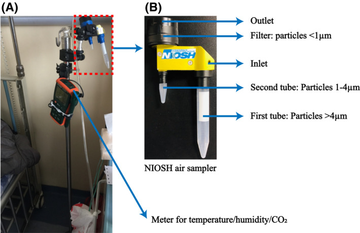
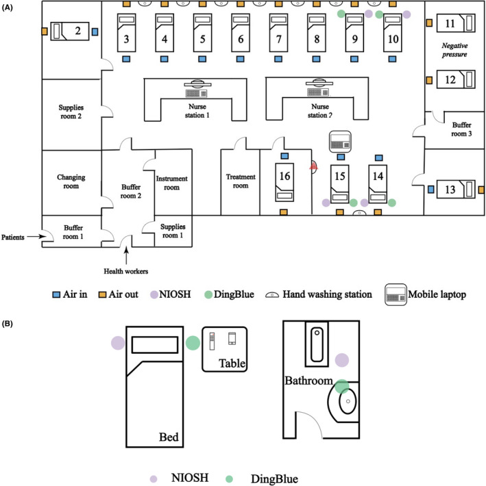
Methods: We collected air and surface samples in a hospital caring for critical and severe COVID-19 cases from common areas and areas proximal to patients.

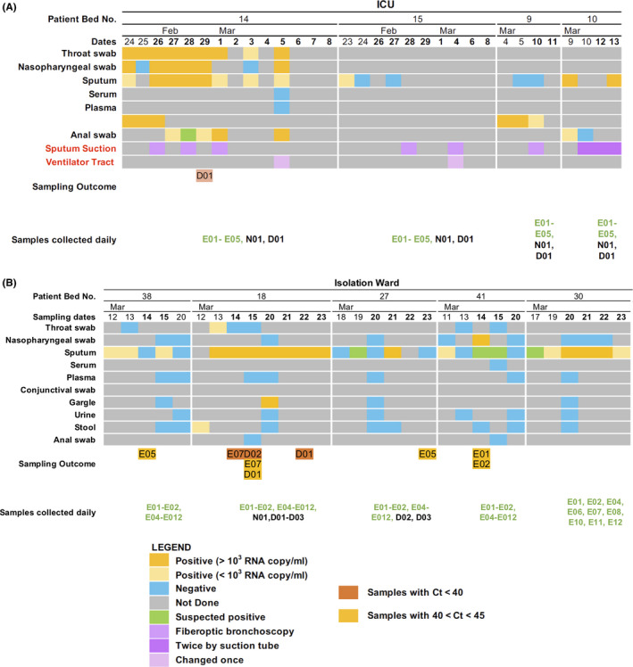
Results: Of the 218 ICU samples, an air sample contained SARS-CoV-2 RNA. Of the 182 isolation ward samples, nine contained SARS-CoV-2 RNA. These were collected from a facemask, the floor, mobile phones, and the air in the patient room and bathroom. Serum antibodies against SARS-CoV-2 were detected in these patients at the beginning of the study.

Conclusions: While there is a perception of increased risk in the ICU, our study demonstrates that isolation wards may pose greater risks to healthcare workers and exposure risks remain with clinically improved patients, weeks after their initial diagnoses. As these patients had serum antibodies, further studies may be warranted to study the utility of serum antibodies as a surrogate of viral clearance in allowing people to return to work. We recommend continued vigilance even with patients who appear to have recovered from COVID-19.

Keywords: COVID-19; SARS-CoV-2; coronavirus; intensive care unit; transmission.


Figures

Figure 1
Figure 1
Arrangement of the NIOSH cyclonic bioaerosol sampler. A, Arrangement of the NIOSH sampler on a tripod next to the head of the patient's bed with the temperature, humidity, and carbon dioxide monitor used in this study. B, NIOSH sampler showing collection tubes and collection filter and the particle sizes collected in each
Figure 2
Figure 2
Summary of viral loads in patient swabs, clinical care procedures performed during sampling, and outcomes of environmental sampling. Timeline shows samples collected from patients and testing results for SARS‐CoV‐2 RNA by quantitative real‐time PCR in the (A) intensive care unit and (B) in the isolation ward. Environmental samples that tested positive for SARS‐CoV‐2 RNA by quantitative real‐time PCR are also shown
Figure 3
Figure 3
Layouts of hospital rooms. Samples were collected in the intensive care unit (ICU) (A) and in an isolation ward (B). In the ICU, airflow originated in the ceiling above the foot of each patient bed and was extracted through vents in the wall at bed height. Placement of the NIOSH and DingBlue air samplers and bed numbers are shown. The red triangle indicates the handwashing station that was sampled. In the isolation ward, each patient was isolated in different rooms with their own bathrooms. The locations of the NIOSH and DingBlue air samplers are shown in their positions relative to the patient's bed, toilet, and bathroom sink. Diagrams are not to scale. Refer to Table 1 for a list of air and environmental samples collected

References

    1. Guo ZD, Wang ZY, Zhang SF, et al. Aerosol and surface distribution of severe acute respiratory syndrome coronavirus 2 in hospital wards, Wuhan, China, 2020. Emerg Infect Dis. 2020;26(7):1583‐1591. - PMC - PubMed
    1. Ong SWX, Tan YK, Chia PY, et al. Air, surface environmental, and personal protective equipment contamination by severe acute respiratory syndrome coronavirus 2 (SARS‐CoV‐2) from a symptomatic patient. JAMA. 2020;323(16):1610. - PMC - PubMed
    1. Ou X, Liu Y, Lei X, et al. Characterization of spike glycoprotein of SARS‐CoV‐2 on virus entry and its immune cross‐reactivity with SARS‐CoV. Nat Commun. 2020;11(1):1620. - PMC - PubMed
    1. Tang AN, Tong Z‐D, Wang H‐L, et al. Detection of novel coronavirus by RT‐PCR in stool specimen from asymptomatic child, China. Emerg Infect Dis. 2020;26(6).1337–1339. - PMC - PubMed
    1. To K‐W, Tsang O‐Y, Yip C‐Y, et al. Consistent detection of 2019 novel coronavirus in saliva. Clin Infect Dis. 2020. Epub ahead of print. - PMC - PubMed

Publication types

MeSH terms