. 2020 Jan-Dec:29:963689720934418.
doi: 10.1177/0963689720934418.

miR-26b-5p/TCF-4 Controls the Adipogenic Differentiation of Human Adipose-derived Mesenchymal Stem Cells

Affiliations

miR-26b-5p/TCF-4 Controls the Adipogenic Differentiation of Human Adipose-derived Mesenchymal Stem Cells

Yadong Luo et al. Cell Transplant. 2020 Jan-Dec.

Abstract

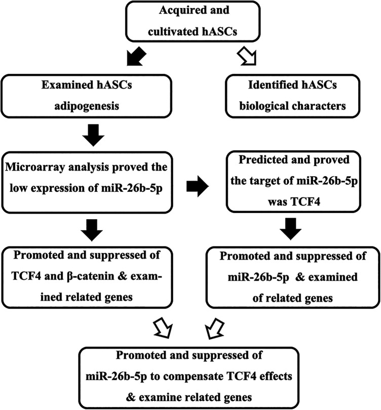
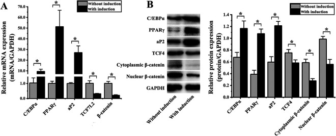
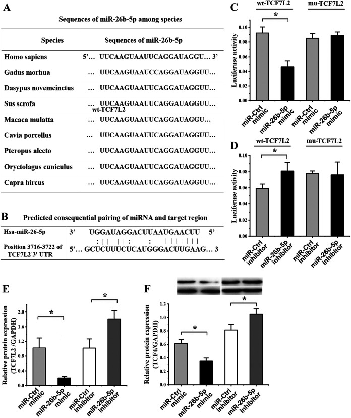
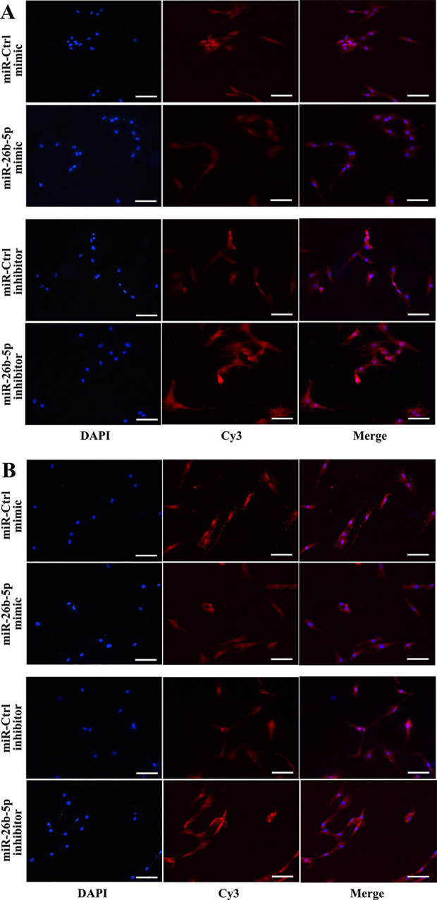
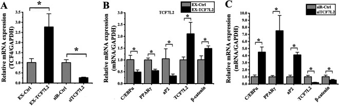
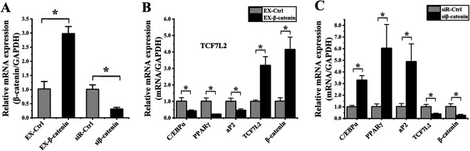
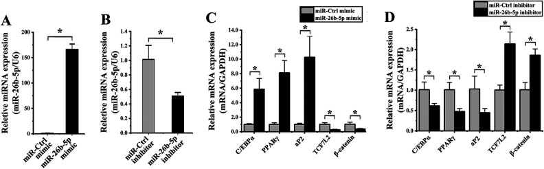
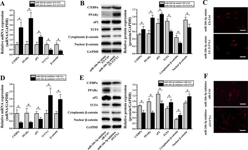
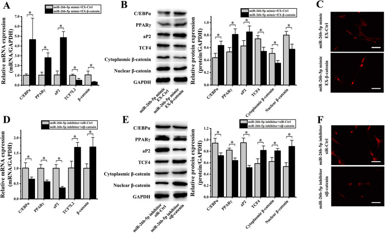
In this study, we assessed the ability of miR-26b-5p to regulate T cell factor 4 (TCF-4) expression and thereby control human adipose-derived mesenchymal stem cell (hADMSC) adipogenic differentiation. Adipogenic medium was used to induce hADMSC differentiation over a 6-d period. The ability of miR-26b-5p to interact with the TCF-4 mRNA was confirmed through both predictive bioinformatics analyses and luciferase reporter assays. Immunofluorescent staining was used to visualize the impact of miR-26b-5p inhibition or overexpression on TCF-4 and β-catenin levels in hADMSCs. Further functional analyses were conducted by transfecting these cells with siRNAs specific for TCF-4 and β-catenin. Adipogenic marker and Wnt/β-catenin pathway gene expression levels were assessed via real-time polymerase chain reaction and western blotting. β-catenin localization was assessed via immunofluorescent staining. As expected, our adipogenic media induced the adipocytic differentiation of hADMSCs. In addition, we confirmed that TCF-4 is an miR-26b-5p target gene in these cells, and that protein levels of both TCF-4 and β-catenin were reduced when these cells were transfected with miR-26b-5p mimics. Overexpression of this microRNA also enhanced hADMSC adipogenesis, whereas TCF-4 and β-catenin overexpression inhibited this process. The enhanced hADMSC adipogenic differentiation that was observed following TCF-4 or β-catenin knockdown was partially reversed when miR-26b-5p expression was inhibited. We found that miR-26b-5p serves as a direct negative regulator of TCF-4 expression within hADMSCs, leading to inactivation of the Wnt/β-catenin pathway and thereby promoting the adipogenic differentiation of these cells in vitro.

Keywords: TCF-4; Wnt/β-catenin pathway; adipogenic differentiation; hADMSCs; miRNA-26b-5p.


Conflict of interest statement

Declaration of Conflicting Interests: The author(s) declared no potential conflicts of interest with respect to the research, authorship, and/or publication of this article.

Figures

Figure 1.
Figure 1.
Study overview.
Figure 2.
Figure 2.
Assessment of hADMSC adipogenic differentiation (A) quantitative polymerase chain reaction confirmed that the expression of C/EBPα, PPARγ, and aP2 was significantly increased in hADMSCs following 4 d of adipogenic differentiation, whereas the expression of TCF7L2 and β-catenin was significantly reduced. (B) Western blotting confirmed that the protein level expression of C/EBPα, PPARγ, and aP2 was significantly increased in hADMSCs following adipogenic differentiation, whereas TCF-4, cytoplasmic β-catenin, and nuclear β-catenin levels were decreased at this same time point. *P < 0.05. GAPDH: glyceraldehyde 3-phosphate dehydrogenase; hADMSC: human adipose-derived mesenchymal stem cell; PPARγ: peroxisome proliferator-activated receptor γ; TCF-4: T cell factor 4.
Figure 3.
Figure 3.
TCF7L2 is an miR-26b-5p target gene. (A) Aligned miR-26b-5p sequences from different species. (B) Sequence complementarity between miR-26b-5p and the TCF7L2 3′-UTR. (C) A luciferase reporter assay was used to confirm interactions between miR-26b-5p and TCF7L2, revealing that miR-26b-5p mimic transfection significantly reduced the activity of the WT but not the MUT reporter construct relative to cells transfected with a control. (D) A second luciferase reporter assay revealed that miR-26b-5p inhibitor transfection was associated with a significant increase in the activity of the WT but not the MUT TCF7L2 3′-UTR reporter construct. (E) The expression of TCF7L2 was assessed by quantitative polymerase chain reaction in hADMSCs following miR-26b-5p mimic or inhibitor transfection, resulting in significant decreases and increases in this expression, respectively, relative to cells transfected with control constructs. (F) TCF-4 protein levels were assessed by western blotting in hADMSCs transfected with miR-26b-5p mimics and inhibitors, revealing significant decreases and increases, respectively, in these protein levels relative to cells transfected with control constructs. GAPDH: glyceraldehyde 3-phosphate dehydrogenase; hADMSC: human adipose-derived mesenchymal stem cell; TCF-4: T cell factor 4; UTR: untranslated region; WT: wild type.
Figure 4.
Figure 4.
miR-26b-5p reduces intracellular TCF-4 protein levels in hADMSCs. (A) Intracellular TCF-4 levels, as assessed by immunofluorescent microscopy, were reduced in hADMSCs following miR-26b-5p mimic transfection. (B) Intracellular TCF-4 levels were increased in hADMSCs following miR-26b-5p inhibitor transfection (scale bar = 100 μm). hADMSC: human adipose-derived mesenchymal stem cell; TCF-4: T cell factor 4.
Figure 5.
Figure 5.
TCF-4 influences hADMSC adipogenic differentiation. (A) The expression of TCF7L2 was assessed by qPCR in cells transfected with the EX-TCF7L2 or siTCF7L2 vectors, resulting in significant increases and decreases in the expression of this gene, respectively, relative to cells transfected with control constructs. (B) Cells overexpressing TCF7L2 were evaluated via qPCR, revealing significant decreases in the expression of C/EBP, PPARγ, and aP2, and significant increases in the expression of TCF7L2 and β-catenin relative to cells transfected with the control construct. (C) Cells in which TCF7L2 had been knocked down were evaluated via qPCR, revealing significant increases in the expression of C/EBP, PPARγ, and aP2, and significant decreases in the expression of TCF7L2 and β-catenin relative to cells transfected with the control siRNA construct. *P < 0.05. GAPDH: glyceraldehyde 3-phosphate dehydrogenase; hADMSC: human adipose-derived mesenchymal stem cell; PPARγ: peroxisome proliferator-activated receptor γ; qPCR: quantitative polymerase chain reaction; TCF-4: T cell factor 4.
Figure 6.
Figure 6.
β-catenin influences hADMSC adipogenic differentiation. (A) The expression of β-catenin was assessed by qPCR in cells transfected with the EX-β-catenin or siβ-catenin vectors, resulting in significant increases and decreases in the expression of this gene, respectively, relative to cells transfected with control constructs. (B) Cells overexpressing β-catenin were evaluated via qPCR, revealing significant decreases in the expression of C/EBP, PPARγ, and aP2, and significant increases in the expression of TCF7L2 and β-catenin relative to cells transfected with the EX-Ctrl construct. (C) Cells in which β-catenin had been knocked down were evaluated via qPCR, revealing significant increases in the expression of C/EBP, PPARγ, and aP2, and significant decreases in the expression of TCF7L2 and β-catenin relative to cells transfected with the control siRNA construct. *P < 0.05. GAPDH: glyceraldehyde 3-phosphate dehydrogenase; hADMSC: human adipose-derived mesenchymal stem cell; PPARγ: peroxisome proliferator-activated receptor γ; qPCR: quantitative polymerase chain reaction.
Figure 7.
Figure 7.
miR-26b-5p alters hADMSC adipogenic differentiation. (A) When hADMSCs were transfected with an miR-26b-5p mimic, significant increases in miR-26b-5p expression were observed relative to cells transfected with an miR-Ctrl construct, whereas this expression was significantly reduced in cells transfected with an miR-26b-5p inhibitor relative to corresponding controls. (B) Cells transfected with an miR-26b-5p mimic were evaluated via qPCR, revealing significant increases in the expression of C/EBP, PPARγ, and aP2, and significant decreases in the expression of TCF7L2 and β-catenin relative to cells transfected with the mimic control construct. (C) Cells transfected with an miR-26b-5p inhibitor were evaluated via qPCR, revealing significant increases in the expression of C/EBP, PPARγ, and aP2, and significant decreases in the expression of TCF7L2 and β-catenin relative to cells transfected with the inhibitor control construct. *P < 0.05. GAPDH: glyceraldehyde 3-phosphate dehydrogenase; hADMSC: human adipose-derived mesenchymal stem cell; PPARγ: peroxisome proliferator-activated receptor γ; qPCR: quantitative polymerase chain reaction.
Figure 8.
Figure 8.
miR-26b-5p regulates TCF-4 so as to influence hADMSCs’ adipogenic differentiation. (A) qPCR revealed that when hADMSCs were co-transfected with an miR-26b-5p mimic and EX-TCF7L2, they exhibited significant increases in the expression of C/EBP, PPARγ, and aP2, and significant decreases in the expression of TCF7L2 and β-catenin relative to cells co-transfected with miR-26b-5p and EX-Ctrl constructs. (B) Western blotting revealed that when hADMSCs were co-transfected with an miR-26b-5p mimic and EX-TCF7L2, they exhibited significant increases in C/EBP, PPARγ, and aP2 levels, and significant decreases in levels of TCF-4, cytoplasmic β-catenin, and nuclear β-catenin. (C) Nuclear β-catenin staining intensity was reduced in cells transfected with an miR-26b-5p mimic and EX-TCF7L2. (D) qPCR revealed that when hADMSCs were co-transfected with an miR-26b-5p inhibitor and siTCF7L2 they exhibited significant decreases in the expression of C/EBP, PPARγ, and aP2, and significant increases in the expression of TCF7L2 and β-catenin relative to cells co-transfected with an miR-26b-5p inhibitor and siR-Ctrl constructs. (E) Western blotting revealed that when hADMSCs were co-transfected with an miR-26b-5p inhibitor and siTCF7L2 they exhibited significant decreases in C/EBP, PPARγ, and aP2 levels, as well as significant increases in levels of TCF-4, cytoplasmic β-catenin, and nuclear β-catenin. (F) hADMSCs transfected with miR-26b-5p inhibitor and siTCF7L2 exhibited significant decreases nuclear β-catenin fluorescence intensity. *P < 0.05. GAPDH: glyceraldehyde 3-phosphate dehydrogenase; hADMSC: human adipose-derived mesenchymal stem cell; PPARγ: peroxisome proliferator-activated receptor γ; qPCR: quantitative polymerase chain reaction; TCF-4: T cell factor 4.
Figure 9.
Figure 9.
β-catenin miR-26b-5p regulates TCF-4 so as to influence hADMSCs’ adipogenic differentiation. (A) qPCR revealed that when hADMSCs were co-transfected with an miR-26b-5p mimic and EX-β-catenin, they exhibited significant increases in the expression of C/EBP, PPARγ, and aP2, and significant decreases in the expression of TCF7L2 and β-catenin relative to cells co-transfected with miR-26b-5p and EX-Ctrl constructs. (B) Western blotting revealed that when hADMSCs were co-transfected with an miR-26b-5p mimic and EX-β-catenin, they exhibited significant increases in C/EBP, PPARγ, and aP2 levels, and significant decreases in levels of TCF-4, cytoplasmic β-catenin, and nuclear β-catenin. (C) Nuclear β-catenin staining intensity was reduced in cells transfected with an miR-26b-5p mimic and EX- β-catenin. (D) qPCR revealed that when hADMSCs were co-transfected with an miR-26b-5p inhibitor and siβ-catenin they exhibited significant decreases in the expression of C/EBP, PPARγ, and aP2, and significant increases in the expression of TCF7L2 and β-catenin relative to cells co-transfected with an miR-26b-5p inhibitor and siR-Ctrl constructs. (E) Western blotting revealed that when hADMSCs were co-transfected with an miR-26b-5p inhibitor and siβ-catenin they exhibited significant decreases in C/EBP, PPARγ, and aP2 levels, as well as significant increases in levels of TCF-4, cytoplasmic β-catenin, and nuclear β-catenin. (F) hADMSCs transfected with miR-26b-5p inhibitor and siβ-catenin exhibited significant decreases in nuclear β-catenin fluorescence intensity. *P < 0.05.GAPDH: glyceraldehyde 3-phosphate dehydrogenase; hADMSC: human adipose-derived mesenchymal stem cell; PPARγ: peroxisome proliferator-activated receptor γ; qPCR: quantitative polymerase chain reaction; TCF-4: T cell factor 4.

Similar articles

Cited by

References

    1. Bradford BD, Lee JW. Reconstruction of the forehead and scalp. Facial Plast Surg Clin North Am. 2019;27(1):85–94. - PubMed
    1. Saban Y. Rhinoplasty: Lessons from “errors”: from anatomy and experience to the concept of sequential primary rhinoplasty. HNO. 2018;66(1):15–25. - PubMed
    1. Kim RY, Sokoya M, Ducic Y, Williams F. Free-flap reconstruction of the mandible. Semin Plast Surg. 2019;33(1):46–53. - PMC - PubMed
    1. Stephan S, Reinisch J. Auricular reconstruction using porous polyethylene implant technique. Facial Plast Surg Clin North Am. 2018;26(1):69–85. - PubMed
    1. Denadai R, Raposo-Amaral CA, Raposo-Amaral CE. Fat grafting in managing craniofacial deformities. Plast Reconstr Surg. 2019;143(5):1447–1455. - PubMed

Publication types