. 2020 Aug 15;31(18):2048-2056.
doi: 10.1091/mbc.E19-10-0589. Epub 2020 Jun 24.

Nuclear body phase separation drives telomere clustering in ALT cancer cells

Affiliations

Nuclear body phase separation drives telomere clustering in ALT cancer cells

Huaiying Zhang et al. Mol Biol Cell. .

Abstract

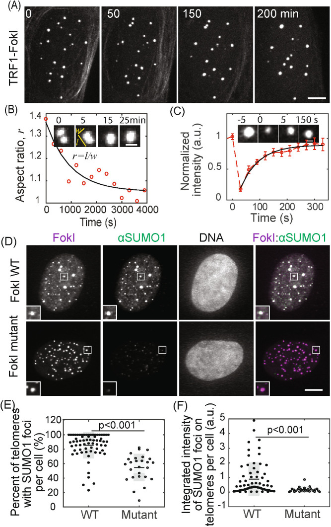
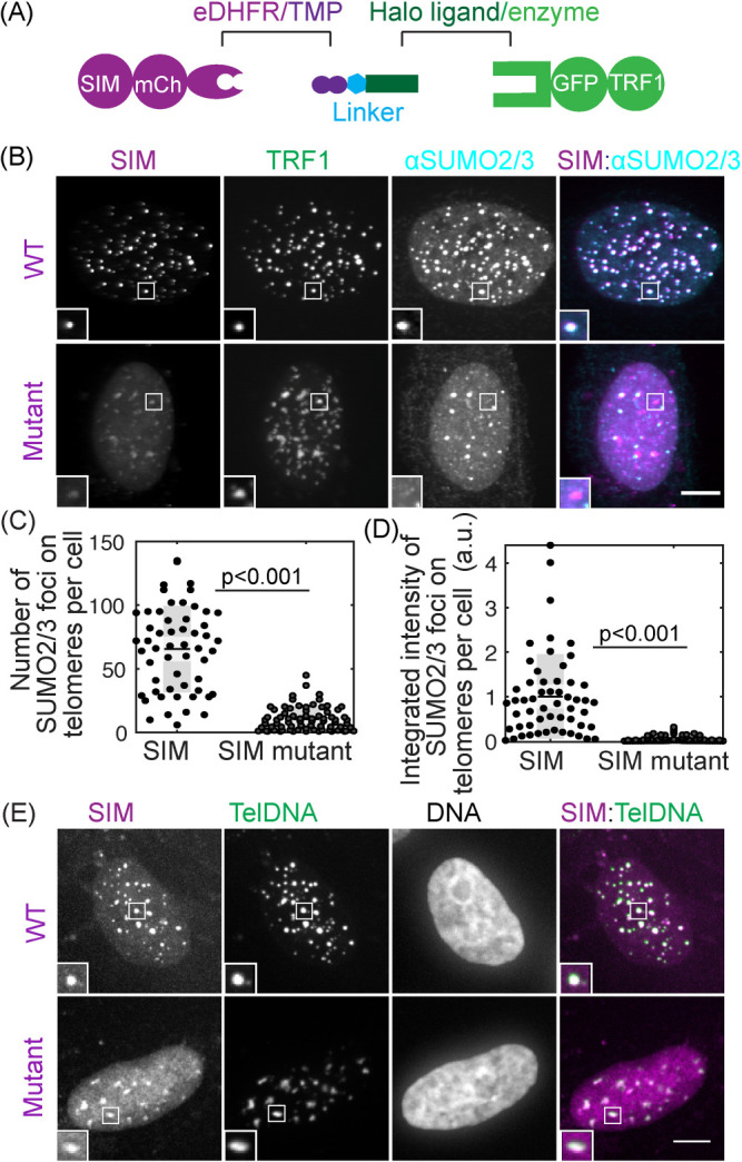
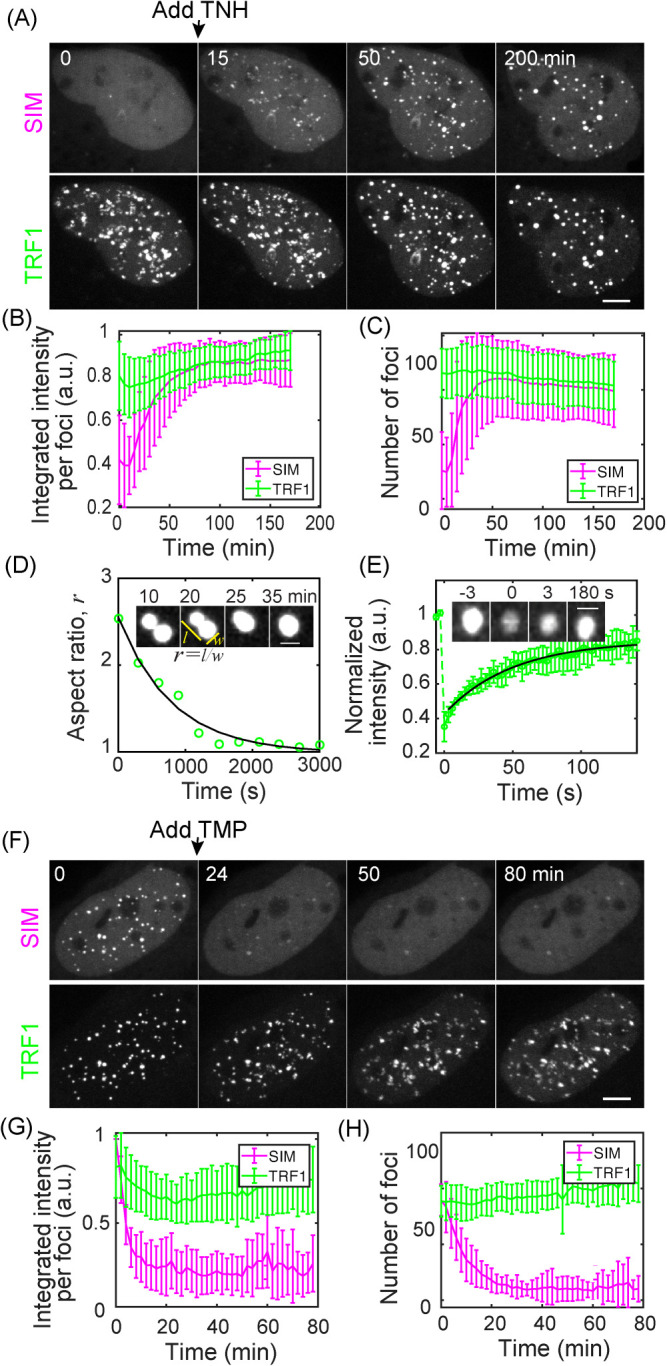
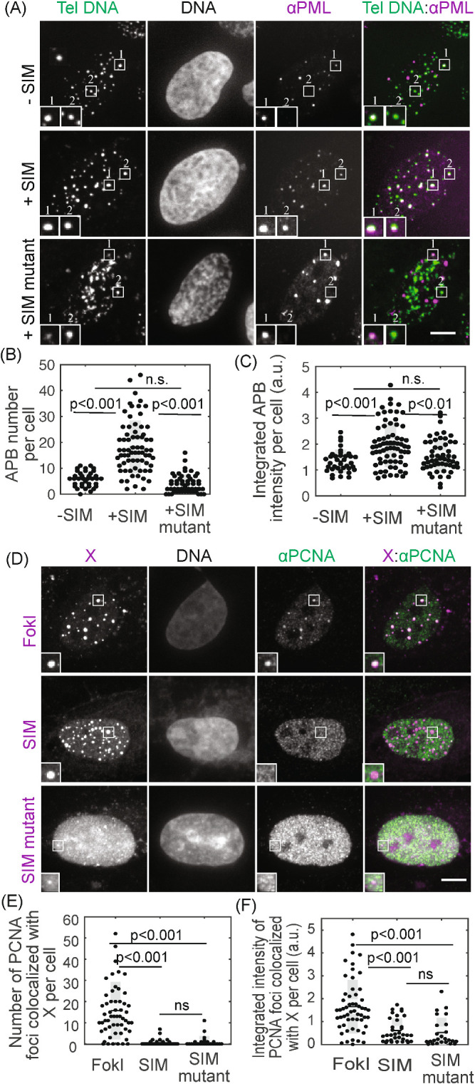
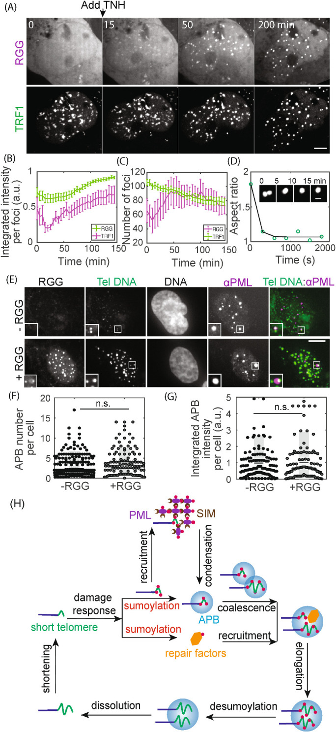
Telomerase-free cancer cells employ a recombination-based alternative lengthening of telomeres (ALT) pathway that depends on ALT-associated promyelocytic leukemia nuclear bodies (APBs), whose function is unclear. We find that APBs behave as liquid condensates in response to telomere DNA damage, suggesting two potential functions: condensation to enrich DNA repair factors and coalescence to cluster telomeres. To test these models, we developed a chemically induced dimerization approach to induce de novo APB condensation in live cells without DNA damage. We show that telomere-binding protein sumoylation nucleates APB condensation via interactions between small ubiquitin-like modifier (SUMO) and SUMO interaction motif (SIM), and that APB coalescence drives telomere clustering. The induced APBs lack DNA repair factors, indicating that APB functions in promoting telomere clustering can be uncoupled from enriching DNA repair factors. Indeed, telomere clustering relies only on liquid properties of the condensate, as an alternative condensation chemistry also induces clustering independent of sumoylation. Our findings introduce a chemical dimerization approach to manipulate phase separation and demonstrate how the material properties and chemical composition of APBs independently contribute to ALT, suggesting a general framework for how chromatin condensates promote cellular functions.


Figures

FIGURE 1:
FIGURE 1:
APBs exhibit liquid behavior and concentrate SUMO. APB formation was induced by creating DNA damage on telomeres with TRF1-FokI. (A, B) Cells were imaged live starting 1 h after triggering mCherry-TRF1-FokI import into the nucleus. Images show clustering of TRF1 foci (A) and fusion (B, insets), quantified by change in aspect ratio (defined as length/width) over time (exponential fit: 15 min half time). Time 0 is defined as the time point when two foci touch. (C) Fluorescence recovery after photobleaching (FRAP) of TRF1-FokI-mCherry, representing DNA damage-induced APBs. Insets shows a single TRF1 foci, intensity normalized to the first time point, exponential fit: 44 ± 17 s recovery half time from 14 events. Error bars STD. (D–F) SUMO1 IF for cells expressing TRF1-FokI or a nuclease-dead mutant. The overlay of FokI (purple) and SUMO1 (green) appears white (D, insets two times enlarged). Graphs show the percentage of telomeres with SUMO1 foci and the integrated intensity of SUMO1 foci on telomeres. Each data point represents one cell from two biological replicates, black lines mean, gray bars 95% confidence interval. Scale bars, 5 μm (A, D) or 1 μm (B, C). See also Figure 1–Supplemental Figure S1.
FIGURE 2:
FIGURE 2:
Recruiting SUMO to telomeres through SIM with a chemical dimerizer. (A) Dimerization schematic: SIM is fused to mCherry and eDHFR, and TRF1 is fused to Halo and GFP. The dimerizer is TNH: TMP-NVOC (6-nitroveratryl oxycarbonyl)-Halo (Zhang et al., 2017). (B–D) Cells expressing SIM-mCherry-DHFR (WT) or a SIM mutant that cannot interact with SUMO, together with Halo-GFP-TRF1, were incubated with TNH before fixing and staining for SUMO2/3. The overlay of SIM (purple) and SUMO2/3 (cyan) appears white (B, insets two times enlarged). Graphs show the number of telomeres with SUMO2/3 foci and the integrated intensity of SUMO2/3 foci on telomeres. Note that the integrated intensity of SUMO2/3 foci on telomeres in the SIM mutant is small compared with WT but not zero because of endogenous telomere sumoylation in U2OS cells. Each data represents one cell from two biological replicates, black lines mean, gray bars 95% confidence interval. (E) Telomere FISH images after recruiting SIM or SIM mutant to telomeres. The overlay of SIM (purple) and telomere DNA FISH (green) appears white. The dim SIM mutant foci on telomeres, relatively to the signal in the nucleoplasm, are due to the inability of the SIM mutant to enrich on telomeres through phase separation, combined with reduced protein fluorescent intensity in the FISH experiment. Scale bars, 5 μm. See also Figure 2–Supplemental Figure S1.
FIGURE 3:
FIGURE 3:
SUMO–SIM interactions drive liquid condensation and telomere clustering. (A–D) TNH was added to U2OS cells expressing SIM-mCherry-DHFR and Halo-GFP-TRF1 after the first time point to induce dimerization. Graphs show mean integrated intensity per TRF1 and SIM foci (B) and number of TRF1 and SIM foci (C) over time; 36 cells from four duplicates; error bars STD. P value between first and last time point for TRF1 foci intensity <0.001, SIM foci intensity <0.001, TRF1 foci number <0.03, SIM foci number <0.001. Insets (D) show an example of a fusion event, with the change in aspect ratio quantified (exponential fit, decay time 13 min). The time when two foci touch is defined as time 0. (E) FRAP of dimerization-induced condensates by bleaching TRF1. Intensity is normalized to the first time point, exponential fit: 35 ± 12 s recovery half time for 12 events. (F–H) After dimerization induced by TNH in U2OS cells expressing SIM-mCherry-DHFR and Halo-GFP-TRF1, TMP was added to release SIM from telomeres; 12 cells from two duplicates, error bars STD. P value between first and last time point for TRF1 foci intensity n.s., SIM foci intensity <0.001, TRF1 foci number <0.02, SIM foci number <0.001. Scale bars, 5 μm. See also Figure 3–Supplemental Figures S1 and S2.
FIGURE 4:
FIGURE 4:
Condensates contain APB scaffold components but not DNA repair factors. (A–C) FISH of telomere DNA and IF of PML for cells with or without SIM recruited to telomeres or with SIM mutant recruited to telomeres. The overlay of PML (purple) and telomere DNA (green) appears white (A, insets two times enlarged), indicating APBs with PML nuclear bodies localized to telomeres. Graphs show APB number and integrated APB intensity per cell. (D–F) Immunofluorescence of PCNA for cells with FokI-induced damage or with SIM or SIM mutant recruited. In representative images (D, insets two times enlarged), X indicates FokI, SIM, or SIM mutant, and colocalization with PCNA appears white in overlay images (right panels). Graphs show the number of PCNA foci colocalized with FokI, SIM, or SIM mutant and integrated intensity. Each data point (B, C, E, F) represents one cell from two biological replicates, black line mean, gray bar 95% confidence interval. Scale bars, 5 μm. See also Figure 4–Supplemental Figures S1–S3.
FIGURE 5:
FIGURE 5:
Non-APB condensation on telomeres drives telomere clustering. (A–D) TNH was added to cells expressing RGG-mCherry-RGG-eDHFR and Halo-GFP-TRF1 to induce dimerization and condensation. Graphs show integrated intensity per TRF1 and RGG foci (B) and the number of TRF1 and RGG foci (C) over time; 15 cells from two duplicates, error bars SEM. P value between first and last time point for TRF1 foci intensity <0.001, RGG foci intensity n.s., TRF1 foci number <0.001, RGG foci number n.s. Insets (D) show an example of a fusion event, with the change in aspect ratio quantified (exponential fit, decay time 6 min). (E–G) FISH of telomere DNA and IF of PML for cells with or without RGG recruitment. In representative images (E) the overlay of PML (purple) and telomere DNA (green) appears white, indicating APBs with PML nuclear bodies localized to telomeres. Insets (two times enlarged) show two telomere foci, one with an APB and one without, indicating the basal level of APBs in these cells. Graphs show APB number per cell and integrated APB intensity per cell. Each data point (F, G) represents one cell from two biological replicates, black line mean, gray bar 95% confidence interval. (H) Model for APB condensation and function. Telomere shortening (or replication stress) triggers a DNA damage response, where telomere sumoylation nucleates APB condensation and drives telomere clustering while another aspect of the damage response pathway recruits DNA repair factors to APB condensates. Together the clustered telomeres and enriched DNA repair factors within APBs lead to homology-directed telomere synthesis in ALT. Scale bars, 5 μm.

Similar articles

Cited by

References

    1. Altmeyer M, Neelsen KJ, Teloni F, Pozdnyakova I, Pellegrino S, Grøfte M, Druedahl Rask M-B, Streicher W, Jungmichel S, Nielsen ML, et al (2015). Liquid demixing of intrinsically disordered proteins is seeded by poly(ADP-ribose). Nat Commun , 8088. - PMC - PubMed
    1. Ballister ER, Aonbangkhen C, Mayo AM, Lampson MA, Chenoweth DM. (2014). Localized light-induced protein dimerization in living cells using a photocaged dimerizer. Nat Commun , 5475. - PMC - PubMed
    1. Banani SF, Lee HO, Hyman AA, Rosen MK. (2017). Biomolecular condensates: organizers of cellular biochemistry. Nat Rev Mol Cell Biol , 285–298. - PMC - PubMed
    1. Banani SF, Rice AM, Peeples WB, Lin Y, Jain S, Parker R, Rosen MK. (2016). Compositional control of phase-separated cellular bodies. Cell , 651–663. - PMC - PubMed
    1. Berry J, Weber SC, Vaidya N, Haataja M, Brangwynne CP. (2015). RNA transcription modulates phase transition-driven nuclear body assembly. Proc Natl Acad Sci USA , E5237–E5245. - PMC - PubMed

Publication types

MeSH terms

LinkOut - more resources