. 2020 Jul 21;31(16):1735-1743.
doi: 10.1091/mbc.E20-01-0049. Epub 2020 Jun 24.

Touch-induced mechanical strain in somatosensory neurons is independent of extracellular matrix mutations in Caenorhabditis elegans

Affiliations

Touch-induced mechanical strain in somatosensory neurons is independent of extracellular matrix mutations in Caenorhabditis elegans

Adam L Nekimken et al. Mol Biol Cell. .

Abstract

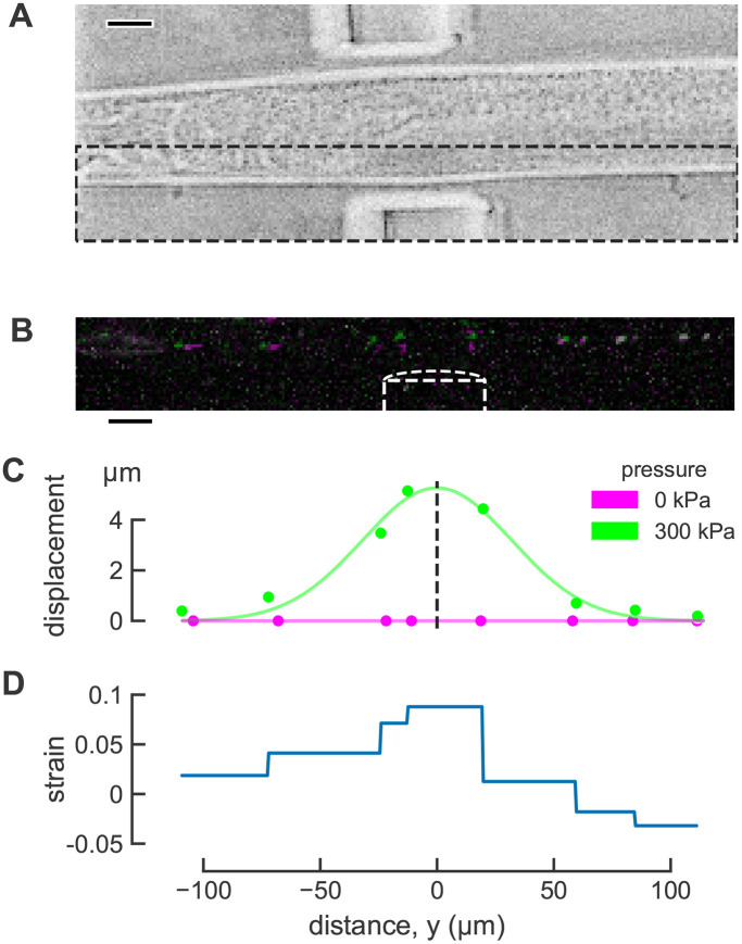
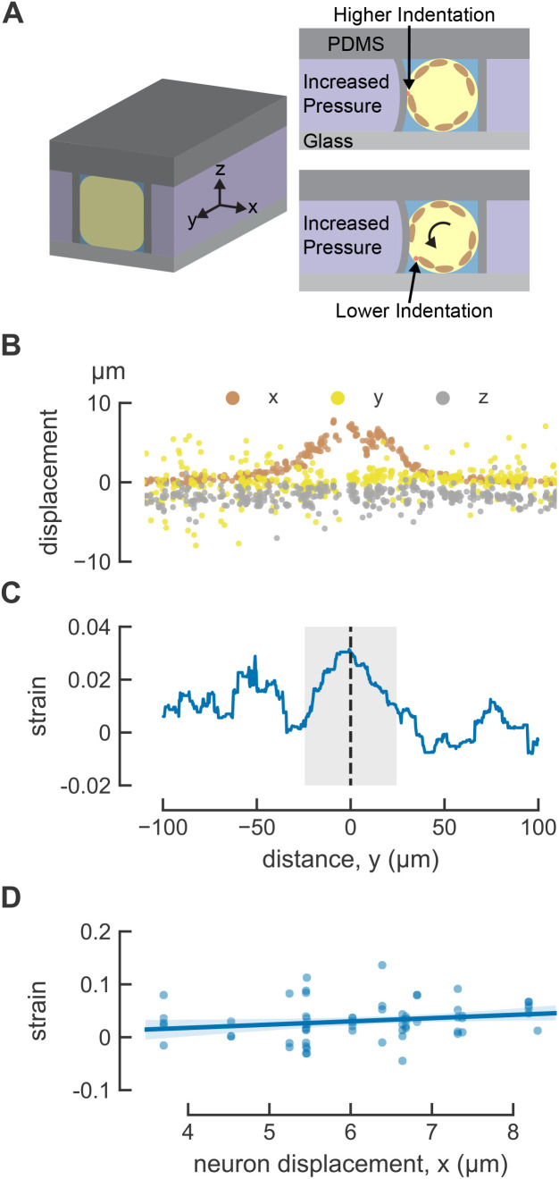
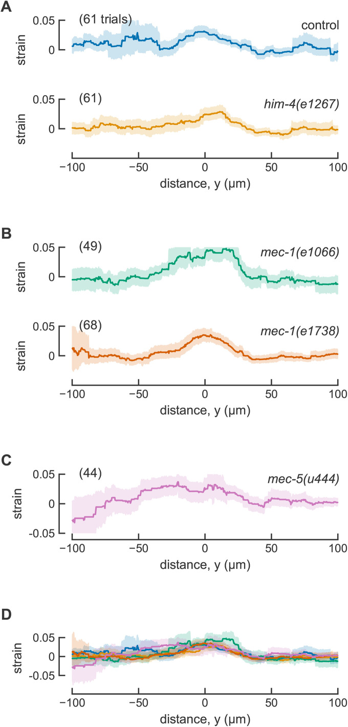
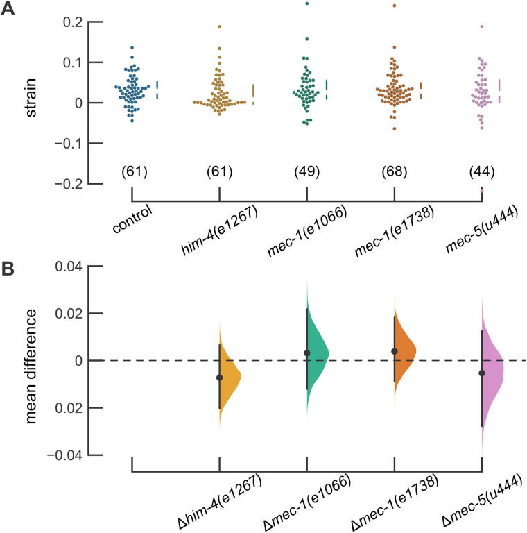
Cutaneous mechanosensory neurons are activated by mechanical loads applied to the skin, and these stimuli are proposed to generate mechanical strain within sensory neurons. Using a microfluidic device to deliver controlled stimuli to intact animals and large, immobile, and fluorescent protein-tagged mitochondria as fiducial markers in the touch receptor neurons (TRNs), we visualized and measured touch-induced mechanical strain in Caenorhabditis elegans worms. At steady state, touch stimuli sufficient to activate TRNs induce an average strain of 3.1% at the center of the actuator and this strain decays to near zero at the edges of the actuator. We also measured strain in animals carrying mutations affecting links between the extracellular matrix (ECM) and the TRNs but could not detect any differences in touch-induced mechanical strain between wild-type and mutant animals. Collectively, these results demonstrate that touching the skin induces local mechanical strain in intact animals and suggest that a fully intact ECM is not essential for transmitting mechanical strain from the skin to cutaneous mechanosensory neurons.


Figures

FIGURE 1:
FIGURE 1:
Touch-induced mechanical strain measured using mitochondria as fiducial markers in C. elegans TRNs in vivo. (A) Brightfield image of worm in the microfluidic device. Scale bar 20 μm. (B) Maximum projection image of TRN mitochondria before (magenta) and during (green) mechanical stimulus. Mitochondria that did not move appreciably appear white due to the overlap of magenta and green. Image intensity and contrast was adjusted to improve visualization. Scale bar 20 μm. (C) Displacement in the direction of actuation before (magenta) and during (green) stimulation. The smooth line (green) is a Gaussian fit used to infer the center of actuator. (D) Distribution of touch-induced strain in the TRN for a single actuation trial.
FIGURE 2:
FIGURE 2:
Indentation induces local longitudinal strain in C. elegans TRNs. (A) Three-dimensional diagram of the positioning of the worm in the microfluidics trap, the animal-centric coordinate system used to characterize strain, and the consequences of trapping animals in different orientations. TRN is red, and muscles are included in brown as visual aid. (B) Touch-induced displacement in three dimensions. (C) Touch-induced strain, anterior is to the left and posterior is to the right; y = 0 is the center of the actuator; n = 61 trials from N = 15 worms. (D) Mechanical strain at y = 0 as a function of maximum neuron displacement. The line is a linear fit to the data: ε = 0.0061x − 0.0067, where ε is the strain and x is the maximum displacement. The shaded area indicates 95% confidence intervals of the fit. R2 = 0.04.
FIGURE 3:
FIGURE 3:
Touch-induced mechanical strain profiles are similar in control animals and ECM mutants. (A) Spatially averaged strain in control ALM neurons with normal TRN attachment (15 animals) and him-4(e1267) with attachment defects (16 animals). The data for control animals are the same data as Figure 2. (B) Spatially averaged strain in mec-1 mutants. 14 independent animals for mec-1(1066) and 16 animals for mec-1(e1738). (C) Spatially averaged strain in mec-5(u444) mutants (17 animals). (D) Overlay of all spatially averaged strain profiles. Smooth lines are the averages across all trials and shaded areas show the error (95% confidence intervals). One ALM neuron was tested in each animal.
FIGURE 4:
FIGURE 4:
Strain at the center of the actuator is similar in control animals and ECM mutants. (A) Strain at the center of the actuator. Each point is the result of a single trial. The vertical lines next to the swarm plots indicate the median and quartiles of the data. Data were collected from 15, 16, 14, 16, and 17 animals (left to right). (B) Difference in the mean strain between control and each mutant with a bootstrapped resampled distribution of the data. Estimation plots generated using the DABEST plotting package (Ho et al., 2019).

Comment in

  • Editorial introduction.
    Yap AS. Yap AS. Mol Biol Cell. 2020 Jul 21;31(16):1651-1653. doi: 10.1091/mbc.E20-06-0414. Mol Biol Cell. 2020. PMID: 32692641 Free PMC article.

Similar articles

Cited by

References

    1. Allan DB, Caswell T, Keim NC, van der Wel CM. (2018). trackpy: Trackpy v0.4.1. 10.5281/ZENODO.1226458. - DOI
    1. Bewick GS, Banks RW. (2015). Mechanotransduction in the muscle spindle. Pflügers Arch Eur J Physiol , 175–190. - PMC - PubMed
    1. Boothe T, Hilbert L, Heide M, Berninger L, Huttner WB, Zaburdaev V, Vastenhouw NL, Myers EW, Drechsel DN, Rink JC. (2017). A tunable refractive index matching medium for live imaging cells, tissues and model organisms. Elife . 10.7554/eLife.27240. - DOI - PMC - PubMed
    1. Bounoutas A, O’Hagan R, Chalfie M. (2009). The multipurpose 15-protofilament microtubules in C. elegans have specific roles in mechanosensation. Curr Biol , 10.1016/j.cub.2009.06.036. - DOI - PMC - PubMed
    1. Chalfie M, Au M. (1989). Genetic control of differentiation of the Caenorhabditis elegans touch receptor neurons. Science , 1027–1033. - PubMed

Publication types

MeSH terms

Substances

LinkOut - more resources